Podcast
Questions and Answers
At what point does the cardiovascular system begin to beat after fertilization?
At what point does the cardiovascular system begin to beat after fertilization?
- 20 to 21 days
- 18 to 19 days
- 22 to 23 days (correct)
- 24 to 25 days
What are the first structures formed in the cardiovascular system during development?
What are the first structures formed in the cardiovascular system during development?
- Endocardial heart tubes (correct)
- Cardiogenic plate
- Heart muscles
- Angiogenic cell clusters
Which end of the endocardial heart tube is considered the venous end?
Which end of the endocardial heart tube is considered the venous end?
- Common atrium
- Bulbus cordis
- Sinus venosus (correct)
- Truncus arteriosus
What type of mesoderm is responsible for the development of the heart?
What type of mesoderm is responsible for the development of the heart?
What is the effect of lateral folding in embryo development?
What is the effect of lateral folding in embryo development?
During cardiac looping, which direction does the primitive ventricle move?
During cardiac looping, which direction does the primitive ventricle move?
Which structures are formed from the partitioning of the truncus arteriosus?
Which structures are formed from the partitioning of the truncus arteriosus?
What is the sequence of structures in the primitive heart tube?
What is the sequence of structures in the primitive heart tube?
What causes the enlargement of the right horn of the sinus venosus?
What causes the enlargement of the right horn of the sinus venosus?
Which structure becomes the coronary sinus?
Which structure becomes the coronary sinus?
What forms the smooth part of the right atrium?
What forms the smooth part of the right atrium?
What happens to the later stages of the left venous valves?
What happens to the later stages of the left venous valves?
Which structure is formed from the cranial part of the right valve?
Which structure is formed from the cranial part of the right valve?
What do both the umbilical and vitelline veins lose connection with?
What do both the umbilical and vitelline veins lose connection with?
What is the function of the venous valves at the sinu-atrial orifice?
What is the function of the venous valves at the sinu-atrial orifice?
What is formed by the left common cardinal vein?
What is formed by the left common cardinal vein?
What are the four heart malformations included in Tetralogy of Fallot?
What are the four heart malformations included in Tetralogy of Fallot?
What is the primary result of transposition of great arteries (TGA)?
What is the primary result of transposition of great arteries (TGA)?
Which arch contributes to the formation of the pulmonary arteries?
Which arch contributes to the formation of the pulmonary arteries?
What role does the ductus arteriosus play in fetal circulation?
What role does the ductus arteriosus play in fetal circulation?
What physiological change occurs when the ductus arteriosus closes at birth?
What physiological change occurs when the ductus arteriosus closes at birth?
Which artery arises from the fourth aortic arch on the left side?
Which artery arises from the fourth aortic arch on the left side?
How does fetal circulation differ from adult circulation in terms of blood flow?
How does fetal circulation differ from adult circulation in terms of blood flow?
What initiates the mixing of oxygenated blood with deoxygenated blood in fetal circulation?
What initiates the mixing of oxygenated blood with deoxygenated blood in fetal circulation?
What primarily contributes to the formation of the right ventricle?
What primarily contributes to the formation of the right ventricle?
What is the role of the aorticopulmonary septum?
What is the role of the aorticopulmonary septum?
Which sequence correctly outlines the steps in the formation of the ventricular septum?
Which sequence correctly outlines the steps in the formation of the ventricular septum?
What is an outcome of the absence of the membranous part of the interventricular septum?
What is an outcome of the absence of the membranous part of the interventricular septum?
In which week does the proliferation of mesenchymal cells appear to form the aorticopulmonary septum?
In which week does the proliferation of mesenchymal cells appear to form the aorticopulmonary septum?
Which part of the heart does the bulbus cordis primarily form?
Which part of the heart does the bulbus cordis primarily form?
What is a characteristic of Atrial Septal Defects (ASD)?
What is a characteristic of Atrial Septal Defects (ASD)?
Where does the conus arteriosus lead?
Where does the conus arteriosus lead?
What structure develops primarily from the left atrium during heart formation?
What structure develops primarily from the left atrium during heart formation?
What is the role of the septum primum during the development of the atrial septum?
What is the role of the septum primum during the development of the atrial septum?
Which of the following correctly describes the origin of the right atrium?
Which of the following correctly describes the origin of the right atrium?
What happens to the foramen primum just before it fully closes?
What happens to the foramen primum just before it fully closes?
Which of the following structures is formed from the fusion of the endocardial cushions during heart development?
Which of the following structures is formed from the fusion of the endocardial cushions during heart development?
What results from the absorption of the single pulmonary vein in the left atrium?
What results from the absorption of the single pulmonary vein in the left atrium?
What area of the heart does the bulbus cordis lie next to initially during development?
What area of the heart does the bulbus cordis lie next to initially during development?
What does the left sinus horn become during heart development?
What does the left sinus horn become during heart development?
What is the primary reason for bypassing the liver in fetal circulation?
What is the primary reason for bypassing the liver in fetal circulation?
How does blood shunt from the right atrium to the left atrium in a fetus?
How does blood shunt from the right atrium to the left atrium in a fetus?
What happens to the ductus arteriosus after birth?
What happens to the ductus arteriosus after birth?
What effect does increased pO2 have on the ductus arteriosus at birth?
What effect does increased pO2 have on the ductus arteriosus at birth?
Why is the foramen ovale able to close after birth?
Why is the foramen ovale able to close after birth?
What anatomical changes occur when the umbilical cord is cut?
What anatomical changes occur when the umbilical cord is cut?
What is the final state of the foramen ovale after it closes post-birth?
What is the final state of the foramen ovale after it closes post-birth?
Flashcards
What is the first functional organ to develop in the embryo?
What is the first functional organ to develop in the embryo?
The first functional organ to develop in the embryo. It arises from the cardiogenic area in the splanchnic mesoderm of the yolk sac, a location cranial to the developing mouth and nervous system and ventral to the pericardial sac.
What are cardiogenic fields?
What are cardiogenic fields?
Paired regions of mesoderm tissue located near the cranial end of the embryo, responsible for the formation of the heart.
What are endocardial heart tubes?
What are endocardial heart tubes?
Two hollow tubes that fuse together to form the single heart tube. They are derived from the cardiogenic fields and are formed by the canalization of angioplastic cords.
What is lateral folding in embryo development?
What is lateral folding in embryo development?
Signup and view all the flashcards
What is cephalocaudal folding in embryo development?
What is cephalocaudal folding in embryo development?
Signup and view all the flashcards
What is the primitive heart tube?
What is the primitive heart tube?
Signup and view all the flashcards
What is the sinus venosus?
What is the sinus venosus?
Signup and view all the flashcards
What is the truncus arteriosus?
What is the truncus arteriosus?
Signup and view all the flashcards
Sinus venosus
Sinus venosus
Signup and view all the flashcards
Right and left horns of the sinus venosus
Right and left horns of the sinus venosus
Signup and view all the flashcards
Veins draining into the sinus venosus
Veins draining into the sinus venosus
Signup and view all the flashcards
Sinu-atrial orifice
Sinu-atrial orifice
Signup and view all the flashcards
Atrial development
Atrial development
Signup and view all the flashcards
Septum spurium
Septum spurium
Signup and view all the flashcards
Fate of the right venous valve
Fate of the right venous valve
Signup and view all the flashcards
Fate of the common cardinal veins
Fate of the common cardinal veins
Signup and view all the flashcards
Right Atrium Development
Right Atrium Development
Signup and view all the flashcards
Sinus Horn Development
Sinus Horn Development
Signup and view all the flashcards
Left Atrium Development
Left Atrium Development
Signup and view all the flashcards
Pulmonary Vein Development
Pulmonary Vein Development
Signup and view all the flashcards
Atrioventricular Canal
Atrioventricular Canal
Signup and view all the flashcards
Septum Intermedium Division
Septum Intermedium Division
Signup and view all the flashcards
Septum Primum Development
Septum Primum Development
Signup and view all the flashcards
Septum Secundum Development
Septum Secundum Development
Signup and view all the flashcards
Ventricular Septation
Ventricular Septation
Signup and view all the flashcards
Primary Interventricular Septum
Primary Interventricular Septum
Signup and view all the flashcards
Primary Interventricular Foramen
Primary Interventricular Foramen
Signup and view all the flashcards
Membranous Interventricular Septum
Membranous Interventricular Septum
Signup and view all the flashcards
Conus Arteriosus (Infundibulum)
Conus Arteriosus (Infundibulum)
Signup and view all the flashcards
Aortic Vestibule
Aortic Vestibule
Signup and view all the flashcards
Truncus Arteriosus
Truncus Arteriosus
Signup and view all the flashcards
Aorticopulmonary Septum
Aorticopulmonary Septum
Signup and view all the flashcards
Tetralogy of Fallot
Tetralogy of Fallot
Signup and view all the flashcards
Transposition of Great Arteries (TGA)
Transposition of Great Arteries (TGA)
Signup and view all the flashcards
Formation of Pulmonary Arteries
Formation of Pulmonary Arteries
Signup and view all the flashcards
Formation of Aortic Arch and Right Subclavian
Formation of Aortic Arch and Right Subclavian
Signup and view all the flashcards
Fetal Blood Flow: Oxygenated Blood Entry
Fetal Blood Flow: Oxygenated Blood Entry
Signup and view all the flashcards
Fetal Circulation Overview
Fetal Circulation Overview
Signup and view all the flashcards
Fetal Circulation: Lung Bypass
Fetal Circulation: Lung Bypass
Signup and view all the flashcards
What is the role of the ductus venosus?
What is the role of the ductus venosus?
Signup and view all the flashcards
What is the role of the foramen ovale?
What is the role of the foramen ovale?
Signup and view all the flashcards
What is the role of the ductus arteriosus?
What is the role of the ductus arteriosus?
Signup and view all the flashcards
What happens to the ductus arteriosus after birth?
What happens to the ductus arteriosus after birth?
Signup and view all the flashcards
What happens to the foramen ovale after birth?
What happens to the foramen ovale after birth?
Signup and view all the flashcards
What happens to the ductus venosus after birth?
What happens to the ductus venosus after birth?
Signup and view all the flashcards
What are the primary triggers for changes in fetal circulation after birth?
What are the primary triggers for changes in fetal circulation after birth?
Signup and view all the flashcards
What is the overall function of fetal circulatory adaptations?
What is the overall function of fetal circulatory adaptations?
Signup and view all the flashcards
Study Notes
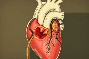
Early Development of the Cardiovascular System
- The cardiovascular system begins developing as two regions near the embryo's cranial end, derived from the mesoderm layer.
- The heart primordium is evident at 18 days.
- The heart is the first functional organ to develop, beating around 22-23 days from fertilization.
- Splanchnic mesoderm in the yolk sac develops the cardiogenic area, which is cranial to the mouth and nervous system.
- Angiogenic cells develop from the cardiogenic plate to form the right and left endocardial heart tubes.
- Lateral folding fuses the embryo's two lateral sides, bringing the cardiogenic fields to the midline for fusion into a primitive heart tube.
- Cephalocaudal folding brings cardiogenic fields from the cranial end to the thoracic region, where the heart will develop.
- The heart tube initially has five parts.
- Sinus venosus
- Truncus arteriosus
- Bulbus cordis
- Common ventricle
- Common atrium
- The endocardial tubes have two ends: the venous end connected to the sinus venosus and the arterial end connected to the truncus arteriosus.
Cardiac Looping
- The heart tube grows longer than the pericardial sac, causing it to loop.
- The primitive ventricle moves ventrally and to the right, while the primitive atrium moves dorsally and to the left.
- This re-orients the inflow and outflow portions of the heart (veins/atria and ventricles/arteries) to their mature positions and orientations.
Fate of Sinus Venosus
- The sinus venosus consists of a body and two horns (right and left).
- Each horn receives blood from the vitelline, umbilical, and common cardinal veins.
- The right horn expands, and the left horn regresses, due to a left-to-right blood shunt.
- The sinus venosus body is absorbed into the primitive atrium, forming part of the right atrium's smooth posterior wall.
- The septum spurium and left venous valves fuse with the interatrial septum.
- The left horn develops into the coronary sinus.
- Parts of the common cardinal veins become the superior or inferior vena cava.
Development of the Atria
- The atrioventricular canal divides into two halves by endocardial cushions.
- The primitive common atrium divides into right and left halves due to the development of the interatrial septum.
- The right horn of the sinus venosus and parts of the primitive atrium become the right atrium.
- The pulmonary veins are incorporated into the left atrium.
Development of the Ventricles
- The interventricular septum develops, dividing the common bulboventricular chamber into the right and left ventricles.
- The right ventricle primarily develops from the bulbus cordis and partly from the primitive ventricle.
- The left ventricle primarily forms from the primitive ventricle and partly from the bulbus cordis.
Partition of the Truncus Arteriosus
- The truncus arteriosus, along with the bulbus cordis, forms a single outflow tube from the heart.
- A spiral septum (aorticopulmonary septum) develops in the 5th week, dividing the truncus arteriosus into the aorta and pulmonary trunk on three different levels.
Major Cardiac Anomalies
- Atrial Septal Defects (ASD): Absence/incomplete development of the septum primum or septum secundum, or a large foramen ovale.
- Ventricular Septal Defects (VSD): Absence of the membranous part of the interventricular septum leading to a hole between the ventricles.
- Tetralogy of Fallot: Four cardiac abnormalities: VSD, pulmonary stenosis, overriding aorta, and right ventricular hypertrophy.
- Transposition of the Great Vessels (TGA): Malformed aorticopulmonary septum causing the right ventricle to connect to the aorta and the left ventricle to the pulmonary trunk.
Fetal Circulation
- Oxygenated blood from the placenta travels via the umbilical vein.
- Blood is shunted from the right atrium to the left atrium through the foramen ovale and through the ductus arteriosus to the aorta.
- The foramen ovale and ductus arteriosus close after birth.
- The ductus venosus, umbilical vein, and umbilical arteries transform into ligaments after birth.
Changes to Circulation After Birth
- Increased oxygen initiates closure of the foramen ovale and ductus arteriosus.
- Ductus arteriosus becomes the ligamentum arteriosum.
- Increased left atrial pressure closes the foramen ovale to the fossa ovalis.
- The umbilical vein and ductus venosus become the ligamentum teres and ligamentum venosum, respectively.
Studying That Suits You
Use AI to generate personalized quizzes and flashcards to suit your learning preferences.