Podcast
Questions and Answers
What do the left and right sides of aortic arch 4 develop into?
What do the left and right sides of aortic arch 4 develop into?
- Both sides form the arch of aorta
- Both sides form the proximal subclavian arteries
- Left forms part of the arch of aorta; right forms proximal right subclavian artery (correct)
- Left forms the arch of aorta; right forms the pulmonary trunk
What is the fate of aortic arch 5 during development?
What is the fate of aortic arch 5 during development?
- It persists and forms collateral circulation
- It forms completely and aids in the development of the pulmonary arteries
- It forms the distal part of the right subclavian artery
- It either never forms or regresses incompletely (correct)
Which of the following describes a patent ductus arteriosus (PDA)?
Which of the following describes a patent ductus arteriosus (PDA)?
- It is common in full-term infants only
- It can cause increased demand for cardiac output leading to heart failure (correct)
- A small shunt from aorta to pulmonary artery is always significant
- It is always symptomatic immediately after birth
What does the ductus arteriosus become after functional closure shortly after birth?
What does the ductus arteriosus become after functional closure shortly after birth?
Which structure develops from the right side of aortic arch 6?
Which structure develops from the right side of aortic arch 6?
What initiates the formation of the three germ layers during embryonic development?
What initiates the formation of the three germ layers during embryonic development?
Which part of the heart is primarily developed from the secondary heart field?
Which part of the heart is primarily developed from the secondary heart field?
At what approximate day does the heart begin beating and pumping blood in embryonic development?
At what approximate day does the heart begin beating and pumping blood in embryonic development?
Which germ layer gives rise to the heart and blood vessels?
Which germ layer gives rise to the heart and blood vessels?
What is the role of the splanchnic layer of the lateral plate mesoderm in cardiogenesis?
What is the role of the splanchnic layer of the lateral plate mesoderm in cardiogenesis?
What does the primary heart field primarily develop into?
What does the primary heart field primarily develop into?
Which structure is NOT formed from the endoderm?
Which structure is NOT formed from the endoderm?
What is the cardiogenic area known for during early embryogenesis?
What is the cardiogenic area known for during early embryogenesis?
How does the inner cell mass differentiate during early development?
How does the inner cell mass differentiate during early development?
What is the result of invagination during gastrulation?
What is the result of invagination during gastrulation?
What is formed by the proximal 1/3 of the bulbus cordis?
What is formed by the proximal 1/3 of the bulbus cordis?
Which structure is responsible for the formation of the smooth-walled portion of the right atrium?
Which structure is responsible for the formation of the smooth-walled portion of the right atrium?
What characterizes dextrocardia?
What characterizes dextrocardia?
What is the order of development for the primitive heart chambers?
What is the order of development for the primitive heart chambers?
Which part of the heart tube contributes to the outflow tracts of both ventricles?
Which part of the heart tube contributes to the outflow tracts of both ventricles?
What happens to the entrance of the sinus venosus during development?
What happens to the entrance of the sinus venosus during development?
What is the fate of the left horn of the sinus venosus?
What is the fate of the left horn of the sinus venosus?
What is formed by the truncus arteriosus?
What is formed by the truncus arteriosus?
On which day does the heart tube bend to form the cardiac loop?
On which day does the heart tube bend to form the cardiac loop?
What is indicated by situs inversus?
What is indicated by situs inversus?
What is the role of the crista terminalis in the right atrium?
What is the role of the crista terminalis in the right atrium?
During the development of the cardiac septa, which structures participate in the formation of the interatrial septum?
During the development of the cardiac septa, which structures participate in the formation of the interatrial septum?
What critical event allows blood to pass between the left and right atrium during fetal development?
What critical event allows blood to pass between the left and right atrium during fetal development?
What is patency of the foramen ovale, and why is it clinically significant?
What is patency of the foramen ovale, and why is it clinically significant?
What are the main vessels that arise through vasculogenesis during early blood vessel development?
What are the main vessels that arise through vasculogenesis during early blood vessel development?
What is the consequence of having a large ventricular septal defect (VSD)?
What is the consequence of having a large ventricular septal defect (VSD)?
Which heart condition is characterized by pulmonary stenosis and an interventricular septal defect?
Which heart condition is characterized by pulmonary stenosis and an interventricular septal defect?
At what stage does the interventricular septum form its membranous portion?
At what stage does the interventricular septum form its membranous portion?
What role do neural crest cells play in cardiac development?
What role do neural crest cells play in cardiac development?
What does the closure of the ostium primum signify in heart development?
What does the closure of the ostium primum signify in heart development?
What structures ultimately form the aorticopulmonary septum?
What structures ultimately form the aorticopulmonary septum?
What happens to the pulmonary vein as the left atrium expands?
What happens to the pulmonary vein as the left atrium expands?
How does the formation of the septum secundum differ from the septum primum?
How does the formation of the septum secundum differ from the septum primum?
What is the functional significance of the fossa ovalis in adults?
What is the functional significance of the fossa ovalis in adults?
What causes the recurrent laryngeal nerves to take their recurrent course?
What causes the recurrent laryngeal nerves to take their recurrent course?
What happens to blood entering the right atrium from the inferior vena cava during fetal development?
What happens to blood entering the right atrium from the inferior vena cava during fetal development?
What is the primary role of the ductus venosus in fetal circulation?
What is the primary role of the ductus venosus in fetal circulation?
What major change occurs in circulation at birth?
What major change occurs in circulation at birth?
What embryological layer contributes to the epithelium lining of the trachea and lungs?
What embryological layer contributes to the epithelium lining of the trachea and lungs?
What initiates the formation of the lung buds during embryological development?
What initiates the formation of the lung buds during embryological development?
What consequence does a tracheoesophageal defect typically have on swallowing?
What consequence does a tracheoesophageal defect typically have on swallowing?
What eventually forms the diaphragm after embryonic development?
What eventually forms the diaphragm after embryonic development?
How do the primary bronchi develop from the lung bud?
How do the primary bronchi develop from the lung bud?
Why do congenital diaphragmatic hernias occur?
Why do congenital diaphragmatic hernias occur?
Which structure serves as a bypass for placental circulation in the fetus?
Which structure serves as a bypass for placental circulation in the fetus?
What happens to the umbilical arteries after birth?
What happens to the umbilical arteries after birth?
During lung bud formation, what is the role of the transcription factor TBX4?
During lung bud formation, what is the role of the transcription factor TBX4?
What structure is formed from the left side of aortic arch 4?
What structure is formed from the left side of aortic arch 4?
What is the primary fate of aortic arch 5 during development?
What is the primary fate of aortic arch 5 during development?
What condition is characterized by an increased demand for cardiac output due to a shunt?
What condition is characterized by an increased demand for cardiac output due to a shunt?
Which nerve course is influenced by the descent of the heart and the regression of aortic arches?
Which nerve course is influenced by the descent of the heart and the regression of aortic arches?
What structure does the ductus arteriosus transform into following its functional closure?
What structure does the ductus arteriosus transform into following its functional closure?
What is the primary component that gives rise to the cardiovascular system in early embryogenesis?
What is the primary component that gives rise to the cardiovascular system in early embryogenesis?
What major change signifies the establishment of the three germ layers?
What major change signifies the establishment of the three germ layers?
During which week of development does the cardiovascular system begin to function?
During which week of development does the cardiovascular system begin to function?
What structures are developed primarily from the primary heart field?
What structures are developed primarily from the primary heart field?
The mesoderm gives rise to which of the following tissues?
The mesoderm gives rise to which of the following tissues?
At what stage does the heart tube begin to bend to form the cardiac loop?
At what stage does the heart tube begin to bend to form the cardiac loop?
Which layer ultimately forms the epithelium lining of the trachea?
Which layer ultimately forms the epithelium lining of the trachea?
What are the progenitor heart cells in early cardiogenesis initially adjacent to?
What are the progenitor heart cells in early cardiogenesis initially adjacent to?
What developmental structure contributes to the formation of the diaphragm?
What developmental structure contributes to the formation of the diaphragm?
Which embryonic structure directly influences the formation of blood cells?
Which embryonic structure directly influences the formation of blood cells?
What structure primarily develops from the bulbus cordis?
What structure primarily develops from the bulbus cordis?
Which part of the heart tube gives rise to the trabeculated parts of both the left and right atrium?
Which part of the heart tube gives rise to the trabeculated parts of both the left and right atrium?
What is the primary consequence of a heart looping to the left instead of the right?
What is the primary consequence of a heart looping to the left instead of the right?
What happens to the entrance of the sinus venosus during its development?
What happens to the entrance of the sinus venosus during its development?
Which embryonic day is marked by the completion of the U-shaped cardiac loop formation?
Which embryonic day is marked by the completion of the U-shaped cardiac loop formation?
What contributes to the smooth-walled portion of the right atrium?
What contributes to the smooth-walled portion of the right atrium?
Which of the following structures is formed by the proximal third of the bulbus cordis?
Which of the following structures is formed by the proximal third of the bulbus cordis?
What happens to the left horn of the sinus venosus as it develops?
What happens to the left horn of the sinus venosus as it develops?
Which structure is primarily responsible for forming the outflow tracts of both ventricles?
Which structure is primarily responsible for forming the outflow tracts of both ventricles?
What clinical condition may result from abnormal cardiac looping during development?
What clinical condition may result from abnormal cardiac looping during development?
What is the role of BMPs in cardiogenesis?
What is the role of BMPs in cardiogenesis?
What is the consequence of inhibiting WNT signaling during heart development?
What is the consequence of inhibiting WNT signaling during heart development?
How do BMP activity and WNT inhibition interact in heart formation?
How do BMP activity and WNT inhibition interact in heart formation?
What is the initial position of the cardiogenic region in relation to other embryonic structures?
What is the initial position of the cardiogenic region in relation to other embryonic structures?
What occurs when the lateral body folds move medially during heart development?
What occurs when the lateral body folds move medially during heart development?
What are the consequences of inhibited lengthening of the heart tube?
What are the consequences of inhibited lengthening of the heart tube?
Which structures produce inhibitors of WNT proteins necessary for heart development?
Which structures produce inhibitors of WNT proteins necessary for heart development?
What is the role of the SHF in heart tube elongation?
What is the role of the SHF in heart tube elongation?
What leads to the recurrent pathway of the recurrent laryngeal nerves during development?
What leads to the recurrent pathway of the recurrent laryngeal nerves during development?
Which of the following structures is formed from the distal part of the 6th aortic arch on the left side?
Which of the following structures is formed from the distal part of the 6th aortic arch on the left side?
How does oxygenated blood enter the left atrium during fetal circulation?
How does oxygenated blood enter the left atrium during fetal circulation?
What critical change occurs in the circulatory system at birth?
What critical change occurs in the circulatory system at birth?
What is the role of retinoic acid in the formation of lung buds?
What is the role of retinoic acid in the formation of lung buds?
What abnormality results from issues in partitioning the esophagus and trachea?
What abnormality results from issues in partitioning the esophagus and trachea?
What role do pleuroperitoneal membranes have during lung development?
What role do pleuroperitoneal membranes have during lung development?
Which structure in fetal circulation directs most blood from the pulmonary trunk to the aorta?
Which structure in fetal circulation directs most blood from the pulmonary trunk to the aorta?
What is a common consequence of congenital diaphragmatic hernias?
What is a common consequence of congenital diaphragmatic hernias?
What anatomical feature serves as a guide for the muscles of the diaphragm during development?
What anatomical feature serves as a guide for the muscles of the diaphragm during development?
What forms from the fusion of the tracheoesophageal ridges?
What forms from the fusion of the tracheoesophageal ridges?
What prevents the mixing of pulmonary and systemic blood after birth?
What prevents the mixing of pulmonary and systemic blood after birth?
What anatomical change occurs to the lung bud during development?
What anatomical change occurs to the lung bud during development?
How does blood from the placenta bypass the fetal liver?
How does blood from the placenta bypass the fetal liver?
Flashcards are hidden until you start studying
Study Notes
Cardiovascular Development
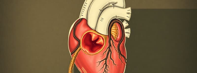
- The cardiovascular system is the first functional system to develop in the embryo.
- Heartbeat begins around day 21 and blood pumping begins around day 24-25.
- Progenitor heart cells originate in the epiblast and migrate through the primitive streak into the splanchnic layer of the lateral plate mesoderm.
- The cardiogenic area that develops contains the primary heart field (PHF), which forms part of the atria and the left ventricle.
- The secondary heart field (SHF), which forms the right ventricle, the outflow tract, and part of the atria.
Formation of Heart Tube and Cardiac Loop
- The heart tube formation results in a series of constrictions and dilations creating the sinus venosus, primitive atrium, primitive ventricle, and bulbus cordis.
- The bulbus cordis has three parts: the proximal third becomes the trabeculated part of the right ventricle, the middle part becomes the conus cordis, and the distal part becomes the truncus arteriosus.
- The cardiac loop forms as the heart tube grows and elongates.
- The cephalic portion bends ventrally, caudally, and to the right, while the atrial portion shifts dorsocranially and to the left.
Development of the Sinus Venosus
- The sinus venosus is the most caudal dilation at the venous end of the heart tube, with both a right and left horn.
- The entrance of the sinus venosus shifts to the right, leading to the left horn becoming the coronary sinus.
- The right sinus horn enlarges, becomes incorporated into the right atrium, and creates the smooth-walled part of the right atrium known as the sinus venarum.
Formation of Cardiac Septa
- Septation of the atrium, atrioventricular canal, and ventricle occurs between days 27 and 37, forming the four chambers of the heart.
- The septa form through two main methods: a) the growth of endocardial cushions, and b) differences in the rate of growth of different tissues.
Endocardial Cushions
- Endocardial cushions are swellings of connective tissue in the atrioventricular canal.
- They fuse and divide the AV canal into right and left canals, contributing to the formation of the mitral and tricuspid valves, interatrial septum, and membranous portion of the interventricular septum.
Septum Formation in the Atria
- The septum primum grows downward from the roof of the common atrium, leaving an opening known as the ostium primum.
- The closure of the ostium primum is followed by the formation of the ostium secundum and the septum secundum.
- The septum primum ultimately forms a “flaplike” valve for the foramen ovale, allowing right to left blood flow.
- After birth, the pressure gradient changes, closing the foramen ovale and leaving the fossa ovalis.
Formation of the Pulmonary Vein
- The pulmonary vein develops within the dorsal mesenchymal protrusion (DMP).
- It integrates into the posterior wall of the left atrium, resulting in four separate openings.
Interventricular Septum
- The primitive interventricular septum grows from the primitive ventricle.
- The interventricular foramen remains open until the end of the 7th week, when it is closed by the membranous portion of the interventricular septum.
Atrioventricular Valves
- The fusion of superior and inferior endocardial cushions divides the AV canal.
- Mesenchymal tissue around the AV canals proliferates and hollows out, forming the AV valves.
Septation of the Outflow Tracts
- Bulbar ridges in the conus cordis and truncus arteriosus fuse and spiral, forming the aorticopulmonary septum.
- The aorticopulmonary septum divides the outflow tracts into the pulmonary trunk and aorta.
Semilunar Valves
- The semilunar valves develop around the aortic and pulmonary orifices, forming three cusps.
Neural Crest Cells
- Neural crest cells migrate to the outflow region of the heart, contributing to the formation of endocardial cushions.
- Defects in neural crest cell migration can lead to abnormalities in both the heart and craniofacial development.
Ventricular Septal Defects (VSDs)
- VSDs are the most common type of congenital heart malformation, often resulting from a defect in the muscular region of the ventricular septum.
- Small defects may close spontaneously, while large defects can lead to left-to-right shunts, left ventricular dilation, and congestive heart failure.
Tetralogy of Fallot
- Tetralogy of Fallot is a conotruncal abnormality characterized by four defects: pulmonary stenosis, ventricular septal defect, overriding aorta, and right ventricular hypertrophy.
- It can lead to cyanosis due to insufficient oxygen supply to the tissues.
Formation of Blood Cells
- Blood cells arise from mesoderm.
- Blood islands form in the yolk sac mesoderm, providing the first blood cells.
- Definitive hematopoietic stem cells are derived from mesoderm surrounding the aorta, ultimately colonizing the bone marrow.
Vascular Development
- Vasculogenesis refers to the formation of vessels by coalescence of angioblasts.
- Angiogenesis refers to the sprouting of vessels from existing vessels.
- Blood islands along the primitive streak form paired vessels, leading to the dorsal aortae.
Aortic Arches
- Aortic arches develop from the aortic sac during the formation of pharyngeal arches.
- Each arch receives an artery and a cranial nerve.
- They terminate in the dorsal aortae, eventually fusing to form the descending aorta.
Aortic Arch Development
- Aortic arch 1 forms but mostly disappears, with a small portion becoming the maxillary artery.
- Aortic arch 2 forms and disappears, with remnants becoming the hyoid and stapedial arteries.
- Aortic arch 3 forms the common carotid artery, external carotid artery, and proximal internal carotid artery.
- Aortic arch 4 persists on both sides, forming the part of the aortic arch (left side), and the proximal part of the right subclavian artery (right side).
- Aortic arch 5 regresses.
- Aortic arch 6 becomes the proximal part of the right pulmonary artery and the left pulmonary artery and ductus arteriosus.
Ductus Arteriosus
- The ductus arteriosus functionally closes shortly after birth, forming the ligamentum arteriosum.
- Anatomical closure takes 1-3 months.
- A patent ductus arteriosus (PDA) can lead to a shunt from the aorta to the pulmonary artery, potentially leading to heart problems if severe.
Aortic Arches and Recurrent Laryngeal Nerve
- The descent of the heart and disappearance of portions of aortic arches leads to different courses of the right and left recurrent laryngeal nerves.
- The right recurrent laryngeal nerve loops beneath the right subclavian artery, while the left recurrent laryngeal nerve loops beneath the aortic arch.
Recurrent Laryngeal Nerve
- The recurrent laryngeal nerve hooks around the 6th aortic arch as the heart descends.
- On the right side, the 6th and 5th aortic arches disappear, causing the nerve to ascend and hook around the right subclavian artery.
- On the left side, the nerve does not move up because the distal part of the 6th aortic arch persists as the ductus arteriosus which later forms the ligamentum arteriosum.
Fetal Circulation
- Oxygenated blood from the placenta enters the fetus via the umbilical vein.
- Most of the blood bypasses the liver, passing through the ductus venosus directly into the inferior vena cava.
- Deoxygenated blood from the lower limbs mixes with the placental blood in the inferior vena cava.
- Blood bypasses the right ventricle as the lungs are not yet functioning, entering the left atrium via the foramen ovale.
- Blood from the superior vena cava enters the right atrium, passes to the right ventricle, and then into the pulmonary trunk.
- Most of this blood goes to the aorta via the ductus arteriosus.
- Partially oxygenated blood in the aorta returns to the placenta via the umbilical arteries that arise from the internal iliac arteries.
Postnatal Circulation
- At birth, pulmonary respiration begins, causing pulmonary blood pressure to fall.
- The foramen ovale and ductus arteriosus close, eliminating the fetal left-to-right shunts.
- Pulmonary and systemic circuits are now separate.
- The umbilical arteries (median umbilical ligaments), umbilical vein (round ligament of liver), and ductus venosus (ligamentum venosum) occlude.
- Blood to be metabolized now passes through the liver.
Respiratory System Development
Respiratory Development
- The internal lining of the larynx, trachea, bronchi, and lungs develop from endoderm.
- The cartilaginous, muscular, and connective tissue components of the trachea and lungs come from mesoderm.
Formation of Lung Buds
- The respiratory diverticulum (lung bud) appears as an outgrowth from the ventral wall of the foregut during the beginning of week 4.
- Increased retinoic acid, produced by adjacent mesoderm, influences the formation and location of the lung bud.
- Increased retinoic acid upregulates the transcription factor TBX4 in the gut tube endoderm at the site of the respiratory diverticulum.
- TBX4 is responsible for the formation and differentiation of the lungs.
Formation of Lung Buds
- The lung bud is initially in open communication with the foregut.
- Two longitudinal ridges, the tracheoesophageal ridges, separate the lung bud from the foregut.
- These ridges fuse to form the tracheoesophageal septum, dividing the foregut into dorsal (esophagus) and ventral (trachea) portions.
Tracheoesophageal Defects
- Abnormalities in the partitioning of the esophagus and trachea by the tracheoesophageal septum results in esophageal atresia with or without tracheoesophageal fistulas.
- In 90% of these defects, the upper esophagus ends in a blind pouch while the lower segment forms a fistula with the trachea.
- Swallowing is not possible and milk or saliva may spill out of the mouth or get inhaled into the lungs.
Trachea & Bronchi
- The lung bud forms the trachea and two lateral outpocketings (primary bronchial buds).
- These buds enlarge to form the right and left primary bronchi during the beginning of week 5.
- The right primary bronchus forms 3 secondary bronchi and the left primary bronchus forms 2.
Pericardioperitoneal Canals
- The lungs expand into the body cavity, growing in caudal and lateral directions.
- The pericardioperitoneal canals, which are located on each side of the foregut, serve as spaces for lung expansion.
Pericardioperitoneal Canals
- The pleural cavities communicate openly with the peritoneal (abdominal) cavity via the pericardioperitoneal canals
- The pleuroperitoneal folds partially close this communication by covering the septum transversum.
Pleuroperitoneal Membranes
- The pleuroperitoneal membranes form the central tendon of the diaphragm.
- The periphery develops into connective tissue acting as a scaffold for migrating myoblasts, which form the musculature of the diaphragm from cervical segments C3-C5.
- Innervation is via the phrenic nerves, which are derived from spinal nerves originating from C3-C5.
Diaphragmatic Hernias
- Congenital diaphragmatic hernias are a common malformation (1/2,000 births).
- Most occur due to a lack of muscle cell development in the pleuroperitoneal membranes, causing a weakened area and subsequent herniation of abdominal organs into the thoracic cavity.
Cardiovascular Development
- Cardiovascular system begins developing in the middle of the 3rd week of development due to the embryo's growing needs for nutrition.
- Progenitor heart cells originate in the epiblast.
- Primary Heart Field (PHF) forms the part of the atria and the left ventricle.
- Secondary Heart Field (SHF) forms the right ventricle, outflow tract (conus cordis and truncus arteriosus), and part of the atria.
- Bone Morphogenic Proteins (BMPs) 2 & 4, secreted by the endoderm and lateral plate mesoderm, upregulate the expression of fibroblast growth factor 8 (FGF8) which is important for the expression of cardiac-specific proteins.
- WNT proteins, secreted by the neural tube, inhibit heart development.
- CRESCENT and CERBERUS, inhibitors of WNT proteins, are produced by endoderm cells adjacent to the heart-forming mesoderm in the anterior half of the embryo.
- The combination of BMP activity and WNT inhibition leads to the expression of NKX2.5, the master gene for heart development.
- As the embryo grows and folds, the heart moves from the cervical region to the thorax.
- The right and left limbs of the first heart field fuse to form a single tube called the primary heart tube.
- The heart tube elongates as cells are added from the SHF to its cranial end.
- The heart tube bends on itself to form a U-shaped cardiac loop between days 23-28.
- Dextrocardia is a condition where the heart is located on the right side of the thorax instead of the left, caused by the heart looping to the left instead of the right.
- The sinus venosus is the most caudal dilation at the venous end of the heart tube, contributing to portions of the right atrium and coronary sinus.
- The bulbus cordis forms the trabeculated part of the right ventricle, the outflow tracts of both ventricles, and contributes to the pulmonary trunk and aorta.
Aortic Arches
- Aortic arch 3 forms the common carotid artery, external carotid artery, and proximal part of the internal carotid artery.
- Aortic arch 4 persists on both sides, forming part of the aortic arch on the left, and the proximal part of the right subclavian artery on the right.
- Aortic arch 5 either does not form or regresses.
- Aortic arch 6 develops into the proximal part of the right pulmonary artery on the right side and the left pulmonary artery and ductus arteriosus on the left side.
- The ductus arteriosus closes anatomically after birth, forming the ligamentum arteriosum.
- Patent ductus arteriosus (PDA) is a common abnormality of the great vessels, where the ductus arteriosus remains open.
- The descent of the heart and disappearance of various portions of aortic arches affects the course of the recurrent laryngeal nerves.
Fetal Circulation
- Oxygenated blood from the placenta enters the fetus via the umbilical vein.
- Most of the blood flows through the ductus venosus to the inferior vena cava, bypassing the liver.
- Blood bypasses the right ventricle through the foramen ovale to enter the left atrium.
- Blood from the superior vena cava enters the right ventricle and then to the pulmonary trunk, most of which enters the aorta through the ductus arteriosus.
- Partially oxygenated blood is then returned to the placenta via the umbilical arteries.
Postnatal Circulation
- With the start of pulmonary respiration, the blood pressure in the pulmonary trunk decreases.
- The foramen ovale and ductus arteriosus close, eliminating the fetal shunts.
- The umbilical arteries and vein, and the ductus venosus close, and blood now passes through the liver.
Respiratory System Development
- The respiratory diverticulum (lung bud) appears as an outgrowth from the ventral wall of the foregut.
- The formation of the lung bud is dependent on an increase in retinoic acid produced by adjacent mesoderm, which upregulates the expression of TBX4.
- The lung bud separates from the foregut, forming the trachea and two primary bronchial buds.
- The pericardioperitoneal canals are spaces on each side of the foregut that are filled by the expanding lungs.
- Pleuroperitoneal folds partially close the communication between the pleural and peritoneal cavities.
- The pleuroperitoneal membranes, formed by the pleuroperitoneal folds, fuse to cover the septum transversum.
- The central region of the pleuroperitoneal membranes forms the central tendon of the diaphragm.
- Myoblasts migrate from the cervical segments C3-C5 to form the musculature of the diaphragm, which is innervated by the phrenic nerves.
- Congenital diaphragmatic hernias occur when muscle cells fail to populate the pleuroperitoneal membranes, resulting in a weakened area and herniation of abdominal organs.
Studying That Suits You
Use AI to generate personalized quizzes and flashcards to suit your learning preferences.


