Podcast
Questions and Answers
What does the P wave on an ECG represent?
What does the P wave on an ECG represent?
- Atrial repolarization
- Atrial depolarization (correct)
- Ventricular depolarization
- Ventricular repolarization
Which component of the ECG is associated with ventricular contraction?
Which component of the ECG is associated with ventricular contraction?
- QRS complex (correct)
- P wave
- PR interval
- T wave
What is the function of the AV nodal delay represented in the ECG?
What is the function of the AV nodal delay represented in the ECG?
- Preventing atrial contraction
- Limiting blood flow to the aorta
- Speeding up heart rate
- Allowing time for ventricular filling (correct)
Tachycardia is characterized by which of the following?
Tachycardia is characterized by which of the following?
Which of the following does NOT contribute to the formation of the ECG?
Which of the following does NOT contribute to the formation of the ECG?
What cardiac event does the T wave on an ECG indicate?
What cardiac event does the T wave on an ECG indicate?
Which ECG abnormality is characterized by an irregular heart rhythm?
Which ECG abnormality is characterized by an irregular heart rhythm?
During diastole, how does the heart fill with blood?
During diastole, how does the heart fill with blood?
What occurs during diastole's active filling phase?
What occurs during diastole's active filling phase?
What initiates the period of isovolumic contraction?
What initiates the period of isovolumic contraction?
Which valves are closed during the period of ejection in systole?
Which valves are closed during the period of ejection in systole?
During which phase do the semilunar valves close?
During which phase do the semilunar valves close?
What is the primary characteristic of laminar flow during passive ventricular filling?
What is the primary characteristic of laminar flow during passive ventricular filling?
What happens to the AV valves during the phase of isovolumic contraction?
What happens to the AV valves during the phase of isovolumic contraction?
Which phase follows the period of ejection?
Which phase follows the period of ejection?
What is the state of the semilunar valves during ventricular filling?
What is the state of the semilunar valves during ventricular filling?
What happens to the AV valves at the onset of ventricular systole?
What happens to the AV valves at the onset of ventricular systole?
What occurs during passive ventricular filling?
What occurs during passive ventricular filling?
What is the state of the semilunar valves during isovolumic relaxation?
What is the state of the semilunar valves during isovolumic relaxation?
During which phase do the AV valves open to allow ventricular filling?
During which phase do the AV valves open to allow ventricular filling?
What primarily causes the closing of the semilunar valves during diastole?
What primarily causes the closing of the semilunar valves during diastole?
What is the effect of ventricular contraction on the AV valves?
What is the effect of ventricular contraction on the AV valves?
Which processes primarily take place during active ventricular filling?
Which processes primarily take place during active ventricular filling?
Which of the following best describes the ECG?
Which of the following best describes the ECG?
What does the QRS complex primarily represent in the ECG?
What does the QRS complex primarily represent in the ECG?
Which ECG abnormality is characterized by a fast heart rate?
Which ECG abnormality is characterized by a fast heart rate?
In the context of ECG, what does the P wave indicate?
In the context of ECG, what does the P wave indicate?
Which component of the cardiac cycle occurs after ventricular contraction?
Which component of the cardiac cycle occurs after ventricular contraction?
Which of the following leads is used to represent the difference between the left arm and the left leg?
Which of the following leads is used to represent the difference between the left arm and the left leg?
What is the main significance of the ST segment in an ECG?
What is the main significance of the ST segment in an ECG?
What occurs during the diastolic phase of the cardiac cycle?
What occurs during the diastolic phase of the cardiac cycle?
Flashcards
What does an ECG record?
What does an ECG record?
An ECG doesn't directly record electrical activity in the heart; it measures the impulses that reach the body surface.
What does the ECG show?
What does the ECG show?
The ECG shows changes in voltage detected between two points on the skin, not the actual electrical potential in the heart.
What does the P wave represent?
What does the P wave represent?
The P wave on an ECG represents the electrical signal that starts the contraction of the atria.
What does the QRS complex represent?
What does the QRS complex represent?
Signup and view all the flashcards
What does the T wave represent?
What does the T wave represent?
Signup and view all the flashcards
What is tachycardia?
What is tachycardia?
Signup and view all the flashcards
What is an extrasystole (premature beat)?
What is an extrasystole (premature beat)?
Signup and view all the flashcards
What is a complete heart block?
What is a complete heart block?
Signup and view all the flashcards
Diastole
Diastole
Signup and view all the flashcards
Systole
Systole
Signup and view all the flashcards
Passive Ventricular Filling
Passive Ventricular Filling
Signup and view all the flashcards
Active Ventricular Filling
Active Ventricular Filling
Signup and view all the flashcards
Atrioventricular (AV) Valves
Atrioventricular (AV) Valves
Signup and view all the flashcards
Semilunar Valves
Semilunar Valves
Signup and view all the flashcards
Isovolumic Contraction
Isovolumic Contraction
Signup and view all the flashcards
Ejection Phase
Ejection Phase
Signup and view all the flashcards
Isovolumic Relaxation
Isovolumic Relaxation
Signup and view all the flashcards
What does ECG show?
What does ECG show?
Signup and view all the flashcards
What is an extrasystole?
What is an extrasystole?
Signup and view all the flashcards
Study Notes
Cardiac Physiology 2
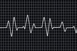
- Electrocardiogram (ECG): Records the electrical impulses that reach the body surface during cardiac activity, not a direct recording of the heart's electrical activity.
- ECG shows the overall cardiac activity during depolarization and repolarization.
- The recording is the comparison of voltages detected by electrodes on the skin, not actual potentials.
- ECG waves correlate to specific cardiac events:
- P wave: Represents atrial depolarization.
- QRS complex: Represents ventricular depolarization.
- T wave: Represents ventricular repolarization.
Limb Leads
- ECG uses electrodes placed on different parts of the body to measure the electrical activity.
- Limb leads measure the electrical potential differences between limbs (arms and legs).
- Lead I: Right arm to left arm
- aVR: Right arm
- aVL: Left arm
- Lead II: Right arm to left leg
- aVF: Left leg
- Lead III: Left arm to left leg
- A ground electrode is also used.
Chest Leads
- Chest leads (V1-V6) are placed on the chest wall to measure the electrical activity.
- The placement locations are numbered as seen in the diagrams.
Cardiac Arrhythmias
- Tachycardia: A rapid heart rate.
- Extrasystole (premature beat): An extra heartbeat that occurs before the next normal heartbeat.
- Ventricular fibrillation: Disorganized electrical activity in the ventricles.
- Complete heart block: A condition where the electrical signals from the atria do not reach the ventricles.
Cardiac Myopathies
- Myocardial infarction: A heart attack, where blood flow to the heart muscle is blocked.
- Characterized by specific ECG patterns.
Cardiac Cycle
- Diastole: The period of relaxation of the heart chambers, divided into different phases (Passive ventricular filling, and Active ventricular filling).
-
During Diastole, the AV valves open, allowing blood to passively fill the ventricles.
-
During Active ventricular filling, the AV valves remain closed and the atria contract to complete ventricular filling.
-
- Systole: The period of contraction of the heart chambers, which can be further divided into:
- Isovolumetric contraction: Ventricular contraction causes the AV valves to close, beginning ventricular systole. The semilunar valves remain closed during this period.
- Period of ejection: Continued ventricular contraction pushes blood out of the ventricles, causing the semilunar valves to open.
- Isovolumetric relaxation: Blood flowing back toward the relaxing ventricles closes the semilunar valves, marking the start of ventricular diastole. The AV valves also close during this period.
Heart Sounds
- Heart sounds are produced by the turbulent flow of blood through the heart valves.
- The first heart sound (S1) is caused by the closure of the AV valves.
- The second heart sound (S2) is caused by the closure of the semilunar valves.
- There are instances where no audible sound is produced during the various phases of the cardiac cycle.
Heart Valve Disorders and Murmurs
- Murmurs are abnormal heart sounds caused by turbulent blood flow through damaged or narrowed heart valves.
- Murmur patterns and timings with stethoscope correlate to heart valve disorders.
- Different types of murmurs (e.g., systolic, diastolic) and associated valve defects (stenotic, insufficient) are linked to specific murmur characteristics.
Studying That Suits You
Use AI to generate personalized quizzes and flashcards to suit your learning preferences.