Podcast
Questions and Answers
Why is the anterior surface of the heart more accessible for auscultation and palpation compared to the posterior surface?
Why is the anterior surface of the heart more accessible for auscultation and palpation compared to the posterior surface?
The anterior surface is more superficial and has fewer overlying structures, making it easier to feel the cardiac impulse and hear valve sounds.
During ventricular contraction, what is the role of the chordae tendineae and why is this function important?
During ventricular contraction, what is the role of the chordae tendineae and why is this function important?
The chordae tendineae prevent the atrioventricular valves from prolapsing into the atria, ensuring unidirectional blood flow.
If a patient has a prominent cardiac impulse that is palpated laterally to the typical location, what might this suggest?
If a patient has a prominent cardiac impulse that is palpated laterally to the typical location, what might this suggest?
It may suggest left ventricular enlargement (hypertrophy) or displacement of the heart.
Describe the structural difference between semilunar valves and atrioventricular valves and how this relates to their function.
Describe the structural difference between semilunar valves and atrioventricular valves and how this relates to their function.
In which intercostal space would you typically auscultate the aortic valve?
In which intercostal space would you typically auscultate the aortic valve?
If a patient has an enlarged right ventricle, which anatomical structure on the anterior surface of the heart would be most prominent during palpation?
If a patient has an enlarged right ventricle, which anatomical structure on the anterior surface of the heart would be most prominent during palpation?
Why would you want to avoid using the posterior region of the heart to auscultate for heart valve abnormalities?
Why would you want to avoid using the posterior region of the heart to auscultate for heart valve abnormalities?
A patient is diagnosed with mitral valve prolapse. Explain how this condition affects the valve's function and what anatomical structures are involved.
A patient is diagnosed with mitral valve prolapse. Explain how this condition affects the valve's function and what anatomical structures are involved.
A patient presents with a heart murmur best heard at the 2nd intercostal space along the left sternal border. Which heart valve is most likely affected?
A patient presents with a heart murmur best heard at the 2nd intercostal space along the left sternal border. Which heart valve is most likely affected?
Why is the fifth intercostal space at the midclavicular line the optimal location for palpating the point of maximal impulse (PMI)?
Why is the fifth intercostal space at the midclavicular line the optimal location for palpating the point of maximal impulse (PMI)?
During a physical exam, a doctor wants to listen specifically to the aortic valve. Where should the stethoscope be placed on the patient's chest?
During a physical exam, a doctor wants to listen specifically to the aortic valve. Where should the stethoscope be placed on the patient's chest?
Why is the Angle of Louis a clinically important landmark for locating the 2nd intercostal space?
Why is the Angle of Louis a clinically important landmark for locating the 2nd intercostal space?
A patient's chart indicates they have an enlarged left ventricle. How might this affect the location of the PMI, and where would you expect to palpate it?
A patient's chart indicates they have an enlarged left ventricle. How might this affect the location of the PMI, and where would you expect to palpate it?
During a physical exam, you need to auscultate the aortic valve. Describe the surface anatomy landmark you would use to locate the optimal auscultation point.
During a physical exam, you need to auscultate the aortic valve. Describe the surface anatomy landmark you would use to locate the optimal auscultation point.
A patient has a mass located in the 4th intercostal space along the midclavicular line. Which lobe of the lung is most likely directly deep to this mass?
A patient has a mass located in the 4th intercostal space along the midclavicular line. Which lobe of the lung is most likely directly deep to this mass?
When performing auscultation, you identify a murmur originating between the 4th and 5th intercostal spaces along the right sternal border. This sound is most indicative of which valve dysfunction, and why?
When performing auscultation, you identify a murmur originating between the 4th and 5th intercostal spaces along the right sternal border. This sound is most indicative of which valve dysfunction, and why?
A clinician notes that a patient's Point of Maximal Impulse (PMI) is difficult to palpate. What body habitus variations might make the PMI harder to detect and why?
A clinician notes that a patient's Point of Maximal Impulse (PMI) is difficult to palpate. What body habitus variations might make the PMI harder to detect and why?
Explain how knowledge of surface anatomy aids in the placement of electrodes for an electrocardiogram (ECG).
Explain how knowledge of surface anatomy aids in the placement of electrodes for an electrocardiogram (ECG).
What is the clinical significance of identifying the intercostal spaces during a cardiac exam, and how does this relate to accurate valve auscultation?
What is the clinical significance of identifying the intercostal spaces during a cardiac exam, and how does this relate to accurate valve auscultation?
List the structures that form the right border of the heart.
List the structures that form the right border of the heart.
How does understanding the surface anatomy of the heart aid in the diagnosis of specific valvular abnormalities during a physical examination?
How does understanding the surface anatomy of the heart aid in the diagnosis of specific valvular abnormalities during a physical examination?
List the structures that form the inferior surface of the heart.
List the structures that form the inferior surface of the heart.
During the cardiac cycle, where would you palpate for the point of maximal impulse (PMI) and what anatomical structure creates it?
During the cardiac cycle, where would you palpate for the point of maximal impulse (PMI) and what anatomical structure creates it?
Flashcards
Atrioventricular valves
Atrioventricular valves
Valves that control blood flow between atria and ventricles; larger and floppy.
Semilunar valves
Semilunar valves
Valves that allow blood flow from ventricles to arteries; smaller and tighter without chordae tendinae.
Chordae tendinae
Chordae tendinae
Fibrous cords that anchor atrioventricular valves to papillary muscles, preventing prolapse.
Papillary muscles
Papillary muscles
Signup and view all the flashcards
Point of Maximal Impulse (PMI)
Point of Maximal Impulse (PMI)
Signup and view all the flashcards
Anterior surface of heart
Anterior surface of heart
Signup and view all the flashcards
Posterior surface of heart
Posterior surface of heart
Signup and view all the flashcards
Valves' function
Valves' function
Signup and view all the flashcards
Base of the heart
Base of the heart
Signup and view all the flashcards
Auscultation location
Auscultation location
Signup and view all the flashcards
Aortic valve location
Aortic valve location
Signup and view all the flashcards
Left ventricle sound
Left ventricle sound
Signup and view all the flashcards
Left AV valve location
Left AV valve location
Signup and view all the flashcards
PMI palpation
PMI palpation
Signup and view all the flashcards
Pulmonic valve
Pulmonic valve
Signup and view all the flashcards
Aortic valve
Aortic valve
Signup and view all the flashcards
Superficial Anatomical Landmarks
Superficial Anatomical Landmarks
Signup and view all the flashcards
Angle of Louis
Angle of Louis
Signup and view all the flashcards
2nd Intercostal Space
2nd Intercostal Space
Signup and view all the flashcards
Deep Structures Localization
Deep Structures Localization
Signup and view all the flashcards
Right Border of the Heart
Right Border of the Heart
Signup and view all the flashcards
Inferior Surface of the Heart
Inferior Surface of the Heart
Signup and view all the flashcards
Left Border of the Heart
Left Border of the Heart
Signup and view all the flashcards
Palpation in Physical Exams
Palpation in Physical Exams
Signup and view all the flashcards
Study Notes
Physiology 1.03 - Clinical Physiology
- Clinical Physiology is the fundamental physiologic basis of vital signs.
- This course is part of BMS 100.
The Cardiac Exam
- Heart Anatomy: Includes surface anatomy, chambers, valves, and great vessels.
- Basic Cardiac Physiology: Covers the cardiac cycle and valvular movements.
- The Cardiac Physical Exam: Outlines surface anatomy, phases of the cardiac cycle, heart sounds, murmurs, and locations of major pulses.
The Heart Chambers - Anatomy Review
- Chambers: The locations of the left and right atria and ventricles are detailed, particularly the interventricular septum.
- Great Vessels: Locations of the pulmonary trunk, pulmonary arteries, aorta, superior/inferior vena cavae, and pulmonary veins are noted.
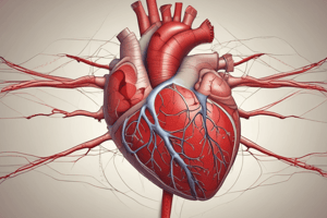
Basic Heart Anatomy
- A diagram illustrating the heart's structure and associated major vessels is presented.
The Heart - Valves
- Atrioventricular Valves: These valves, between the atria and ventricles, prevent backflow.
- Semilunar Valves: Located between the ventricles and great arteries, these also prevent backflow.
The Heart - Valves (detailed)
- Atrioventricular valves are larger and floppier, anchored by chordae tendineae.
- Semilunar valves are smaller and tighter, needing no chordae tendineae.
Basic Heart Anatomy (detailed)
- Additional structural details of the heart and associated vessels are illustrated in a diagram.
The Heart Chambers - Anatomy Review (detailed)
- Key anatomical locations of heart chambers and great vessels are reviewed. Questions relating the great vessels to connected chambers.
The Heart - Valves (detailed 2)
- Different types of heart valves and how they function are described. It emphasizes how they ensure one-way blood flow.
Basic Heart Anatomy - More Details
- Detailed anatomical structures of the heart are described. Areas for locating and examining are covered.
Anterior And Posterior Views Of The Heart
- Anterior and posterior views of the heart are visually presented, with labeled structures on both illustrations.
Anterior vs. Posterior Surfaces
- The anterior side of the heart is easier to examine and feel than the posterior.
- Key structures found on the anterior surface and why the PMI is an important location are elucidated.
Valves - The Superior Aspect
- Diagrams and detailed description of the superior aspect of the heart valves, highlighting different phases of the cardiac cycle.
Valves - The Superior Aspect (detailed)
- Detailed information regarding the superior aspect of heart valves during the cardiac cycle's various phases is provided. Focuses on the different phases.
The Heart - Surface Anatomy
- Description of how surface anatomy is used during physical exams to locate structures within the chest.
- Uses for physical exam maneuvers covered.
Bones Of The Thoracic Cage
- The location of various bones within the thoracic cage is described, specifically focusing on landmarks used to locate the heart during observation.
The Heart - Surface Anatomy (detailed)
- A diagram highlighting location of the heart in relation to important bones and structures in the thoracic cavity.
The Heart - Surface Anatomy (detailed 2)
- The heart's borders and what structures make them are detailed, focusing on the relationships to surrounding structures.
The Heart - Surface Anatomy (detailed 3)
- Provides a detailed overview of key areas for auscultation and palpation of the heart. It specifies regions associated with sounds produced by various heart valves.
The Heart - Surface Anatomy (more detailed)
- Explains preferred regions to hear the heart's specific sounds.
Another View - Surface Anatomy
- A diagram shows the anatomical locations of the valves in the chest, using colored areas to emphasize where the cardiac sounds will be heard best.
The Systemic Pressures And The Cardiac Cycle
- The tracing of pressures in the aorta, left ventricle, and left atrium is covered in relation to heart sounds.
Systemic Pressures
- A diagram illustrates the relationship between the cardiac cycle's various phases and pressure changes in the aorta, ventricles and atria.
Systemic Pressures – The Left Atrium
- Tracing of pressures within the left atrium illustrating relationships to heart valves and sounds.
Systemic Pressures – The Left Ventricle
- Detailed information about the relationships between pressure changes in the left ventricle and the operation of the heart's valves and their associated cardiac sounds.
Systemic Pressures – The Aorta
- A pressure tracing illustrating the relationship between the cardiac cycle, pressures in the aorta and its associated heart sounds.
Types of Heart Sounds
- Valve closing sounds are normal. AV valves produce a "Lub" sound (lower frequency), semilunar valves a "Dub" (higher frequency).
- Valve opening sounds ("opening snaps") are uncommon.
Types of Heart Sounds (detailed)
- Describes laminar and turbulent blood flow patterns and their relation to normal and abnormal heart sounds. Extra heart sounds and murmurs are considered.
Types Of Valvular Abnormalities
- Stenosis: The valve opening is narrowed, requiring greater pressure to move blood through.
- Regurgitation: The valve isn't closing completely, leading to backflow.
Types Of Valvular Abnormalities (detailed)
- These types of conditions affect the heart's valves' functions, potentially making them not close or open appropriately. The consequences of each in terms of heart sounds are explained.
Fill in following chart on the next slide
- Describes a method to determine causes of heart murmurs (an abnormal heart sound).
- Location of the murmur during ventricular systole or diastole helps identify the valve involved.
Location (detailed)
- A table correlating heart murmur locations with suspected valve abnormalities is provided.
Studying That Suits You
Use AI to generate personalized quizzes and flashcards to suit your learning preferences.
Related Documents
Description
An overview of cardiac physiology, covering heart anatomy including chambers, valves, and vessels. It also details the cardiac cycle, valvular movements, physical exam techniques, heart sounds, and pulse locations.