Podcast
Questions and Answers
Which type of bone is characterized as immature and irregular typically found in the fetus?
Which type of bone is characterized as immature and irregular typically found in the fetus?
- Lamellar bone
- Compact bone
- Spongy bone
- Woven bone (correct)
What is the role of osteoclasts in bone physiology?
What is the role of osteoclasts in bone physiology?
- To provide tensile strength to bones
- To form new bone through mineralization
- To resorb bone tissue (correct)
- To produce osteoid
Which structure in the bone is responsible for housing blood vessels and nerves?
Which structure in the bone is responsible for housing blood vessels and nerves?
- Lacunae
- Epiphyseal line
- Volkmann’s canal (correct)
- Osteon
Which statement best describes the process of ossification?
Which statement best describes the process of ossification?
Which type of bone is known for its highly organized structure and is chiefly involved in providing strength?
Which type of bone is known for its highly organized structure and is chiefly involved in providing strength?
Which histopathological feature is characterized by central fibrinoid necrosis surrounded by palisading macrophages?
Which histopathological feature is characterized by central fibrinoid necrosis surrounded by palisading macrophages?
What is a common extra-articular manifestation of rheumatoid arthritis?
What is a common extra-articular manifestation of rheumatoid arthritis?
Which feature is NOT typically associated with rheumatoid arthritis?
Which feature is NOT typically associated with rheumatoid arthritis?
What is the minimum number of clinical features required for a diagnosis of rheumatoid arthritis?
What is the minimum number of clinical features required for a diagnosis of rheumatoid arthritis?
Which of the following is commonly found within the proliferative synovitis seen in rheumatoid arthritis?
Which of the following is commonly found within the proliferative synovitis seen in rheumatoid arthritis?
What is the most common type of osteoporosis?
What is the most common type of osteoporosis?
Which of the following is a characteristic of osteomalacia?
Which of the following is a characteristic of osteomalacia?
What dietary intake is associated with the prevention of primary osteoporosis?
What dietary intake is associated with the prevention of primary osteoporosis?
What effect does corticosteroid use have on bone health?
What effect does corticosteroid use have on bone health?
What role does parathyroid hormone play in hyperparathyroidism?
What role does parathyroid hormone play in hyperparathyroidism?
Which of the following conditions can lead to secondary osteoporosis?
Which of the following conditions can lead to secondary osteoporosis?
What is a primary cause of osteoarthritis?
What is a primary cause of osteoarthritis?
Which of the following is a complication associated with osteomyelitis?
Which of the following is a complication associated with osteomyelitis?
What is a common sign of osteoporosis in vertebrae?
What is a common sign of osteoporosis in vertebrae?
What is a characteristic feature of rickets seen in children?
What is a characteristic feature of rickets seen in children?
Which joints are most commonly affected by osteoarthritis?
Which joints are most commonly affected by osteoarthritis?
What type of organisms are primarily responsible for osteomyelitis?
What type of organisms are primarily responsible for osteomyelitis?
What is a potential consequence of chronic osteomyelitis?
What is a potential consequence of chronic osteomyelitis?
What is the most common clinical feature of arthritis?
What is the most common clinical feature of arthritis?
Which of the following factors is NOT associated with secondary osteoarthritis?
Which of the following factors is NOT associated with secondary osteoarthritis?
What structural change is characteristic of osteoarthritis?
What structural change is characteristic of osteoarthritis?
What condition is characterized by the deposition of calcium pyrophosphate crystals in cartilage and juxta-articular tissues?
What condition is characterized by the deposition of calcium pyrophosphate crystals in cartilage and juxta-articular tissues?
Which type of CPPD is specifically associated with elderly men and presents as pseudo gout?
Which type of CPPD is specifically associated with elderly men and presents as pseudo gout?
What is the primary genetic inheritance pattern associated with Duchenne muscular dystrophy?
What is the primary genetic inheritance pattern associated with Duchenne muscular dystrophy?
Which symptom is commonly observed in boys with Duchenne muscular dystrophy before the age of 5?
Which symptom is commonly observed in boys with Duchenne muscular dystrophy before the age of 5?
What is the major pathological finding in Duchenne muscular dystrophy after one year of age?
What is the major pathological finding in Duchenne muscular dystrophy after one year of age?
What is the role of dystrophin testing in diagnosing Duchenne muscular dystrophy?
What is the role of dystrophin testing in diagnosing Duchenne muscular dystrophy?
Why might negative results in serum creatine kinase tests not exclude the carrier state of Duchenne muscular dystrophy?
Why might negative results in serum creatine kinase tests not exclude the carrier state of Duchenne muscular dystrophy?
By what age do most boys with Duchenne muscular dystrophy require a wheelchair?
By what age do most boys with Duchenne muscular dystrophy require a wheelchair?
Flashcards are hidden until you start studying
Study Notes
Musculoskeletal System
- The musculoskeletal system provides form, support, stability, and movement to the body.
- Composed of: bones, muscles, cartilage, tendons, ligaments, joints, and connective tissue.
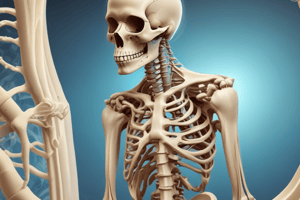
Bone
- Dynamic tissue with osteoblasts (forming bone) and osteoclasts (resorbing bone).
- Osteoblasts produce osteoid, a protein matrix composed of type 1 collagen.
- Calcium and phosphate are incorporated into the matrix, forming calcium hydroxyapatite, which gives bone its hardness.
- Bone anatomy consists of:
- Diaphysis: shaft of the bone
- Metaphysis: wider portion at the end of the diaphysis
- Epiphysis: end of the bone, further divided into proximal and distal epiphyses
- Epiphyseal line: growth plate between the epiphysis and metaphysis
- Periosteum: outer covering of bone
- Compact (cortical) bone: dense outer layer
- Spongy bone: inner layer with trabeculae
- Articular cartilage: smooth surface covering the joint ends
- Medullary cavity: central space within the bone
- Marrow: tissue within the medullary cavity (red marrow produces blood cells)
Types of Bone Tissue
- Woven bone: irregular, immature bone found in the fetus and during fracture repair
- Lamellar bone: regular, mature bone found in adults, further divided into:
- Circumferential lamellae: outer layers
- Concentric lamellae: layers arranged around a central haversian canal
- Interstitial lamellae: remnants of old osteons
- Trabecular lamellae: found in spongy bone
- Compact and spongy bone are both lamellar bone.
- Bone formation can be endochondral (cartilage template) or intramembranous (directly from mesenchymal tissue).
Bone Growth Plate
- Located at the epiphyseal line.
- Responsible for longitudinal bone growth.
- Composed of zones of proliferating chondrocytes (cartilage cells).
Osteoporosis
- Reduction in bone mass per unit of bone volume.
- Metabolic bone disease with normal mineral to matrix ratio.
- Primary osteoporosis: most common, uncertain etiology, common in postmenopausal women and elderly.
- Affects peak bone mass, involves estrogen decline, and aging.
- Calcium intake (800mg/day), exercise, and environmental factors like smoking (decreases estrogen) are important.
- Secondary osteoporosis: caused by other conditions, including:
- Corticosteroids: inhibit osteoblastic activity and impair vitamin D's role in calcium absorption.
- Hematologic malignancies: can impact bone mass
- Malabsorption: GI and liver diseases
- Alcoholism: inhibits osteoblasts and reduces calcium absorption
Osteoporosis Manifestations
- Osteopenia: decrease in bone thickness, especially cortical bone.
- Reduction in the number and size of trabeculae, the thin, bony struts within spongy bone.
- Fractures can be the first sign, particularly compression fractures of vertebrae.
Osteomalacia and Rickets
- Inadequate mineralization of the newly formed bone matrix.
- Osteomalacia: in adults
- Rickets: in children, affecting the open epiphyseal plates and cartilage.
- Characterized by:
- Beaded appearance of costochondral junctions (where ribs meet cartilage)
- Pectus carinatum (pigeon chest)
- Dental abnormalities
- Caused by:
- Vitamin D deficiency
- Phosphate deficiency
- Defects in mineralization process
Osteomalacia and Rickets Manifestations
- Osteopenia
- Exaggeration of osteoid seams (unmineralized bone matrix)
- Poorly localized pain, often in the femoral neck, pubic ramus, spine, and ribs
Hyperparathyroidism
- Excess parathyroid hormone (PTH) due to:
- Parathyroid adenoma (tumor)
- Parathyroid hyperplasia (overgrowth of glands)
- Parathyroid malignancy (rare)
- PTH causes:
- Increased phosphate excretion in urine and stimulation of osteoclastic activity, leading to hypercalcemia (high blood calcium).
- Increased tubular reabsorption of calcium and excretion of phosphate.
- Increased intestinal calcium absorption.
Hyperparathyroidism Manifestations
- Stones: kidney stones
- Bones: brown tumors (bone lesions)
- Psychiatric depression
- GI tract irregularities
Secondary Hyperparathyroidism
- Occurs due to chronic renal failure.
- Leads to:
- Decreased filtration of phosphate, causing hyperphosphatemia (high blood phosphate).
- Impaired active vitamin D production.
- Decreased calcium absorption in GI tract, leading to hypocalcemia (low blood calcium).
- Secondary hyperparathyroidism develops as a response to these abnormalities.
Osteomyelitis
- Inflammation of bone caused by an infectious organism, often bacteria like:
- Staphylococcus
- Streptococcus
- Escherichia coli
- Neisseria gonorrhea
- Haemophilus influenza
- Salmonella
- Infection can occur via:
- Direct penetration: wounds, fractures, surgery
- Hematogenous spread: through the bloodstream (common in metaphyses of long bones, especially the knee, ankle, and hip)
Osteomyelitis Complications
- Septicemia: blood poisoning
- Acute bacterial arthritis
- Pathologic fractures: fractures caused by weakened bone
- Squamous cell carcinoma: a type of skin cancer
- Amyloidosis: abnormal protein deposits
- Chronic osteomyelitis: persistent infection
Joints - Anatomy
- Joints are the points where two or more bones meet
- Different types of joints with varying degrees of movement
- Articular cartilage covers the ends of the bones, providing a smooth surface
- Synovial fluid lubricates the joint
Arthritis
- Inflammation of joints, a common condition.
- Often a site for autoimmune injury due to exposure of heart valves and joints to hidden antigens.
- Can be caused by infections and degeneration.
- Clinical features:
- Pain caused by inflammation of the capsule, synovium, and periosteum.
- Swelling due to inflammation, effusion (fluid accumulation), and synovial proliferation (growth).
- Restricted movement caused by pain, fluid, synovial swelling, and joint damage.
- Deformity caused by mal-alignment, erosion, and ankylosis (joint fusion).
Osteoarthritis
- Most common joint disease, a slow progressive degeneration of articular cartilage.
- Affects weight-bearing joints (hips, knees) and fingers.
- Primary osteoarthritis: defect in cartilage, not inflammatory.
- Secondary osteoarthritis: caused by various factors like trauma, crystal deposits, and infection.
- Narrowing of the joint space is a key sign, indicating loss of cartilage.
- Increased thickness of subchondral bone: the bone beneath the cartilage becomes thickened (eburnation).
- Subchondral cysts: fluid-filled cavities within the bone beneath the cartilage.
- Osteophytes: bony growths (bone spurs), also known as Heberden nodes on the fingers.
Osteoarthritis Causes
- Primary: intrinsic cartilage defect
- Secondary:
- Intra-articular fracture
- Previous infective arthritis
- Rheumatoid arthritis
- Congenital dislocation of hip
- Abnormal stresses (Paget's disease)
- Chronic overuse
- Metabolic and endocrine disorders (hemochromatosis, gout, calcium phosphate deposition)
- Neuropathic disorders (peripheral neuropathy in diabetes mellitus)
- Excess intra-articular corticosteroids
Osteoarthritis Histopathology
- Fibrillation: fraying and roughening of the articular cartilage
- Loose bodies: pieces of cartilage floating in the joint space
- Eburnation: hardened articular bone
- Subarticular cyst formation
- Rice bodies: small, white, rice-shaped bodies in the joint fluid, comprised of fibrin.
- Hyperplastic synovium: thickened synovium
- Pannus: a thick, abnormal layer of tissue that can form in the synovium and over the cartilage.
- Allison-Ghormley bodies: small, round bodies found in the joint fluid.
- Rheumatoid nodules: firm, rubbery lumps that can appear in the skin or other organs.
Rheumatoid Arthritis (RA)
- Autoimmune, chronic inflammatory disease.
- Inflammation of the synovium (lining of the joint), which eventually destroys cartilage.
- Affects small joints, especially in the hands and feet, bilaterally (symmetrically).
RA - Clinical Features
- Morning stiffness that lasts at least an hour.
- Arthritis in at least 3 joint areas.
- Arthritis in the small hand joints.
- Symmetrical arthritis.
- Rheumatoid nodules.
- Positive serum rheumatoid factor (antibody).
- Typical radiographic changes
- At least 4 of these features are required for diagnosis.
RA Pannus
- Hyperplastic and inflamed synovium.
- Infiltrates the joint space, destroying cartilage and bone.
Extra-Articular RA
- Can affect other parts of the body:
- Rheumatoid nodules: firm, rubbery lumps in the skin, caused by inflammation.
- Vasculitis: inflammation of blood vessels, affecting small arteries, especially those in the fingers.
- Pleuritis: inflammation of the pleura (lining of the lungs).
- Pericarditis: inflammation of the pericardium (lining of the heart).
- Tendonitis: inflammation of tendons.
RA Nodule Microscopic Features
- Central fibrinoid necrosis: a central area of dead tissue, often with a granular appearance.
- Palisading macrophages: macrophages (immune cells) arranged around the necrotic area.
Joint Involvement in RA
- Swan neck deformity: a bending of the finger joint at the middle joint (PIP) and straightening of the end joint (DIP), resulting in a swan-neck shape.
Differentiating RA from Osteoarthritis
| Feature | Rheumatoid Arthritis | Osteoarthritis |
|---|---|---|
| Age | Young, often 20-40 years of age | Elderly |
| Joints | Small joints (hands, wrists, feet), symmetrical | Large, weight-bearing joints (hips, knees, spine) |
| Cause | Autoimmune, chronic inflammation of the synovium | Degenerative disease of cartilage |
| Cartilage | Eroded by pannus | Gradual breakdown, wear-and-tear |
Pseudogout (Calcium Pyrophosphate Deposition Disease - CPPD)
- Deposition of calcium pyrophosphate crystals in cartilage and juxta-articular tissues.
Types of CPPD
- Type A (Pseudo Gout): primarily affects the knee of elderly men.
- Type B (Pseudo Rheumatoid): affects multiple joints, causing polyarthritis, similar to RA.
- Type C: Combines features of OA and acute attacks, more common in women.
- Type D: Pseudo OA, acute attacks, primarily affect the hands.
- Type E: Asymptomatic, without symptoms.
- Type F: Pseudoneuropathic, associated with nerve damage, resembling Charcot joint.
CPPD Clinical Features
- Often found in patients with hyperparathyroidism.
- Frequent in elderly.
- Affects joints like the knee.
- Characterized by joint swelling and inflammation, commonly triggered by trauma.
CPPD Histopathology
- Calcium pyrophosphate crystals present in the cartilage, synovium, and joint fluid.
Muscular Dystrophy
- Group of genetic diseases affecting muscle, causing weakness and degeneration.
- Duchenne muscular dystrophy (DMD) is the most common type, X-linked recessive.
Duchenne Muscular Dystrophy (DMD)
- Affects males primarily because the gene is on the X chromosome.
- Onset before age 5: Delayed motor development, waddling gait, difficulty running.
- Gower's manoeuvre: using arms to push the body up from a squatting position due to weakness in pelvic girdle muscles.
- Wheelchairs needed by 13 years old.
- Death from respiratory failure or cardiac arrhythmias in late teens.
DMD Pathological Findings
- Hyaline (prenecrotic) and necrotic muscle fibers with initial good regeneration.
- Regeneration decreases over time.
- Progressive replacement of muscle fibers with adipose and fibrous tissue, causing pseudohypertrophy (muscle feels larger due to fat and connective tissue).
DMD Diagnosis
- Dystrophin testing on muscle biopsy (dystrophin is a protein important for muscle function).
- Serum creatine kinase estimation: Elevated in carriers, but negative results don't rule out the possibility of being a carrier.
Studying That Suits You
Use AI to generate personalized quizzes and flashcards to suit your learning preferences.