Podcast
Questions and Answers
What is the function of the atrial natriuretic factor produced by the heart?
What is the function of the atrial natriuretic factor produced by the heart?
- Regulating blood pressure (correct)
- Forming the epicardium
- Producing cardiac fibrous skeleton
- Stimulating cardiac muscle contraction
What is the thickest layer of the heart wall?
What is the thickest layer of the heart wall?
- Epicardium
- Endocardium
- Pericardium
- Myocardium (correct)
What is the function of the cardiac fibrous skeleton?
What is the function of the cardiac fibrous skeleton?
- Regulating heart rate
- Providing a site for origin and insertion of cardiac muscle cells (correct)
- Forming the pericardium
- Producing atrial natriuretic factor
What is the outermost layer of the heart wall?
What is the outermost layer of the heart wall?
What type of cells makes up the endocardium?
What type of cells makes up the endocardium?
What is homologous with the tunica intima of blood vessels?
What is homologous with the tunica intima of blood vessels?
What type of cells form the inner lining of the blood vessels and heart?
What type of cells form the inner lining of the blood vessels and heart?
What type of filaments provide structural support to endothelial cells?
What type of filaments provide structural support to endothelial cells?
What is the main function of Weibel-Palade bodies in endothelial cells?
What is the main function of Weibel-Palade bodies in endothelial cells?
What is the shape of endothelial cells?
What is the shape of endothelial cells?
What is the origin of endothelial cells?
What is the origin of endothelial cells?
What is the function of the complexes of miosin and actin in endothelial cells?
What is the function of the complexes of miosin and actin in endothelial cells?
What is the function of microvilli in endothelial cells?
What is the function of microvilli in endothelial cells?
What is the location of endothelial cells?
What is the location of endothelial cells?
What is characteristic of blood vessels that carry blood toward the heart?
What is characteristic of blood vessels that carry blood toward the heart?
What is the size range of postcapillary venules in diameter?
What is the size range of postcapillary venules in diameter?
What is a characteristic of small or medium-sized veins?
What is a characteristic of small or medium-sized veins?
What is the function of the valves in veins?
What is the function of the valves in veins?
What is the characteristic of the adventitia layer in large veins?
What is the characteristic of the adventitia layer in large veins?
What is the function of the contraction of muscles compressing veins?
What is the function of the contraction of muscles compressing veins?
What is a characteristic of the intima layer in large veins?
What is a characteristic of the intima layer in large veins?
What is a characteristic of the media layer in small or medium-sized veins?
What is a characteristic of the media layer in small or medium-sized veins?
What is the main function of sinusoidal capillaries?
What is the main function of sinusoidal capillaries?
What type of capillaries are found in the renal glomerulus?
What type of capillaries are found in the renal glomerulus?
What is the role of pericytes in capillaries?
What is the role of pericytes in capillaries?
Which of the following tissues has a poor supply of capillaries?
Which of the following tissues has a poor supply of capillaries?
What is the characteristic of continuous capillaries?
What is the characteristic of continuous capillaries?
What is the function of precapillary sphincters?
What is the function of precapillary sphincters?
What is the characteristic of fenestrated capillaries?
What is the characteristic of fenestrated capillaries?
What is the composition of a capillary bed?
What is the composition of a capillary bed?
What is the outermost layer of blood vessels?
What is the outermost layer of blood vessels?
What is the function of the lymphatic vascular system?
What is the function of the lymphatic vascular system?
What is characteristic of lymphatic vessels?
What is characteristic of lymphatic vessels?
What is the structure that controls the heartbeat?
What is the structure that controls the heartbeat?
What is the pericardium?
What is the pericardium?
What is the characteristic of the endocardium?
What is the characteristic of the endocardium?
Which layer of the arterial wall contains connective tissue, vasa vasorum, and nerves?
Which layer of the arterial wall contains connective tissue, vasa vasorum, and nerves?
What is the main characteristic of the media layer in muscular arteries?
What is the main characteristic of the media layer in muscular arteries?
Which type of fibers are present in the media layer of muscular arteries?
Which type of fibers are present in the media layer of muscular arteries?
What is the characteristic of the intima layer in arterioles?
What is the characteristic of the intima layer in arterioles?
What is the characteristic of the adventitia layer in arterioles?
What is the characteristic of the adventitia layer in arterioles?
What is the main function of smooth muscle cells in the media layer of muscular arteries?
What is the main function of smooth muscle cells in the media layer of muscular arteries?
What is the main characteristic of the tunica intima?
What is the main characteristic of the tunica intima?
What is the function of vascular smooth muscle cells in muscular arteries?
What is the function of vascular smooth muscle cells in muscular arteries?
What is the purpose of staining for elastic arteries?
What is the purpose of staining for elastic arteries?
What is characteristic of the tunica media in muscular arteries?
What is characteristic of the tunica media in muscular arteries?
What can be found in the tunica adventitia?
What can be found in the tunica adventitia?
What is the location of the internal elastic lamina in muscular arteries?
What is the location of the internal elastic lamina in muscular arteries?
What type of arteries have a prominent internal elastic lamina?
What type of arteries have a prominent internal elastic lamina?
What is the function of the internal elastic lamina in muscular arteries?
What is the function of the internal elastic lamina in muscular arteries?
What type of fibers are present in the tunica media of elastic arteries?
What type of fibers are present in the tunica media of elastic arteries?
What is the main component of the tunica adventitia in large vessels?
What is the main component of the tunica adventitia in large vessels?
Which type of cells are present in the tunica media of blood vessels?
Which type of cells are present in the tunica media of blood vessels?
What is the function of the internal elastic lamina in elastic arteries?
What is the function of the internal elastic lamina in elastic arteries?
What is the characteristic of the tunica intima in elastic arteries?
What is the characteristic of the tunica intima in elastic arteries?
What type of fibers are present in the tunica media of muscular arteries?
What type of fibers are present in the tunica media of muscular arteries?
What is the characteristic of elastic fibers in elastic arteries?
What is the characteristic of elastic fibers in elastic arteries?
What is the function of the vasa vasorum in large vessels?
What is the function of the vasa vasorum in large vessels?
Flashcards are hidden until you start studying
Study Notes
Blood Vessel Structure
- A blood vessel is composed of three layers: tunica intima, tunica media, and tunica adventitia.
- Tunica intima consists of a single layer of endothelial cells and a subendothelial layer of loose connective tissue.
- Endothelial cells are polygonal, elongated in the direction of blood flow, and bulged into the lumen.
- They have a mesenchymal origin, contain desmin and vimentin, and lie on the basal lamina.
Endothelial Cells
- Endothelial cells have numerous complexes of miosin and actin, allowing for contraction and relaxation.
- They have Weibel-Palade bodies, which store and release von Willebrand factor and P-selectin.
- They have microvilli, which connect with leukocytes.
Capillaries
- There are three types of capillaries: continuous, fenestrated, and discontinuous sinusoidal.
- Continuous capillaries have no fenestrae, a well-developed basal lamina, and are found in muscle tissue, exocrine glands, and blood-brain barrier.
- Fenestrated capillaries have several circular transcellular openings in endothelial cells and are found in endocrine glands, intestine, and renal glomerulus.
- Sinusoidal capillaries have no basal lamina or a discontinuous one, and are found in liver, spleen, and bone marrow.
Pericytes
- Pericytes are support cells for capillaries and small venules.
- They have their own basal lamina, are of mesenchymal origin, and are located along the outside of capillaries and small venules.
- They possess a small Golgi complex, mitochondria, rER, microtubules, and proteins related to contraction.
Functions of Pericytes
- Not specified in the text.
Capillary Bed
- A capillary bed is an interconnected network of vessels running through tissues.
- It consists of metarterioles, capillaries, and venules.
- Precapillary sphincters regulate the flow of blood to tissues.
Veins
- Veins are formed by the convergence of capillaries into a system of channels.
- They become larger in diameter as they approach the heart.
- They are higher in diameter than arteries and are rather elongated or oval than round in shape.
- Adventitia is the best-developed layer.
Postcapillary Venules
- They are 0.1 to 0.5 mm in diameter.
- They have a thin wall and are collapsed.
- They have tunica intima, endothelium, and a thin subendothelial layer, and pericytes.
Small or Medium-Sized Veins
- They have a muscular wall, are 1-9 mm in diameter, and are collapsed.
- Intima is lined with endothelium, with a thin or no subendothelial layer.
- Media has small bundles of smooth muscle cells intermixed with reticular fibers.
- Adventitia is well-developed, with collagen fibers, fibroblasts, nerves, and vasa vasorum.
Large Veins
- They are big venous trunks, close to the heart.
- Intima is well-developed.
- Media is thin, with few layers of smooth muscle cells, and an abundance of collagenous tissue.
- Adventitia is the thickest, with longitudinal layers of smooth muscle cells.
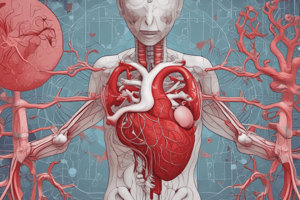
Heart
- The heart is a muscular organ that contracts rhythmically.
- It produces atrial natriuretic factor.
- The structure of the wall consists of endocardium, myocardium, and epicardium.
Heart Structure
- The cardiac fibrous skeleton is a dense connective tissue that forms a central region of the heart.
- It is a site of origin and insertion of cardiac muscle cells.
Endocardium
- It is homologous with the t.intima of blood vessels.
- It consists of a single layer of squamous endothelial cells, a thin subendothelial layer of loose connective tissue, a muscular-elastic layer, and the subendocardial layer.
Myocardium
- It is the thickest layer, homologous with the t.media of blood vessels.
- It consists of cardiac muscle arranged in layers and grouped into three populations.
Epicardium
- It is the serous covering of the heart and forms the visceral layer of the pericardium.
- It is homologous with the t.adventitia of blood vessels.
- It is covered externally by mesothelium.
Lymphatic Vascular System
- It returns the extracellular liquid (lymph) to the bloodstream.
- The lymph circulates in one direction only.
- Lymphatic vessels are found in all organs, are blind-ended, and consist of a single layer of endothelium.
- They have numerous internal valves and no fenestrations in their endothelial cells.
Artery Structure
- Arteries consist of three tunics: tunica intima, tunica media, and tunica adventitia
- Tunica intima is lined with endothelium
- Tunica media is composed of elastic laminae and vascular smooth muscle cells
- Tunica adventitia is made up of connective tissue, vasa vasorum, and nerves
Elastic Arteries
- The largest arteries
- Function: transport blood with oxygen and nutrients to tissues and organs
- Intima: lined with endothelium, subendothelial layer, collagen and elastic fibers, fibroblasts, and internal elastic lamina
- Media: concentric arranged, perforated elastic laminae (30-75), vascular smooth muscle cells, reticular and collagen fibers, proteoglycans, and glycoproteins
- Adventitia: connective tissue, well developed, nerves, and vasa vasorum
- Staining: resorcine-fuchsin and orcein
Muscular Arteries
- Medium-sized arteries
- Control the affluence of blood to organs by contracting or relaxing smooth muscle cells in the media
- Intima: lined with endothelium, subendothelial layer, and internal elastic lamina is prominent, thicker in older people
- Media: concentric layers of helically arranged smooth muscle cells, some elastic fibers, reticular fibers, proteoglycans, and glycoproteins
- Adventitia: fibroblasts, type I collagen fibers, longitudinally oriented elastic fibers, nerves, and adipose tissue
- Function: regulate blood pressure and flow to organs
Arterioles
-
Narrow lumen (less than 0.5 mm in diameter)
-
Intima: lined with endothelium, subendothelial layer is very thin, no internal elastic lamina in very small arterioles
-
Media: 1 or 2 circularly arranged layers of smooth muscle cells, no external elastic lamina
-
Adventitia: very thin
-
Function: regulate blood flow to capillaries
Capillaries
- Single layer of endothelial cells resting on a basal lamina
- Permit metabolic exchange between blood and surrounding tissues
Studying That Suits You
Use AI to generate personalized quizzes and flashcards to suit your learning preferences.