Podcast
Questions and Answers
What type of cell differentiates into a macrophage?
What type of cell differentiates into a macrophage?
Monocyte
Describe the basic structure and primary function of red blood cells (RBCs).
Describe the basic structure and primary function of red blood cells (RBCs).
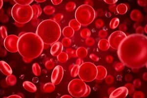
Red blood cells are biconcave discs lacking a nucleus. Their primary function is to transport oxygen bound to hemoglobin.
Which types of blood cells are phagocytic?
Which types of blood cells are phagocytic?
Neutrophils and monocytes (which differentiate into macrophages) are the main phagocytic blood cells.
What is a reticulocyte?
What is a reticulocyte?
Which mature blood cells lack a nucleus?
Which mature blood cells lack a nucleus?
What is the most abundant type of leukocyte (white blood cell)?
What is the most abundant type of leukocyte (white blood cell)?
What is the most abundant plasma protein?
What is the most abundant plasma protein?
What does the gamma globulin fraction of plasma primarily contain?
What does the gamma globulin fraction of plasma primarily contain?
Define Erythropenia.
Define Erythropenia.
Define Leukopenia.
Define Leukopenia.
Define Polycythemia.
Define Polycythemia.
Define Leukocytosis.
Define Leukocytosis.
What are the general causes and characteristics of anemia?
What are the general causes and characteristics of anemia?
What causes sickle cell anemia and what are its main characteristics?
What causes sickle cell anemia and what are its main characteristics?
What is aplastic anemia and what are its potential causes?
What is aplastic anemia and what are its potential causes?
What typically causes nutritional anemia?
What typically causes nutritional anemia?
A low hematocrit level is a common indicator of anemia.
A low hematocrit level is a common indicator of anemia.
Where does myelogenous leukemia originate?
Where does myelogenous leukemia originate?
What antibodies are typically present in the plasma of individuals with Type A, Type B, Type AB, and Type O blood?
What antibodies are typically present in the plasma of individuals with Type A, Type B, Type AB, and Type O blood?
Match the donor blood type (ABO group) with potentially compatible recipients.
Match the donor blood type (ABO group) with potentially compatible recipients.
Which ABO antigens are present on the surface of red blood cells for Type A, Type B, Type AB, and Type O blood?
Which ABO antigens are present on the surface of red blood cells for Type A, Type B, Type AB, and Type O blood?
What is the function of a centrifuge in a laboratory setting, particularly for blood analysis?
What is the function of a centrifuge in a laboratory setting, particularly for blood analysis?
What is the predominant carbohydrate found circulating in blood plasma?
What is the predominant carbohydrate found circulating in blood plasma?
Blood is classified as a type of connective tissue.
Blood is classified as a type of connective tissue.
What does the medical root word 'hemat/o' refer to?
What does the medical root word 'hemat/o' refer to?
List the three layers of the heart wall, from outermost to innermost.
List the three layers of the heart wall, from outermost to innermost.
Name the four chambers and four valves of the human heart.
Name the four chambers and four valves of the human heart.
Describe the pathway of blood flow through the heart, starting with deoxygenated blood entering the right atrium.
Describe the pathway of blood flow through the heart, starting with deoxygenated blood entering the right atrium.
Where do electrical impulses normally originate in the heart, and what is the path they follow?
Where do electrical impulses normally originate in the heart, and what is the path they follow?
Define stroke volume and cardiac output.
Define stroke volume and cardiac output.
Which chamber of the heart has the thickest, strongest muscular wall?
Which chamber of the heart has the thickest, strongest muscular wall?
What is bradycardia?
What is bradycardia?
What is tachycardia?
What is tachycardia?
What occurs during atrial systole?
What occurs during atrial systole?
What events cause the 'lub' (S1) and 'dub' (S2) heart sounds?
What events cause the 'lub' (S1) and 'dub' (S2) heart sounds?
What causes the second heart sound (S2 or 'dub')?
What causes the second heart sound (S2 or 'dub')?
What is endocarditis?
What is endocarditis?
What is an infarct, and what typically causes a myocardial infarct?
What is an infarct, and what typically causes a myocardial infarct?
What is the ductus arteriosus and what happens to it normally after birth?
What is the ductus arteriosus and what happens to it normally after birth?
How does the parasympathetic nervous system affect heart rate?
How does the parasympathetic nervous system affect heart rate?
Compare the general actions of the sympathetic and parasympathetic nervous systems on the heart.
Compare the general actions of the sympathetic and parasympathetic nervous systems on the heart.
What condition is angioplasty primarily used to treat?
What condition is angioplasty primarily used to treat?
Distinguish between modifiable and non-modifiable risk factors for heart disease, providing examples of each.
Distinguish between modifiable and non-modifiable risk factors for heart disease, providing examples of each.
Which major arteries supply blood to the head?
Which major arteries supply blood to the head?
Which arteries run between the ribs?
Which arteries run between the ribs?
Which blood vessels are the primary site of gas exchange in the lungs?
Which blood vessels are the primary site of gas exchange in the lungs?
Compare the general wall thickness of arteries, veins, and capillaries.
Compare the general wall thickness of arteries, veins, and capillaries.
How is arterial blood pressure typically reported, and what does each number represent?
How is arterial blood pressure typically reported, and what does each number represent?
What is considered a normal blood pressure range for adults, and what does diastolic pressure represent?
What is considered a normal blood pressure range for adults, and what does diastolic pressure represent?
In case of severe arterial bleeding in the leg, besides direct pressure on the wound, where is a key pressure point located?
In case of severe arterial bleeding in the leg, besides direct pressure on the wound, where is a key pressure point located?
What is an embolus?
What is an embolus?
Define phlebitis.
Define phlebitis.
Name some common classes of medications used to treat high blood pressure (hypertension).
Name some common classes of medications used to treat high blood pressure (hypertension).
What are the two primary physiological factors that determine arterial blood pressure?
What are the two primary physiological factors that determine arterial blood pressure?
Flashcards
What is a macrophage?
What is a macrophage?
A type of white blood cell that engulfs and digests cellular debris, foreign substances, microbes, cancer cells, and anything else that does not have the types of proteins specific of normal body cells on its surface.
What is a reticulocyte?
What is a reticulocyte?
Red blood cells that are not yet fully mature.
Most abundant type of leukocyte?
Most abundant type of leukocyte?
Neutrophils.
Most abundant plasma protein?
Most abundant plasma protein?
Signup and view all the flashcards
What is anemia?
What is anemia?
Signup and view all the flashcards
What is sickle cell anemia?
What is sickle cell anemia?
Signup and view all the flashcards
What is aplastic anemia?
What is aplastic anemia?
Signup and view all the flashcards
What is nutritional anemia?
What is nutritional anemia?
Signup and view all the flashcards
What is myelogenous leukemia?
What is myelogenous leukemia?
Signup and view all the flashcards
What is a centrifuge?
What is a centrifuge?
Signup and view all the flashcards
Predominant carbohydrate in plasma?
Predominant carbohydrate in plasma?
Signup and view all the flashcards
What is the epicardium?
What is the epicardium?
Signup and view all the flashcards
What is the myocardium?
What is the myocardium?
Signup and view all the flashcards
What is the endocardium?
What is the endocardium?
Signup and view all the flashcards
What is stroke volume?
What is stroke volume?
Signup and view all the flashcards
What is cardiac output?
What is cardiac output?
Signup and view all the flashcards
Strongest chamber of the heart?
Strongest chamber of the heart?
Signup and view all the flashcards
What is bradycardia?
What is bradycardia?
Signup and view all the flashcards
What is tachycardia?
What is tachycardia?
Signup and view all the flashcards
What is atrial systole?
What is atrial systole?
Signup and view all the flashcards
Study Notes
Blood and Blood Components
- Monocytes differentiate into macrophages
Red Blood Cells
- Red blood cells (RBCs) lack a nucleus
- RBCs are also called erythrocytes
- RBCs' primary function is to transport oxygen
- RBCs have a biconcave disc shape to maximize surface area for gas exchange
- Hemoglobin is the oxygen-carrying protein in RBCs
- RBCs are produced in bone marrow through erythropoiesis
Phagocytic Blood Cells
- Neutrophils and monocytes are phagocytic
Reticulocyte
- A reticulocyte is an immature red blood cell
Leukocytes
- Neutrophils are the most abundant type of leukocyte (white blood cell)
- Leukocytes are involved in immune defense
- The gamma globulin fraction contains antibodies
Plasma Protein
- Albumin is the most abundant plasma protein
Blood Cell Level Terminology
- Erythropenia refers to a deficiency in red blood cells
- Leukopenia refers to a deficiency in white blood cells
- Polycythemia refers to an excess of red blood cells
- Leukocytosis refers to an excess of white blood cells
Blood Disorders and Conditions
- Anemia refers to a deficiency in red blood cells or hemoglobin
- Sickle cell anemia is a genetic disorder causing abnormally shaped red blood cells
- Aplastic anemia is a condition where the bone marrow fails to produce enough blood cells
- Nutritional anemia results from dietary deficiencies, such as iron, folate, or vitamin B12
- A low hematocrit indicates anemia
- Myelogenous leukemia originates in the bone marrow
Blood Typing
- Blood type A contains anti-B antibodies
- Blood type B contains anti-A antibodies
- Blood type AB contains neither anti-A nor anti-B antibodies
- Blood type O contains both anti-A and anti-B antibodies
- Type A blood has A antigens on the surface of its re blood cells
- Type B blood has B antigens on the surface of its red blood cells
- Type AB blood has both A and B antigens on the surface of its red blood cells
- Type O blood has neither A nor B antigens on the surface of its red blood cells
Blood Transfusions
- Compatible blood types must be matched for safe transfusions
Blood Testing and Lab Techniques
- A centrifuge separates blood components based on density
- Glucose is the predominant carbohydrate in plasma
- Blood is considered a connective tissue
- The medical root word "hemat/o" refers to blood
Heart Structure
- The heart wall has three layers: epicardium, myocardium, and endocardium
- The heart has four chambers: right atrium, right ventricle, left atrium, and left ventricle
- Valves include the tricuspid, mitral (bicuspid), pulmonary, and aortic valves.
- Blood flow through the heart: right atrium → right ventricle → pulmonary artery → lungs → pulmonary vein → left atrium → left ventricle → aorta
- Electrical impulses originate at the sinoatrial (SA) node and travel through the atrioventricular (AV) node, bundle of His, and Purkinje fibers
- Stroke volume is the amount of blood ejected by the left ventricle per beat
- Cardiac output is the amount of blood pumped by the heart per minute (stroke volume x heart rate)
- The left ventricle is the strongest chamber of the heart
Heart Rhythms
- Bradycardia is a slow heart rate
- Tachycardia is a fast heart rate
- Atrial systole is the contraction of the atria
- The "lub" sound is the closing of the atrioventricular valves (tricuspid and mitral)
- The "dub" sound is the closing of the semilunar valves (aortic and pulmonic)
- The second heart sound (dub) is caused by the closure of the aortic and pulmonic valves
- Endocarditis is inflammation of the heart's inner lining
- An infarct is tissue death due to lack of blood supply
- Ductus arteriosus is a congenital feature where a vessel connects the pulmonary artery and aorta in the fetus
Autonomic Nervous System & The Heart
- The parasympathetic system decreases heart rate
- The sympathetic system increases heart rate, while the parasympathetic system decreases heart rate
Heart Procedures and Risk Factors
- Angioplasty is used to open blocked arteries
- Modifiable risk factors for heart disease: diet, exercise, smoking
- Non-modifiable risk factors for heart disease: age, genetics, sex
Blood Vessels and Circulation
- Carotid arteries supply blood to the head
- Intercostal arteries supply blood to the area between the ribs
- Pulmonary capillaries are located in the lungs
- Arteries have thicker walls than veins and capillaries
- Veins have thinner walls than arteries and transport blood to the heart
- Capillaries are thin-walled vessels for gas exchange
Blood Pressure and First Aid
- Blood pressure is reported as systolic/diastolic
- Normal blood pressure is around 120/80 mm Hg
- Diastolic pressure represents the pressure in the arteries when the heart is at rest
- Apply pressure above the injury (towards the heart) to stop bleeding in the leg
- An embolus is a blood clot, fat globule, or air bubble that travels through the bloodstream and can cause a blockage.
- Phlebitis is vein inflammation
Hypertension and Medication
- Medications used to treat high blood pressure may include diuretics, ACE inhibitors, beta-blockers, and calcium channel blockers
- Physiological contributors to blood pressure include cardiac output and vessel resistance
Studying That Suits You
Use AI to generate personalized quizzes and flashcards to suit your learning preferences.