Podcast
Questions and Answers
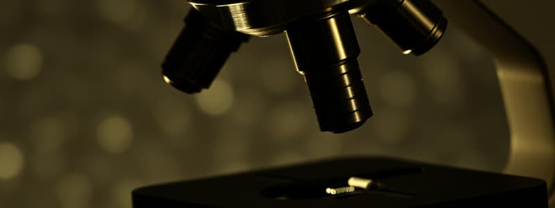
What is the path of light through a compound light microscope?
What is the path of light through a compound light microscope?
Light source (illuminator); condenser; specimen; objective lens; ocular lens.
How is total magnification calculated?
How is total magnification calculated?
The total magnification of an object is calculated by multiplying the magnification of the objective lens by the magnification of the ocular lens (objective lens x ocular lens).
What is the role of the condenser?
What is the role of the condenser?
Focuses the light onto the specimen.
What is the role of the Iris Diaphragm?
What is the role of the Iris Diaphragm?
What are the steps of the bacterial smear?
What are the steps of the bacterial smear?
What is the function of a bacterial smear?
What is the function of a bacterial smear?
Define Resolution.
Define Resolution.
Define Magnification.
Define Magnification.
Define numerical aperture.
Define numerical aperture.
What are the components of Phase Contrast Microscopy?
What are the components of Phase Contrast Microscopy?
What are the Dark Field Microscopy Characteristics?
What are the Dark Field Microscopy Characteristics?
How does the dark field microscope work?
How does the dark field microscope work?
How do we use Fluorescence Microscopy?
How do we use Fluorescence Microscopy?
When do we use Fluorescence Microscopy?
When do we use Fluorescence Microscopy?
What are fluorophores/fluorochromes? And what is excitation and emission?
What are fluorophores/fluorochromes? And what is excitation and emission?
Define the electron microscope.
Define the electron microscope.
What are new components in the electron microscopy?
What are new components in the electron microscopy?
Why does the electron microscope have higher resolution and magnification power than a light microscope?
Why does the electron microscope have higher resolution and magnification power than a light microscope?
Flashcards are hidden until you start studying
Study Notes
Path of Light in a Compound Light Microscope
- Light travels from the light source (illuminator) to the condenser, then to the specimen, through the objective lens, and finally reaches the ocular lens.
Total Magnification Calculation
- Total magnification is found by multiplying the magnification of the objective lens by the magnification of the ocular lens (Objective Lens x Ocular Lens).
Role of the Condenser
- The condenser's purpose is to focus light onto the specimen, enhancing image clarity.
Function of the Iris Diaphragm
- It controls the amount of light that reaches the specimen, allowing for better visualization of details.
Steps of Preparing a Bacterial Smear
- A thin film of the microorganism is spread on a slide.
- The slide is dried using a Bunsen burner.
- Stain is applied and then washed off with water.
- The slide is blotted with absorbent paper and is ready for examination.
Function of a Bacterial Smear
- Fixes microorganisms to the slide, simultaneously killing them and preserving their structure with minimal distortion.
Definition of Resolution
- The ability to distinguish fine details and separate two objects from each other.
Definition of Magnification
- Magnification refers to how much an image is enlarged.
Definition of Numerical Aperture
- A measure of a microscope's ability to gather light and resolve fine specimen details.
Components of Phase Contrast Microscopy
- Highlights internal structures of living cells using light that passes through an annular diaphragm.
- Combines direct and diffracted light rays at the viewer's eye, with direct rays appearing red and reflected/diffracted rays appearing blue.
Characteristics of Dark Field Microscopy
- Used to observe living organisms that are invisible with standard light microscopy.
- Ideal for specimens that cannot be stained without distortion.
Working Principle of Dark Field Microscopy
- Utilizes a special condenser with an opaque disk to block direct light, allowing only light scattered by the specimen to enter the objective lens.
- The specimen appears bright against a dark background.
Use of Fluorescence Microscopy
- Specimens are first stained with fluorochromes and then observed using ultraviolet light through a compound microscope.
Applications of Fluorescence Microscopy
- Primarily used in diagnostic methods such as fluorescent-antibody (FA) technique or immunofluorescence.
Fluorophores and Excitation/Emission
- Fluorophores (or fluorochromes) absorb light and become excited, then emit fluorescent light as they return to a lower energy state.
Definition of Electron Microscope
- An electron microscope examines viruses and internal cell structures using beams of electrons instead of light.
Components of Electron Microscopy
- Uses electromagnetic lenses to focus an electron beam on the specimen, with electrons traveling in waves providing better resolution due to their shorter wavelengths.
Higher Resolution and Magnification in Electron Microscopes
- Electron beams possess higher energy and shorter wavelengths than visible light, yielding significantly improved resolution (0.2 micrometers, 100,000 times smaller than light wavelengths).
Studying That Suits You
Use AI to generate personalized quizzes and flashcards to suit your learning preferences.


