Podcast
Questions and Answers
What is a consequence of accidental ligation of the ureter during oophorectomy or hysterectomy?
What is a consequence of accidental ligation of the ureter during oophorectomy or hysterectomy?
- Reduction in renal stones
- Post-operative renal impairment (correct)
- Enhanced bladder function
- Increased urinary flow
Where does the ureter become the narrowest part?
Where does the ureter become the narrowest part?
- At the ischial spine (correct)
- At the tip of the transverse process of L2
- At the bifurcation of the common iliac artery
- At the renal pelvis junction
Which structure supplies the abdominal part of the ureter with arterial blood?
Which structure supplies the abdominal part of the ureter with arterial blood?
- Internal iliac artery
- Uterine artery
- Common iliac artery
- Gonadal artery (correct)
What is the primary function of the intramural portion of the ureter?
What is the primary function of the intramural portion of the ureter?
What is the approximate length of the ureter?
What is the approximate length of the ureter?
What is the primary draining lymph node for the lower abdominal part of the ureter?
What is the primary draining lymph node for the lower abdominal part of the ureter?
Which of the following correctly describes the nerve supply to the ureters?
Which of the following correctly describes the nerve supply to the ureters?
Which of the following best describes the abdominal part of the ureter's relations?
Which of the following best describes the abdominal part of the ureter's relations?
Which artery is NOT associated with the right ureter?
Which artery is NOT associated with the right ureter?
What is a key characteristic of the blood supply in the ureters?
What is a key characteristic of the blood supply in the ureters?
What structure does the ureter NOT cross in the pelvic part?
What structure does the ureter NOT cross in the pelvic part?
At which point does the ureter cross the bifurcation of the common iliac artery?
At which point does the ureter cross the bifurcation of the common iliac artery?
What key difference is noted between the ureters in males and females?
What key difference is noted between the ureters in males and females?
Which of the following signifies an important relation of the ureter during surgical procedures in females?
Which of the following signifies an important relation of the ureter during surgical procedures in females?
Which nerve is associated with the relations of both the right and left ureters?
Which nerve is associated with the relations of both the right and left ureters?
What is the significance of understanding the arterial supply of the ureter?
What is the significance of understanding the arterial supply of the ureter?
Flashcards
Ureteropelvic Junction
Ureteropelvic Junction
The point where the ureter enters the renal pelvis.
Intramural Ureter
Intramural Ureter
The portion of the ureter that runs through the wall of the bladder.
Ureteric Constrictions
Ureteric Constrictions
Constrictions in the ureter can make it more susceptible to obstruction from kidney stones.
Ureteral Blood Supply
Ureteral Blood Supply
Signup and view all the flashcards
Ureteral Lymphatic Drainage
Ureteral Lymphatic Drainage
Signup and view all the flashcards
Ureteral Nerve Supply
Ureteral Nerve Supply
Signup and view all the flashcards
Ureteral Injury During Surgery
Ureteral Injury During Surgery
Signup and view all the flashcards
Ureteric Constrictions and Kidney Stones
Ureteric Constrictions and Kidney Stones
Signup and view all the flashcards
What is the ureter?
What is the ureter?
Signup and view all the flashcards
What are the three parts of the ureter?
What are the three parts of the ureter?
Signup and view all the flashcards
What are the key relations of the ureter in the pelvic part?
What are the key relations of the ureter in the pelvic part?
Signup and view all the flashcards
Describe the relations of the right ureter in the abdominal part.
Describe the relations of the right ureter in the abdominal part.
Signup and view all the flashcards
Describe the relations of the left ureter in the abdominal part.
Describe the relations of the left ureter in the abdominal part.
Signup and view all the flashcards
What are the constrictions of the ureter?
What are the constrictions of the ureter?
Signup and view all the flashcards
What is the medial relation of the right and left ureters?
What is the medial relation of the right and left ureters?
Signup and view all the flashcards
Describe the arterial supply of the ureter.
Describe the arterial supply of the ureter.
Signup and view all the flashcards
Study Notes
Quranic Verse
- A verse from Surah Adh-Dhariyat, verse 21, is quoted.
- The English translation of the verse is provided.
Renal Module
- Course code: IMP/07/20318
- Phase I
- Second year/semester 3
- Course duration: 5 weeks
Anatomy and Embryology Professor
- Professor Olfat Anwar Abd El Aty
- Email: [email protected]
Anatomy of the Ureter
- Length: 25-30 cm
- Thickness: thick-walled, narrow lumen
- Diameter: 3 mm
- Beginning: funnel-shaped renal pelvis within the hilum of the kidney at L1 level.
- Termination: at the postero-superior angles of the base of the urinary bladder.
Intended Learning Outcomes (ILOs)
- Describe the anatomy of the ureter, including shape, length, and parts.
- State the location and normal constrictions of the ureter.
- Differentiate between the relations of the right and left ureters
- List the arterial supply of the ureter and its significance.
- Describe the lymphatic drainage of the ureter.
- Describe the renal nerve plexus and renal innervations.
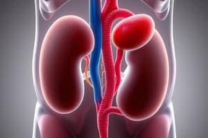
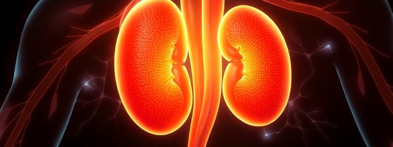
The Ureters
- Two muscular tubes that convey urine from the kidneys to the urinary bladder.
- Peristaltic contractions move urine.
Course
- Each ureter descends downward behind the peritoneum.
- Opposite the transverse processes of vertebrae L2, 3, 4, and 5.
- In front of the psoas major muscle.
- Enters the pelvis by crossing the bifurcation of the common iliac artery at the sacroiliac joint.
- Passes downwards and backwards along the internal iliac artery.
- Curves medially at the level of the ischial spine to open into the postero-superior angle of the urinary bladder.
Relation of the Psoas Major Muscle
- The relationship between the psoas major muscle and the ureters is crucial for locating the ureters in computed tomography (CT) scans.
Parts of the Ureter
- Abdominal part
- Pelvic part
- Intramural part
1-The Abdominal Part of the Ureter
- Medial border of the psoas major and minor muscles
- Tips of the transverse processes of vertebrae L2, 3, 4, and 5
- Genito-femoral nerve
- Bifurcation of the common iliac artery.
- Two/three parts of the duodenum
- Three vessels (Right Colic, Ileocolic, and Right Gonadal vessels)
- Three mesenteric related structures (Root of the mesentery, Superior mesenteric vessels, and coils of the small intestine)
- Three vessels (Left Colic, Sigmoidal, and Left Gonadal vessels)
- Apex of the sigmoid mesocolon
- Medial relation (Inferior vena cava, Inferior mesenteric vein, Left colic vessels, and Sigmoidal vessels).
2- Pelvic Part of the Ureter
- Relations: external iliac artery and vein, obturator nerve, obturator artery, and obturator vein.
- Male: ureters crossed by vas deferens.
- Female: ureters pass above the lateral fornix of the vagina.
Female Relation and Removal of Organs
- The relationship between the ureter and ovarian/uterine blood vessels during oophorectomy and hysterectomy is important.
- Ligation of organs' blood supply during organ removal.
- Potential for ureteral injury.
- Immediate surgical intervention may be necessary.
3- Intramural Part of the Ureter
- The ureters pierce the postero-superior angles of the base of the urinary bladder.
- Run obliquely through the bladder wall.
- Opening: ureteric orifices.
- Length: 1.5-2.0 cm within the bladder wall.
- Prevention of urine reflux into the ureter when the bladder is full.
- Intramural ureters thought to be occluded during bladder pressure increases.
Normal Ureter Constrictions
- Three constrictions along the ureter's course.
- First: where the ureter joins the renal pelvis (at the tip of the transverse process of L2).
- Second: at the brim of the lesser pelvis (where it crosses the bifurcation of the common iliac artery).
- Third: within the urinary bladder wall (at the ischial spine).
- These sites are a common location for renal stones.
MCQs
- Multiple-choice questions on ureter anatomy and location.
- Presented as examples for students.
Arterial Blood Supply of the Ureter
- Abdominal part: supplied by arteries medial to the ureter (renal artery, gonadal artery, branches of the abdominal aorta).
- Pelvic part: supplied by vessels lateral to the ureter (common iliac, internal iliac, vesicle and uterine arteries.)
- Venous drainage generally follows the arterial supply.
- Anastomosis (connection) exists between arterial branches.
- Safe transection (cutting) is possible because of the arterial connections.
Lymphatic Drainage of the Ureter
- Upper abdominal part: collecting lymph vessels join the renal or lateral aortic nodes near the gonadal artery
- Lower abdominal part: common iliac nodes
- Pelvic part: common, external, or internal iliac nodes.
Nerve Supply to the Ureters
- Supplied via the renal, testicular/ovarian, and hypogastric plexuses.
- Sympathetic plexus: nerve cells lie in T12, L1, L2 spinal segments.
- Parasympathetic plexus: S2, S3, S4 spinal segments.
- Nerves reach the kidneys through the pelvic splanchnic nerves.
Contact Information
- Contact information is provided for assistance.
Thank You
- A general "thank you" message.
Studying That Suits You
Use AI to generate personalized quizzes and flashcards to suit your learning preferences.