Podcast
Questions and Answers
Which muscles are responsible for hip extension, abduction, and external rotation?
Which muscles are responsible for hip extension, abduction, and external rotation?
- Hamstrings
- Gluteal muscles (correct)
- Calves (gastrocnemius and soleus)
- Quadriceps
Which nerve originates from the L4-S3 spinal nerve roots and is the largest nerve in the body?
Which nerve originates from the L4-S3 spinal nerve roots and is the largest nerve in the body?
- Femoral nerve
- Obturator nerve
- Tibial nerve
- Sciatic nerve (correct)
Which muscle group is responsible for knee extension and hip flexion, essential for walking and running?
Which muscle group is responsible for knee extension and hip flexion, essential for walking and running?
- Gluteal muscles
- Calves (gastrocnemius and soleus)
- Hamstrings
- Quadriceps (correct)
Which muscles are responsible for plantarflexion, or pointing the toes downward, crucial for walking and maintaining balance?
Which muscles are responsible for plantarflexion, or pointing the toes downward, crucial for walking and maintaining balance?
Which nerve originates from the L2-L4 spinal nerve roots and is responsible for innervating the quadriceps and hip flexors?
Which nerve originates from the L2-L4 spinal nerve roots and is responsible for innervating the quadriceps and hip flexors?
Which artery originates from the aorta and supplies blood to the hip, thigh, and knee?
Which artery originates from the aorta and supplies blood to the hip, thigh, and knee?
Which bone is responsible for connecting the hip joint to the knee joint?
Which bone is responsible for connecting the hip joint to the knee joint?
What type of joint is formed by the articulation of the femur, tibia, and fibula?
What type of joint is formed by the articulation of the femur, tibia, and fibula?
Which nerve originates from the L4-S3 spinal nerve roots and supplies the muscles of the lower leg and foot?
Which nerve originates from the L4-S3 spinal nerve roots and supplies the muscles of the lower leg and foot?
What is the main artery that branches from the popliteal artery and supplies blood to the foot and toes?
What is the main artery that branches from the popliteal artery and supplies blood to the foot and toes?
Flashcards are hidden until you start studying
Study Notes
Anatomy of the Lower Limb: A Comprehensive Exploration of Muscles, Nerves, Blood Supply, Bones, and Joints
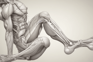
The lower limb, encompassing the hip, thigh, knee, leg, ankle, and foot, is a complex and intricate part of the human body. Understanding its anatomy is essential for maintaining good health, preventing injuries, and treating ailments. In this article, we'll focus on the lower limb's essential components: muscles, nerves, blood supply, bones, and joints.
Muscles
The lower limb contains numerous muscles, each with specific functions and roles. Key muscles for locomotion and stability include:
- Quadriceps: responsible for knee extension and hip flexion, essential for walking and running.
- Hamstrings: responsible for knee flexion and hip extension, important for walking and maintaining posture.
- Gluteal muscles: responsible for hip extension, abduction, and external rotation, critical for stabilizing the pelvis and providing power during running.
- Calves (gastrocnemius and soleus): responsible for plantarflexion, or pointing the toes downward, crucial for walking and maintaining balance.
Nerves
The lower limb's nerves originate from the spinal cord and innervate muscles and sensory structures. Key nerves include:
- Sciatic nerve: originates from the L4-S3 spinal nerve roots and is the largest nerve in the body. It supplies the muscles of the lower leg and foot.
- Femoral nerve: originates from the L2-L4 spinal nerve roots and is responsible for innervating the quadriceps and hip flexors.
- Tibial nerve: originates from the L4-S3 spinal nerve roots and supplies the muscles of the lower leg and foot, including the calf muscles and intrinsic foot muscles.
Blood Supply
Blood flow to the lower limb is essential for supplying oxygen and nutrients to muscles and maintaining proper tissue function. Key blood vessels include:
- Femoral artery: originates from the aorta and supplies blood to the hip, thigh, and knee.
- Popliteal artery: branches from the femoral artery and supplies blood to the knee and lower leg.
- Tibial artery: branches from the popliteal artery and supplies blood to the foot and toes.
Bones
The lower limb consists of 29 bones, including:
- Femur: the longest bone in the body, responsible for connecting the hip joint to the knee joint.
- Tibia and fibula: the lower leg bones that connect the knee joint to the ankle joint.
- Patella: a sesamoid bone found in the knee joint, which protects the knee joint and improves the mechanical efficiency of the quadriceps muscle.
Joints
The lower limb contains various joints, each with unique functions:
- Hip joint: a ball-and-socket joint formed by the head of the femur and the acetabulum of the pelvis.
- Knee joint: a hinge and pivot joint formed by the articulation of the femur, tibia, and fibula.
- Ankle joint: a complex joint formed by the articulation of the talus with the tibia and fibula.
- Subtalar joint: a complex joint formed by the articulation of the talus with the calcaneus.
In a healthy lower limb, all these components must work together to maintain mobility, stability, and balance. Understanding their anatomy is essential to preventing injuries, promoting healing, and optimizing overall lower limb function.
Studying That Suits You
Use AI to generate personalized quizzes and flashcards to suit your learning preferences.


