Podcast
Questions and Answers
What are the three main segments of the leg?
What are the three main segments of the leg?
- Femur, tibia, fibula
- Thigh, leg, foot (correct)
- Knee, ankle, foot
- Thigh, knee, foot
Which bone is the longest bone in the body?
Which bone is the longest bone in the body?
- Metatarsal
- Fibula
- Tibia
- Femur (correct)
What primary action does the tibialis anterior muscle perform?
What primary action does the tibialis anterior muscle perform?
- Dorsiflexion of the ankle (correct)
- Plantarflexion of the ankle
- Extension of the knee
- Flexion of the knee
Which of the following is NOT a type of joint in the foot?
Which of the following is NOT a type of joint in the foot?
How many bones are there in the foot?
How many bones are there in the foot?
Which muscles are primarily responsible for plantarflexion of the ankle?
Which muscles are primarily responsible for plantarflexion of the ankle?
Which arch is NOT one of the three main arches of the foot?
Which arch is NOT one of the three main arches of the foot?
What is the main function of intrinsic muscles of the foot?
What is the main function of intrinsic muscles of the foot?
What is the primary function of the quadriceps muscle group?
What is the primary function of the quadriceps muscle group?
Which bones are included in the tarsals of the foot?
Which bones are included in the tarsals of the foot?
Which of the following describes the function of the hamstrings?
Which of the following describes the function of the hamstrings?
What role do the intrinsic muscles of the foot primarily serve?
What role do the intrinsic muscles of the foot primarily serve?
Which muscle is primarily responsible for dorsiflexion of the foot?
Which muscle is primarily responsible for dorsiflexion of the foot?
Which compartment of the leg houses the primary evertors of the foot?
Which compartment of the leg houses the primary evertors of the foot?
What is the primary purpose of the medial longitudinal arch of the foot?
What is the primary purpose of the medial longitudinal arch of the foot?
Which muscle group is primarily involved in maintaining stability during foot movements?
Which muscle group is primarily involved in maintaining stability during foot movements?
What is the primary function of the hamstrings?
What is the primary function of the hamstrings?
Which muscle group is crucial for producing powerful upward movement during jumping?
Which muscle group is crucial for producing powerful upward movement during jumping?
Which intrinsic muscle helps support the arches of the foot?
Which intrinsic muscle helps support the arches of the foot?
How do extrinsic muscles contribute to foot movements?
How do extrinsic muscles contribute to foot movements?
Which aspect is essential for effective movement coordination between intrinsic and extrinsic muscles?
Which aspect is essential for effective movement coordination between intrinsic and extrinsic muscles?
What role does the leg musculature play in maintaining body posture?
What role does the leg musculature play in maintaining body posture?
What action do the calf muscles primarily assist with?
What action do the calf muscles primarily assist with?
Which muscle group is involved in the process of adducting the leg?
Which muscle group is involved in the process of adducting the leg?
What is the origin of the Vastus medialis muscle?
What is the origin of the Vastus medialis muscle?
Which head of the quadriceps femoris muscle inserts into the tibial tuberosity via the patellar tendon?
Which head of the quadriceps femoris muscle inserts into the tibial tuberosity via the patellar tendon?
What movement is primarily facilitated by the gastrocnemius and soleus muscles?
What movement is primarily facilitated by the gastrocnemius and soleus muscles?
Which muscle originates from the ischial tuberosity?
Which muscle originates from the ischial tuberosity?
What role do the hamstrings play during physical activities?
What role do the hamstrings play during physical activities?
In terms of muscle insertion, where does the semimembranosus insert?
In terms of muscle insertion, where does the semimembranosus insert?
Which muscle is crucial for maintaining balance and posture through dorsiflexion of the foot?
Which muscle is crucial for maintaining balance and posture through dorsiflexion of the foot?
What is the primary function of the intrinsic foot muscles?
What is the primary function of the intrinsic foot muscles?
Which muscle originates from the medial and lateral condyles of the femur?
Which muscle originates from the medial and lateral condyles of the femur?
Which of the following muscles inserts into the medial surface of the proximal tibia?
Which of the following muscles inserts into the medial surface of the proximal tibia?
Which muscle group is primarily involved in knee flexion and hip extension?
Which muscle group is primarily involved in knee flexion and hip extension?
What is the insertion point for the gastrocnemius and soleus muscles?
What is the insertion point for the gastrocnemius and soleus muscles?
Which of the following statements accurately describes the function of the tibialis anterior muscle?
Which of the following statements accurately describes the function of the tibialis anterior muscle?
The primary extensor of the knee is which muscle group?
The primary extensor of the knee is which muscle group?
Which of the following muscles primarily maintains posture during endurance activities?
Which of the following muscles primarily maintains posture during endurance activities?
What is the origin of the biceps femoris long head?
What is the origin of the biceps femoris long head?
Flashcards
Segments of the Leg
Segments of the Leg
The leg is divided into three parts: thigh, leg (shin), and foot.
Thigh Anatomy
Thigh Anatomy
Contains the femur, the longest bone in the body, crucial for weight-bearing.
Leg Bones
Leg Bones
The leg comprises two bones: tibia (shinbone) and fibula (provides stability).
Knee Joint Function
Knee Joint Function
Signup and view all the flashcards
Ankle Joint Function
Ankle Joint Function
Signup and view all the flashcards
Foot Structure
Foot Structure
Signup and view all the flashcards
Tarsal Bones
Tarsal Bones
Signup and view all the flashcards
Metatarsals
Metatarsals
Signup and view all the flashcards
Phalanges
Phalanges
Signup and view all the flashcards
Foot Arches
Foot Arches
Signup and view all the flashcards
Quadriceps
Quadriceps
Signup and view all the flashcards
Hamstrings
Hamstrings
Signup and view all the flashcards
Calf Muscles
Calf Muscles
Signup and view all the flashcards
Tibialis Anterior
Tibialis Anterior
Signup and view all the flashcards
Intrinsic Foot Muscles
Intrinsic Foot Muscles
Signup and view all the flashcards
Extrinsic Foot Muscles
Extrinsic Foot Muscles
Signup and view all the flashcards
Dorsiflexion
Dorsiflexion
Signup and view all the flashcards
Plantarflexion
Plantarflexion
Signup and view all the flashcards
Inversion
Inversion
Signup and view all the flashcards
Eversion
Eversion
Signup and view all the flashcards
Toe Flexion
Toe Flexion
Signup and view all the flashcards
Toe Extension
Toe Extension
Signup and view all the flashcards
Femur
Femur
Signup and view all the flashcards
Patella
Patella
Signup and view all the flashcards
Subtalar Joint
Subtalar Joint
Signup and view all the flashcards
Metatarsophalangeal Joints
Metatarsophalangeal Joints
Signup and view all the flashcards
Medial Longitudinal Arch
Medial Longitudinal Arch
Signup and view all the flashcards
Lateral Longitudinal Arch
Lateral Longitudinal Arch
Signup and view all the flashcards
Transverse Arch
Transverse Arch
Signup and view all the flashcards
Study Notes
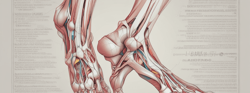
Anatomy of the Leg and Foot
Leg Anatomy
- Segments: Divided into three parts: thigh, leg (shin), and foot.
- Thigh: Contains the femur, the longest bone in the body.
- Leg: Composed of two bones – tibia (shinbone) and fibula.
- Knee Joint: Connects the thigh to the leg; allows flexion and extension.
- Ankle Joint: Connects the leg to the foot; allows dorsiflexion and plantarflexion.
Foot Anatomy
- Bones: 26 bones in the foot, including:
- Tarsals (7): Includes calcaneus (heel), talus, navicular, and cuboid.
- Metatarsals (5): Long bones in the midfoot.
- Phalanges (14): Bones of the toes (3 per toe, 2 in the big toe).
- Arches: Three main arches (medial, lateral, and transverse) provide stability and shock absorption.
- Joints: Numerous joints facilitate movement, including:
- Subtalar joint (between talus and calcaneus).
- Metatarsophalangeal joints (between metatarsals and phalanges).
Muscle Function of the Leg and Foot
Leg Muscles
- Quadriceps: Front thigh muscles; extend the knee.
- Hamstrings: Back thigh muscles; flex the knee and extend the hip.
- Calf Muscles: Includes gastrocnemius and soleus; responsible for plantarflexion of the ankle.
- Tibialis Anterior: Located in the front of the leg; dorsiflexes the ankle.
Foot Muscles
- Intrinsic Muscles: Located within the foot; provide fine motor control and support arches.
- Flexor digitorum brevis: Flexes toes.
- Abductor hallucis: Abducts the big toe.
- Extrinsic Muscles: Originate in the leg and affect foot movement.
- Flexor hallucis longus: Flexes the big toe.
- Extensor digitorum longus: Extends toes.
Muscle Actions
- Dorsiflexion: Raising the foot upwards; primarily by tibialis anterior.
- Plantarflexion: Pointing the toes downwards; primarily by gastrocnemius and soleus.
- Inversion/Eversion: Inversion (turning the foot inward) and eversion (turning the foot outward) involve several intrinsic and extrinsic muscles.
- Toe Flexion/Extension: Flexing and extending the toes for balance and movement control.
Leg Anatomy
- Divided into three segments: thigh, leg (shin), and foot.
- Thigh contains the femur, the longest bone in the body, essential for weight-bearing and movement.
- The leg consists of two bones: tibia (weight-bearing shinbone) and fibula (provides stability).
- The knee joint connects the thigh to the leg, allowing for flexion and extension to facilitate walking and running.
- The ankle joint connects the leg to the foot, permitting dorsiflexion (raising the foot) and plantarflexion (pointing the toes).
Foot Anatomy
- Comprises 26 bones, divided into three categories: tarsals, metatarsals, and phalanges.
- Tarsals include 7 bones such as the calcaneus (heel) and talus, which play crucial roles in movement and balance.
- Metatarsals are 5 long bones in the midfoot, supporting weight distribution.
- Phalanges consist of 14 bones for the toes (3 per toe, 2 in the big toe), enabling toe movement and grip.
- The foot features three main arches (medial, lateral, and transverse) that enhance stability and shock absorption during activities.
- Numerous joints facilitate foot movement, including the subtalar joint (allows inversion and eversion) and metatarsophalangeal joints (connect metatarsals to phalanges).
Muscle Function of the Leg and Foot
Leg Muscles
- Quadriceps are the front thigh muscles responsible for knee extension, essential for standing and squatting.
- Hamstrings are located at the back of the thigh, functioning to flex the knee and extend the hip, crucial for running and jumping.
- Calf muscles, including gastrocnemius and soleus, enable plantarflexion of the ankle, important for walking, running, and jumping.
- Tibialis anterior, situated in the front of the leg, is responsible for dorsiflexing the ankle, helping in walking and preventing dragging of the toes.
Foot Muscles
- Intrinsic muscles reside within the foot, providing fine motor control and supporting arches for stability.
- Flexor digitorum brevis is responsible for flexing the toes, aiding in balance.
- Abductor hallucis functions to abduct the big toe, important for lateral stability.
- Extrinsic muscles originate in the leg and primarily influence foot movement.
- Flexor hallucis longus flexes the big toe, critical for propulsion during walking.
- Extensor digitorum longus extends the toes, aiding in balance and movement.
Muscle Actions
- Dorsiflexion raises the foot upwards, primarily executed by the tibialis anterior.
- Plantarflexion points the toes downwards, mainly driven by gastrocnemius and soleus.
- Inversion involves turning the foot inward, while eversion turns it outward, both requiring coordination between intrinsic and extrinsic muscles.
- Toe flexion and extension are essential for balance and movement control, allowing for adjustments during locomotion.
Leg Anatomy
- Femur: Longest bone in the body, located in the thigh.
- Patella: Protects the knee joint.
- Tibia: Larger shin bone, responsible for weight bearing.
- Fibula: Smaller bone alongside the tibia, aids in stability.
Joints
- Knee Joint: Connects femur, tibia, and patella; facilitates flexion and extension.
- Ankle Joint: Connects tibia and fibula with the talus; allows dorsiflexion (lifting foot) and plantarflexion (pointing foot).
Regions
- Thigh: Contains critical muscles and major blood vessels.
- Leg: Divided into anterior, posterior, and lateral compartments.
- Foot: Comprised of tarsals, metatarsals, and phalanges.
Foot Anatomy
- Tarsals: Seven bones including the talus and calcaneus, foundational for foot structure.
- Metatarsals: Five bones forming the midfoot region.
- Phalanges: Fourteen bones in the toes, allowing flexibility and movement.
Foot Arches
- Medial Longitudinal Arch: Supports body weight and absorbs shock.
- Lateral Longitudinal Arch: Contributes to stability and balance.
- Transverse Arch: Helps maintain the shape of the foot.
Leg Muscle Groups
-
Anterior Compartment:
- Main Muscles: Quadriceps (rectus femoris, vastus lateralis, vastus medialis, vastus intermedius).
- Function: Assists in knee extension and hip flexion.
-
Posterior Compartment:
- Main Muscles: Hamstrings (biceps femoris, semitendinosus, semimembranosus).
- Function: Facilitates knee flexion and hip extension.
-
Lateral Compartment:
- Main Muscles: Fibularis longus and fibularis brevis.
- Function: Aids foot eversion and ankle stability.
Foot Muscle Groups
-
Intrinsic Muscles: Located within the foot for toe movements and stability; includes abductor hallucis, flexor digitorum brevis, and adductor hallucis.
-
Extrinsic Muscles: Originates in the leg but influences foot movement; includes tibialis anterior for dorsiflexion, and gastrocnemius and soleus for plantarflexion.
Overall Functions
- Locomotion: Muscles integrate for walking, running, and jumping motions.
- Balance and Stability: Muscle coordination is essential during dynamic activities and static postures.
- Shock Absorption: Arches work with muscles to mitigate impact during weight-bearing activities.
Major Muscle Groups in the Leg
- Quadriceps: Four muscles located at the front of the thigh; primarily responsible for extending the knee.
- Hamstrings: Comprised of three muscles situated at the back of the thigh; serve the function of flexing the knee.
- Calf Muscles: Include the gastrocnemius and soleus; crucial for plantar flexion and movement of the foot.
- Adductors: Group of muscles located in the inner thigh; responsible for adducting (pulling towards the midline) the leg.
Muscles of the Foot
- Intrinsic Muscles: Fully contained within the foot; responsible for fine motor control and supporting the foot's arches. Examples include abductor hallucis, flexor digitorum brevis, and dorsal interossei.
- Extrinsic Muscles: Originate in the leg and insert into the foot; these muscles facilitate gross movement. Examples include tibialis anterior, peroneus longus, and flexor hallucis longus.
Functions Of Leg Muscles
- Movement:
- Walking and running involve coordinated contractions of leg muscles, enabling locomotion.
- Jumping requires strong contractions from both the quadriceps and calf muscles for vertical elevation.
- Stability and Support:
- Leg muscles contribute to balance, helping maintain stability during standing and dynamic movements.
- They play a vital role in supporting overall body posture and alignment.
- Shock Absorption:
- Leg muscles absorb impact during activities such as running and jumping, protecting joints from stress.
Foot Muscle Coordination
- Intrinsic Coordination:
- Intrinsic muscles stabilize the arches of the foot and enhance balance by adapting to varying surfaces.
- Extrinsic Influence:
- Extrinsic muscles manage larger movements, offering stability during physical activities.
- Effective movement requires a harmonious coordination between intrinsic and extrinsic muscles.
- Neuromuscular Control:
- Feedback from proprioceptors in muscles and joints is essential for proper muscle function.
- Coordination is vital for agility and the ability to make quick directional changes.
Muscle Origins
- Quadriceps Femoris consists of four heads with distinct origins:
- Rectus femoris: Originates from the anterior inferior iliac spine (AIIS) and superior edge of the acetabulum.
- Vastus lateralis: Arises from the greater trochanter and linea aspera of the femur.
- Vastus medialis: Originates from the intertrochanteric line and linea aspera of the femur.
- Vastus intermedius: Emerges from the anterior and lateral surfaces of the femur.
- Hamstrings are a group of three muscles with common origins:
- Biceps femoris: Has a long head originating from the ischial tuberosity and a short head from the linea aspera.
- Semitendinosus and Semimembranosus: Both originate from the ischial tuberosity.
- Calf Muscles play a crucial role in lower leg movement:
- Gastrocnemius: Originates from the medial and lateral condyles of the femur.
- Soleus: Originates from the posterior surfaces of the tibia and fibula.
- Tibialis Anterior originates from the lateral condyle and upper two-thirds of the tibia.
Muscle Insertions
- Quadriceps Femoris heads insert into the patellar tendon, which continues to the tibial tuberosity, enabling effective knee extension.
- Hamstrings insert into different locations:
- Biceps femoris: Inserts at the head of the fibula.
- Semitendinosus: Inserts on the medial surface of the proximal tibia at the pes anserinus.
- Semimembranosus: Inserts on the posterior part of the medial condyle of the tibia.
- Calf Muscles insert into the calcaneus via the Achilles tendon, facilitating foot movement.
- Tibialis Anterior inserts at the medial cuneiform and base of the first metatarsal, playing a role in foot movement.
Functional Anatomy
- Quadriceps Femoris serves as the primary knee extensor, essential for activities like walking, running, and jumping.
- Hamstrings are vital for knee flexion and hip extension, significantly impacting activities such as running and cycling.
- Calf Muscles (gastrocnemius and soleus) work in unison to plantar flex the foot, which is critical for locomotion.
- Tibialis Anterior is responsible for dorsiflexing the foot at the ankle, aiding in balance and posture maintenance.
- Intrinsic Foot Muscles support the arches of the foot and provide fine motor control, enhancing balance and movement.
- Overall Function of leg and foot muscles is to coordinate locomotion, stability, and posture, enabling effective movement.
Muscle Origins
- Quadriceps Femoris
- Rectus Femoris originates from the anterior inferior iliac spine (AIIS) and acetabulum's superior margin.
- Vastus Lateralis arises from the greater trochanter, intertrochanteric line, and linea aspera.
- Vastus Medialis is located at the intertrochanteric line and linea aspera.
- Vastus Intermedius develops from the anterior and lateral surfaces of the femur.
- Hamstrings
- Biceps Femoris has its long head beginning at the ischial tuberosity, while the short head starts from the linea aspera.
- Semitendinosus and Semimembranosus both originate from the ischial tuberosity.
- Calf Muscles
- Gastrocnemius originates from the medial and lateral condyles of the femur.
- Soleus starts at the posterior surfaces of the tibia and fibula.
- Tibialis Anterior
- Arises from the lateral condyle and upper two-thirds of the tibia.
Muscle Insertions
- Quadriceps Femoris
- All heads insert into the patellar tendon, leading to attachment at the tibial tuberosity.
- Hamstrings
- Biceps Femoris inserts at the head of the fibula, while Semitendinosus attaches to the medial surface of the proximal tibia (pes anserinus).
- Semimembranosus connects to the medial condyle of the tibia.
- Calf Muscles
- Gastrocnemius and Soleus insert into the calcaneus via the Achilles tendon.
- Tibialis Anterior
- Inserts at the medial cuneiform and base of the first metatarsal.
Functional Anatomy
- Quadriceps Femoris
- Major extensor of the knee, essential for activities such as walking, running, and jumping.
- Hamstrings
- Serves as primary knee flexors and hip extensors, important for running, cycling, and motions employing hip extension.
- Calf Muscles
- Gastrocnemius contributes to knee flexion and ankle plantarflexion, vital for explosive movements.
- Soleus stabilizes the leg during standing and plays a key role in endurance activities and maintaining posture.
- Tibialis Anterior
- Responsible for foot dorsiflexion and ankle inversion; crucial for walking and preventing foot drop during gait.
- Overall Functionality
- Muscles in the leg and foot collaborate for locomotion, balance, and stability.
- Effective coordination among muscle groups is necessary for optimal movement patterns in various activities.
Studying That Suits You
Use AI to generate personalized quizzes and flashcards to suit your learning preferences.


