Podcast
Questions and Answers
If a patient is unable to plantarflex their foot and has difficulty with toe flexion, which nerve is MOST likely affected?
If a patient is unable to plantarflex their foot and has difficulty with toe flexion, which nerve is MOST likely affected?
- Deep Fibular Nerve
- Sural Nerve
- Superficial Fibular Nerve
- Tibial Nerve (correct)
A ballet dancer is experiencing pain and limited range of motion during plantarflexion. Palpation reveals inflammation around the calcaneal tendon. Which condition is MOST likely causing these symptoms?
A ballet dancer is experiencing pain and limited range of motion during plantarflexion. Palpation reveals inflammation around the calcaneal tendon. Which condition is MOST likely causing these symptoms?
- Metatarsal Stress Fracture
- Achilles Tendinitis (correct)
- Ankle Sprain (ATFL)
- Plantar Fasciitis
Following a severe ankle inversion injury, a patient exhibits instability during weight-bearing and has difficulty with eversion. Which ligament(s) is/are MOST likely compromised?
Following a severe ankle inversion injury, a patient exhibits instability during weight-bearing and has difficulty with eversion. Which ligament(s) is/are MOST likely compromised?
- Posterior Talofibular Ligament (PTFL)
- Calcaneofibular and Anterior Talofibular Ligaments (correct)
- Anterior Talofibular Ligament (ATFL)
- Deltoid Ligament
A patient reports numbness and paresthesia along the lateral aspect of their foot and ankle. Which nerve is MOST likely involved?
A patient reports numbness and paresthesia along the lateral aspect of their foot and ankle. Which nerve is MOST likely involved?
A patient presents with pain in the medial arch of their foot, which worsens with weight-bearing activities. Imaging reveals no acute fracture, but the pain is localized along the plantar aspect of the heel. Which condition is MOST likely?
A patient presents with pain in the medial arch of their foot, which worsens with weight-bearing activities. Imaging reveals no acute fracture, but the pain is localized along the plantar aspect of the heel. Which condition is MOST likely?
During a biomechanical assessment, a physical therapist observes excessive pronation during the loading response of the gait cycle. Which combination of movements is occurring at the subtalar joint?
During a biomechanical assessment, a physical therapist observes excessive pronation during the loading response of the gait cycle. Which combination of movements is occurring at the subtalar joint?
A patient has suffered damage to the deep fibular nerve. Which muscular action would be MOST affected?
A patient has suffered damage to the deep fibular nerve. Which muscular action would be MOST affected?
Which of the following is NOT a primary function of the arches of the foot?
Which of the following is NOT a primary function of the arches of the foot?
A patient struggles to evert their foot against resistance during a manual muscle test. Which muscle group is MOST likely to be weak or impaired?
A patient struggles to evert their foot against resistance during a manual muscle test. Which muscle group is MOST likely to be weak or impaired?
Which artery primarily supplies blood to the anterior compartment of the lower leg, and what does it become in the foot?
Which artery primarily supplies blood to the anterior compartment of the lower leg, and what does it become in the foot?
A patient has limited dorsiflexion following immobilization after a lower leg fracture. Which of the following interventions would MOST effectively address this limitation?
A patient has limited dorsiflexion following immobilization after a lower leg fracture. Which of the following interventions would MOST effectively address this limitation?
Which of the following ligaments primarily resists excessive eversion of the ankle?
Which of the following ligaments primarily resists excessive eversion of the ankle?
During the push-off phase of gait, what combination of movements is occurring at the subtalar joint to create a rigid lever for propulsion?
During the push-off phase of gait, what combination of movements is occurring at the subtalar joint to create a rigid lever for propulsion?
Damage to the superficial fibular nerve would MOST significantly impair which muscular function?
Damage to the superficial fibular nerve would MOST significantly impair which muscular function?
Which joint is classified as a hinge (ginglymus) joint with one degree of freedom, primarily allowing for plantarflexion and dorsiflexion?
Which joint is classified as a hinge (ginglymus) joint with one degree of freedom, primarily allowing for plantarflexion and dorsiflexion?
In a closed chain kinetic movement, what occurs at the ankle and foot complex?
In a closed chain kinetic movement, what occurs at the ankle and foot complex?
Which of the following best describes the primary function of the windlass mechanism?
Which of the following best describes the primary function of the windlass mechanism?
A patient is diagnosed with plantar fasciitis. Which of the following signs and symptoms is LEAST likely to be associated with this condition?
A patient is diagnosed with plantar fasciitis. Which of the following signs and symptoms is LEAST likely to be associated with this condition?
Which combination of bones forms Chopart's joint, contributing significantly to foot inversion and eversion?
Which combination of bones forms Chopart's joint, contributing significantly to foot inversion and eversion?
Which of the following scenarios would MOST likely result in a stress fracture of the metatarsals?
Which of the following scenarios would MOST likely result in a stress fracture of the metatarsals?
A patient presents with medial ankle pain and reports a recent injury involving forceful eversion. Which ligament is MOST likely injured?
A patient presents with medial ankle pain and reports a recent injury involving forceful eversion. Which ligament is MOST likely injured?
Which nerve innervates the tibialis anterior muscle, responsible for dorsiflexion of the ankle?
Which nerve innervates the tibialis anterior muscle, responsible for dorsiflexion of the ankle?
Which bones primarily form the lateral longitudinal arch of the foot?
Which bones primarily form the lateral longitudinal arch of the foot?
Which ligaments are MOST commonly involved in a lateral ankle sprain?
Which ligaments are MOST commonly involved in a lateral ankle sprain?
What is the primary function of the distal tibiofibular joint?
What is the primary function of the distal tibiofibular joint?
Which movement occurs at the ankle joint during the loading response phase of gait?
Which movement occurs at the ankle joint during the loading response phase of gait?
What is the primary function of the fibularis longus muscle?
What is the primary function of the fibularis longus muscle?
Which of the following muscles is located in the deep posterior compartment of the lower leg?
Which of the following muscles is located in the deep posterior compartment of the lower leg?
Which of the following is a characteristic of the medial longitudinal arch of the foot?
Which of the following is a characteristic of the medial longitudinal arch of the foot?
A patient is diagnosed with a Lisfranc injury. Which joint is affected?
A patient is diagnosed with a Lisfranc injury. Which joint is affected?
Following an injury, a patient has difficulty with plantarflexion and inversion. Which muscle group is MOST likely affected?
Following an injury, a patient has difficulty with plantarflexion and inversion. Which muscle group is MOST likely affected?
The metatarsophalangeal (MTP) joints allowing flexion, extension, abduction, and adduction are classified as what type of joint?
The metatarsophalangeal (MTP) joints allowing flexion, extension, abduction, and adduction are classified as what type of joint?
Which of the following statements best describes the term 'mortise' in relation to the ankle joint?
Which of the following statements best describes the term 'mortise' in relation to the ankle joint?
Which of the following actions would be MOST limited by a restriction in subtalar joint motion?
Which of the following actions would be MOST limited by a restriction in subtalar joint motion?
Which of the following represents the MOST accurate description of pronation?
Which of the following represents the MOST accurate description of pronation?
Where does the anterior tibial artery transition into the dorsalis pedis artery?
Where does the anterior tibial artery transition into the dorsalis pedis artery?
What is the classification of the proximal tibiofibular joint?
What is the classification of the proximal tibiofibular joint?
A patient exhibits a noticeable lack of arch support and experiences pain along the plantar aspect of the foot. Radiographic imaging reveals no fractures, but clinical examination indicates significant laxity in the spring ligament. What is the MOST likely primary consequence of this condition regarding foot biomechanics?
A patient exhibits a noticeable lack of arch support and experiences pain along the plantar aspect of the foot. Radiographic imaging reveals no fractures, but clinical examination indicates significant laxity in the spring ligament. What is the MOST likely primary consequence of this condition regarding foot biomechanics?
Following a traumatic injury to the lower leg, a patient presents with an inability to actively evert the foot and reports a loss of sensation on the lateral aspect of the lower leg and dorsum of the foot, excluding the webspace between the first and second toes. Which nerve is MOST likely injured?
Following a traumatic injury to the lower leg, a patient presents with an inability to actively evert the foot and reports a loss of sensation on the lateral aspect of the lower leg and dorsum of the foot, excluding the webspace between the first and second toes. Which nerve is MOST likely injured?
A long-distance runner reports progressive onset of pain along the distal third of the tibia. Examination reveals localized tenderness and pain with percussion over the bone. A bone scan confirms a stress reaction. Which of the following biomechanical factors is MOST likely contributing to this injury?
A long-distance runner reports progressive onset of pain along the distal third of the tibia. Examination reveals localized tenderness and pain with percussion over the bone. A bone scan confirms a stress reaction. Which of the following biomechanical factors is MOST likely contributing to this injury?
A patient with chronic ankle instability is being evaluated. During gait analysis, the physical therapist observes that the patient's subtalar joint is excessively pronated throughout the stance phase. Which muscle is MOST likely weakened, contributing to this excessive pronation?
A patient with chronic ankle instability is being evaluated. During gait analysis, the physical therapist observes that the patient's subtalar joint is excessively pronated throughout the stance phase. Which muscle is MOST likely weakened, contributing to this excessive pronation?
A patient is diagnosed with tarsal tunnel syndrome. Which combination of signs and symptoms would MOST likely be present?
A patient is diagnosed with tarsal tunnel syndrome. Which combination of signs and symptoms would MOST likely be present?
Flashcards
Tibia
Tibia
Larger, weight-bearing bone of the lower leg.
Fibula
Fibula
Smaller, non-weight-bearing bone located laterally in the lower leg.
Proximal Tibiofibular Joint
Proximal Tibiofibular Joint
A synovial joint that allows slight gliding and rotation between the tibia and fibula.
Distal Tibiofibular Joint
Distal Tibiofibular Joint
Signup and view all the flashcards
Ankle Joint (Talocrural Joint)
Ankle Joint (Talocrural Joint)
Signup and view all the flashcards
Mortise of the Ankle
Mortise of the Ankle
Signup and view all the flashcards
Talocrural Joint Function
Talocrural Joint Function
Signup and view all the flashcards
Subtalar Joint (Talocalcaneal Joint)
Subtalar Joint (Talocalcaneal Joint)
Signup and view all the flashcards
Midfoot Joints
Midfoot Joints
Signup and view all the flashcards
Chopart's Joint
Chopart's Joint
Signup and view all the flashcards
Lisfranc Joint
Lisfranc Joint
Signup and view all the flashcards
Metatarsophalangeal (MTP) Joints
Metatarsophalangeal (MTP) Joints
Signup and view all the flashcards
Interphalangeal (IP) Joints
Interphalangeal (IP) Joints
Signup and view all the flashcards
Medial Longitudinal Arch
Medial Longitudinal Arch
Signup and view all the flashcards
Lateral Longitudinal Arch
Lateral Longitudinal Arch
Signup and view all the flashcards
Transverse Arch
Transverse Arch
Signup and view all the flashcards
Plantar Fascia
Plantar Fascia
Signup and view all the flashcards
Deltoid Ligament
Deltoid Ligament
Signup and view all the flashcards
Anterior Talofibular Ligament (ATFL)
Anterior Talofibular Ligament (ATFL)
Signup and view all the flashcards
Calcaneofibular Ligament (CFL)
Calcaneofibular Ligament (CFL)
Signup and view all the flashcards
Posterior Talofibular Ligament (PTFL)
Posterior Talofibular Ligament (PTFL)
Signup and view all the flashcards
Anterior Compartment Muscles
Anterior Compartment Muscles
Signup and view all the flashcards
Lateral Compartment Muscles
Lateral Compartment Muscles
Signup and view all the flashcards
Superficial Posterior Compartment Muscles
Superficial Posterior Compartment Muscles
Signup and view all the flashcards
Deep Posterior Compartment Muscles
Deep Posterior Compartment Muscles
Signup and view all the flashcards
Open Chain Kinematics
Open Chain Kinematics
Signup and view all the flashcards
Closed Chain Kinematics
Closed Chain Kinematics
Signup and view all the flashcards
Pronation
Pronation
Signup and view all the flashcards
Supination
Supination
Signup and view all the flashcards
Pronation During Gait
Pronation During Gait
Signup and view all the flashcards
Supination During Gait
Supination During Gait
Signup and view all the flashcards
Tibial Nerve
Tibial Nerve
Signup and view all the flashcards
Common Fibular (Peroneal) Nerve
Common Fibular (Peroneal) Nerve
Signup and view all the flashcards
Deep Fibular Nerve
Deep Fibular Nerve
Signup and view all the flashcards
Superficial Fibular Nerve
Superficial Fibular Nerve
Signup and view all the flashcards
Sural Nerve
Sural Nerve
Signup and view all the flashcards
Anterior Tibial Artery
Anterior Tibial Artery
Signup and view all the flashcards
Posterior Tibial Artery
Posterior Tibial Artery
Signup and view all the flashcards
Fibular (Peroneal) Artery
Fibular (Peroneal) Artery
Signup and view all the flashcards
Ankle Sprains
Ankle Sprains
Signup and view all the flashcards
Plantar Fasciitis
Plantar Fasciitis
Signup and view all the flashcards
Achilles Tendinitis
Achilles Tendinitis
Signup and view all the flashcards
Stress Fractures (Foot)
Stress Fractures (Foot)
Signup and view all the flashcards
Study Notes
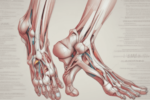
- The leg and foot complex is essential for weight bearing, balance, and locomotion.
- It consists of the lower leg (tibia and fibula), ankle, hindfoot (talus and calcaneus), midfoot (navicular, cuboid, and cuneiforms), and forefoot (metatarsals and phalanges).
- Bones, joints, ligaments, muscles, nerves, and blood vessels all contribute to its function.
Bones of the Lower Leg
- Tibia: The larger, weight-bearing bone of the lower leg.
- Fibula: The smaller, non-weight-bearing bone located laterally.
- The proximal Tibiofibular joint is a synovial joint that allows slight gliding and rotation.
- The distal Tibiofibular joint is a syndesmosis joint connected by strong interosseous ligaments.
Ankle Joint (Talocrural Joint)
- Formed by the articulation of the distal tibia and fibula.
- A mortise is created by the tibia and fibula which surrounds the talus.
- Functions primarily in plantarflexion and dorsiflexion.
- Classified as a hinge (ginglymus) joint with one degree of freedom.
Subtalar Joint (Talocalcaneal Joint)
- Formed by the articulation between the talus and calcaneus.
- Allows for inversion and eversion.
- Important for adapting to uneven surfaces.
Midfoot Joints
- Consists of the navicular, cuboid, and three cuneiform bones.
- Chopart's joint (talonavicular and calcaneocuboid joints) contributes to foot inversion and eversion.
- Lisfranc joint (tarsometatarsal joints) provides stability to the midfoot.
Forefoot Joints
- Metatarsophalangeal (MTP) joints: condyloid joints that allow flexion, extension, abduction, and adduction.
- Interphalangeal (IP) joints: hinge joints that allow flexion and extension of the toes.
Arches of the Foot
- Medial Longitudinal Arch: Formed by the calcaneus, talus, navicular, cuneiforms, and the first three metatarsals.
- Lateral Longitudinal Arch: Formed by the calcaneus, cuboid, and fourth and fifth metatarsals.
- Transverse Arch: Formed by the cuneiforms, cuboid, and metatarsal bases.
- Arches provide shock absorption and distribute weight during weight-bearing activities.
- Plantar fascia supports the arches and contributes to the windlass mechanism.
Ligaments of the Ankle and Foot
- Deltoid Ligament: Located on the medial side of the ankle.
- Resist excessive eversion.
- Anterior Talofibular Ligament (ATFL): Located on the lateral side of the ankle.
- Calcaneofibular Ligament (CFL): Located on the lateral side of the ankle.
- Posterior Talofibular Ligament (PTFL): Located on the lateral side of the ankle.
- ATFL, CFL, and PTFL resist excessive inversion and plantarflexion
Muscles of the Lower Leg
- Anterior Compartment: Includes tibialis anterior, extensor hallucis longus, extensor digitorum longus, and fibularis tertius.
- Responsible for dorsiflexion and toe extension
- Lateral Compartment: Includes fibularis longus and fibularis brevis.
- Responsible for eversion of the foot.
- Superficial Posterior Compartment: Includes gastrocnemius, soleus, and plantaris.
- Responsible for plantarflexion of the ankle.
- Deep Posterior Compartment: Includes tibialis posterior, flexor digitorum longus, and flexor hallucis longus.
- Responsible for plantarflexion, inversion, and toe flexion.
Ankle and Foot Kinematics
- Open Chain: The foot is not in contact with the ground, and the leg rotates.
- Closed Chain: The foot is fixed on the ground, and the leg moves over the foot.
- Pronation: A combination of dorsiflexion, eversion, and abduction.
- Supination: A combination of plantarflexion, inversion, and adduction.
- During the gait cycle, the foot pronates during the loading response to absorb shock and adapts to uneven surfaces.
- During push-off, the foot supinates to provide a rigid lever for propulsion.
Nerves of the Lower Leg and Foot
- Tibial Nerve: Innervates the muscles in the posterior compartment of the lower leg and the plantar muscles of the foot.
- Common Fibular (Peroneal) Nerve: Divides into the superficial and deep fibular nerves.
- Deep Fibular Nerve: Innervates the muscles in the anterior compartment of the lower leg.
- Superficial Fibular Nerve: Innervates the muscles in the lateral compartment of the lower leg.
- Sural Nerve: Sensory innervation to the lateral aspect of the foot and ankle.
Blood Supply
- Anterior tibial artery supplies the anterior compartment of the leg and becomes the dorsalis pedis artery in the foot.
- Posterior tibial artery supplies the posterior compartment of the leg and branches into the medial and lateral plantar arteries in the foot.
- Fibular (Peroneal) artery supplies the lateral compartment of the leg.
Functional Anatomy
- Weight Bearing: The foot and ankle must be able to support the body's weight during standing, walking, and running.
- Balance: The muscles of the lower leg and foot work together to maintain balance and prevent falls.
- Gait: The ankle and foot play a crucial role in the gait cycle, providing shock absorption, stability, and propulsion.
- Adaptability: The joints of the foot allow it to adapt to uneven surfaces and maintain contact with the ground.
Kinesiology of the Ankle and Foot
- Range of Motion: Ankle dorsiflexion is approximately 20 degrees, and plantarflexion is approximately 50 degrees.
- Subtalar joint inversion and eversion range from 20-30 degrees.
- Muscle Actions: The muscles of the lower leg work in synergy to control movement at the ankle and foot.
- During plantarflexion, the gastrocnemius and soleus are the primary movers.
- During dorsiflexion, the tibialis anterior is the primary mover.
- During eversion, the fibularis longus and brevis are the primary movers.
- During inversion, the tibialis posterior and anterior are the primary movers.
Common Injuries
- Ankle Sprains: Lateral ankle sprains are the most common, involving the ATFL, CFL, and PTFL.
- Plantar Fasciitis: Inflammation of the plantar fascia, causing heel pain.
- Achilles Tendinitis: Inflammation of the Achilles tendon, causing pain in the back of the heel.
- Stress Fractures: Common in the metatarsals, caused by repetitive stress and overuse.
Studying That Suits You
Use AI to generate personalized quizzes and flashcards to suit your learning preferences.