Podcast
Questions and Answers
Which ligament is primarily responsible for stabilizing the subtalar joint?
Which ligament is primarily responsible for stabilizing the subtalar joint?
- Deltoid ligament (correct)
- Calcaneofibular ligament
- Anterior talofibular ligament
- Posterior talofibular ligament
Which muscle is primarily responsible for dorsiflexion of the foot?
Which muscle is primarily responsible for dorsiflexion of the foot?
- Tibialis anterior (correct)
- Tibialis posterior
- Soleus
- Flexor hallucis longus
What is the primary function of the plantar arches in the foot?
What is the primary function of the plantar arches in the foot?
- Provide structural support and shock absorption (correct)
- Facilitate nerve transmission
- Aid in blood circulation
- Enhance muscle attachment
Which nerve is primarily responsible for innervating most of the intrinsic muscles of the foot?
Which nerve is primarily responsible for innervating most of the intrinsic muscles of the foot?
Which joint allows for inversion and eversion movements in the foot?
Which joint allows for inversion and eversion movements in the foot?
Which ligament is primarily responsible for stabilizing both the subtalar and talocalcaneonavicular joints?
Which ligament is primarily responsible for stabilizing both the subtalar and talocalcaneonavicular joints?
Which joint is primarily involved with the movement at the calcaneocuboid joint?
Which joint is primarily involved with the movement at the calcaneocuboid joint?
What is another name for the plantar calcaneonavicular ligament?
What is another name for the plantar calcaneonavicular ligament?
Which of the following ligaments is not involved in stabilizing the talocalcaneonavicular joint?
Which of the following ligaments is not involved in stabilizing the talocalcaneonavicular joint?
Which two bones articulate at the subtalar joint?
Which two bones articulate at the subtalar joint?
What is the main function of tendon sheaths?
What is the main function of tendon sheaths?
Which nerve innervates the extensor hallucis brevis muscle?
Which nerve innervates the extensor hallucis brevis muscle?
Which of the following muscles does the extensor digitorum brevis work closely with?
Which of the following muscles does the extensor digitorum brevis work closely with?
What is the origin of the extensor hallucis brevis muscle?
What is the origin of the extensor hallucis brevis muscle?
Which of the following nerves provides cutaneous innervation to the skin on each side of the first interphalangeal space?
Which of the following nerves provides cutaneous innervation to the skin on each side of the first interphalangeal space?
What type of membrane do tendon sheaths contain?
What type of membrane do tendon sheaths contain?
Which nerve is classified as a part of the common fibular nerve?
Which nerve is classified as a part of the common fibular nerve?
What is the primary role of the superficial fibular nerve?
What is the primary role of the superficial fibular nerve?
Which statement about the interossei muscles is true?
Which statement about the interossei muscles is true?
What is the primary role of the extensor hood?
What is the primary role of the extensor hood?
Which nerve is responsible for innervating all interossei muscles?
Which nerve is responsible for innervating all interossei muscles?
What is the anatomical relationship of the posterior tibial artery as it passes the medial malleolus?
What is the anatomical relationship of the posterior tibial artery as it passes the medial malleolus?
Which of the following correctly lists the branches of the tibial nerve?
Which of the following correctly lists the branches of the tibial nerve?
What terms describe the actions of the dorsal and plantar interossei muscles?
What terms describe the actions of the dorsal and plantar interossei muscles?
What structure allows the extensor digitorum tendons to act on multiple joints?
What structure allows the extensor digitorum tendons to act on multiple joints?
How do the posterior tibial artery and nerve initially travel through the leg?
How do the posterior tibial artery and nerve initially travel through the leg?
Which muscle is innervated by the medial plantar nerve?
Which muscle is innervated by the medial plantar nerve?
Which artery forms the deep plantar arch in the foot?
Which artery forms the deep plantar arch in the foot?
What is the primary ligament supporting the medial longitudinal arch?
What is the primary ligament supporting the medial longitudinal arch?
Which muscle is NOT innervated by the lateral plantar nerve?
Which muscle is NOT innervated by the lateral plantar nerve?
Which statement about the plantar arteries is correct?
Which statement about the plantar arteries is correct?
What is a key function of the plantar aponeurosis?
What is a key function of the plantar aponeurosis?
Which muscle is correctly paired with its function?
Which muscle is correctly paired with its function?
Which of the following structures provides arch support in the foot?
Which of the following structures provides arch support in the foot?
Which ligaments are involved in the high ankle sprain?
Which ligaments are involved in the high ankle sprain?
What feature differentiates the talocrural joint's anterior and posterior regions?
What feature differentiates the talocrural joint's anterior and posterior regions?
In relation to the proximal tibiofibular joint, which statement is accurate?
In relation to the proximal tibiofibular joint, which statement is accurate?
What is the primary role of the deltoid ligament at the talocrural joint?
What is the primary role of the deltoid ligament at the talocrural joint?
Which statement correctly describes the role of the distal tibiofibular joint?
Which statement correctly describes the role of the distal tibiofibular joint?
Which structure plays the most significant role in the motion occurring at the subtalar joint?
Which structure plays the most significant role in the motion occurring at the subtalar joint?
What defining characteristic is associated with a grade 1 ankle sprain?
What defining characteristic is associated with a grade 1 ankle sprain?
Which aspects of the talocalcaneonavicular joint are correctly defined?
Which aspects of the talocalcaneonavicular joint are correctly defined?
What is the function of the interosseous membrane in the context of the proximal tibiofibular joint?
What is the function of the interosseous membrane in the context of the proximal tibiofibular joint?
Which ankle joint receives the widest anterior talus during dorsiflexion?
Which ankle joint receives the widest anterior talus during dorsiflexion?
Which joint type allows for the necessary movement of the wider anterior talus during dorsiflexion?
Which joint type allows for the necessary movement of the wider anterior talus during dorsiflexion?
Which grade of ankle sprain typically involves partial tearing of the anterior talofibular ligament?
Which grade of ankle sprain typically involves partial tearing of the anterior talofibular ligament?
In which type of ankle injury does the stress view show involvement of both the anterior talofibular and calcaneofibular ligaments?
In which type of ankle injury does the stress view show involvement of both the anterior talofibular and calcaneofibular ligaments?
What essential motion occurs at the subtalar joint that allows for inversion and eversion of the foot?
What essential motion occurs at the subtalar joint that allows for inversion and eversion of the foot?
What surgical procedure is commonly performed to treat severe ankle fractures requiring internal fixation?
What surgical procedure is commonly performed to treat severe ankle fractures requiring internal fixation?
Which aspect of the talocrural joint contributes to its unique function during full dorsiflexion?
Which aspect of the talocrural joint contributes to its unique function during full dorsiflexion?
What feature distinguishes a grade 3 ankle sprain from lower-grade injuries?
What feature distinguishes a grade 3 ankle sprain from lower-grade injuries?
What is the primary function of tendon sheaths in the context of joint mechanics?
What is the primary function of tendon sheaths in the context of joint mechanics?
Which ligament is least involved in the stabilization of the talocrural joint?
Which ligament is least involved in the stabilization of the talocrural joint?
In the event of an ankle sprain, which type is characterized by a complete tear of ligaments with associated joint instability?
In the event of an ankle sprain, which type is characterized by a complete tear of ligaments with associated joint instability?
Which statement best describes the primary function of the subtalar joint?
Which statement best describes the primary function of the subtalar joint?
Which surgical intervention is most commonly performed for fractures involving the ankle joint?
Which surgical intervention is most commonly performed for fractures involving the ankle joint?
Which ligament primarily supports the ankle during lateral sprains?
Which ligament primarily supports the ankle during lateral sprains?
Which nerve provides the cutaneous innervation to the skin on each side of the first interphalangeal space?
Which nerve provides the cutaneous innervation to the skin on each side of the first interphalangeal space?
Which characteristic of the fibularis longus tendon is crucial for its interaction with the long plantar ligament?
Which characteristic of the fibularis longus tendon is crucial for its interaction with the long plantar ligament?
What type of joint are all intertarsal and tarsometatarsal joints classified as?
What type of joint are all intertarsal and tarsometatarsal joints classified as?
Which anatomical structures are involved in stabilizing the metatarsophalangeal joints?
Which anatomical structures are involved in stabilizing the metatarsophalangeal joints?
What is the primary function of the joint capsules in the intertarsal joints?
What is the primary function of the joint capsules in the intertarsal joints?
Which ligaments are primarily involved in high ankle sprains?
Which ligaments are primarily involved in high ankle sprains?
What role do the sesamoid bones play in the metatarsophalangeal joint of the great toe?
What role do the sesamoid bones play in the metatarsophalangeal joint of the great toe?
Which feature characterizes the mechanism of the subtalar joint in the foot?
Which feature characterizes the mechanism of the subtalar joint in the foot?
Which structure is essential for the surgical intervention of fractures around the ankle region?
Which structure is essential for the surgical intervention of fractures around the ankle region?
Which ligament serves as a major stabilizing structure for the talocrural joint?
Which ligament serves as a major stabilizing structure for the talocrural joint?
What prevents excessive foot dorsiflexion in the ankle joint?
What prevents excessive foot dorsiflexion in the ankle joint?
Which of the following motions primarily occurs at the talocrural joint?
Which of the following motions primarily occurs at the talocrural joint?
What differentiates a grade 2 ankle sprain from a grade 1 sprain?
What differentiates a grade 2 ankle sprain from a grade 1 sprain?
Which ligament contributes significantly to the stability of the ankle joint during inversion?
Which ligament contributes significantly to the stability of the ankle joint during inversion?
What is the primary role of the subtalar joint?
What is the primary role of the subtalar joint?
Which surgical intervention is typically performed for severe ankle fractures?
Which surgical intervention is typically performed for severe ankle fractures?
Which ligament is not typically injured in a low ankle sprain?
Which ligament is not typically injured in a low ankle sprain?
Which of the following ligaments plays a critical role in stabilizing the subtalar joint?
Which of the following ligaments plays a critical role in stabilizing the subtalar joint?
What role does the interosseous membrane play in the proximal tibiofibular joint?
What role does the interosseous membrane play in the proximal tibiofibular joint?
Which aspect of the talocrural joint is most affected during dorsiflexion?
Which aspect of the talocrural joint is most affected during dorsiflexion?
Which type of ankle sprain is characterized by an injury primarily affecting the lateral ligaments?
Which type of ankle sprain is characterized by an injury primarily affecting the lateral ligaments?
What is the primary joint function provided by the talocalcaneonavicular joint?
What is the primary joint function provided by the talocalcaneonavicular joint?
Which joint provides the major articulation for the movement of the foot in relation to the leg?
Which joint provides the major articulation for the movement of the foot in relation to the leg?
Which nerve is primarily involved in proprioception in the foot, especially surrounding the ankle joint?
Which nerve is primarily involved in proprioception in the foot, especially surrounding the ankle joint?
Which ligaments are primarily involved in preventing excessive motion during a high ankle sprain?
Which ligaments are primarily involved in preventing excessive motion during a high ankle sprain?
In surgical interventions for ankle fractures, which of the following structures is often stabilized to restore normal joint function?
In surgical interventions for ankle fractures, which of the following structures is often stabilized to restore normal joint function?
What is the role of the long plantar ligament in the foot?
What is the role of the long plantar ligament in the foot?
Which joint components are primarily involved in triplanar motion of the foot?
Which joint components are primarily involved in triplanar motion of the foot?
What anatomical feature is essential for increasing the surface area of contact between the talus and its adjacent bones?
What anatomical feature is essential for increasing the surface area of contact between the talus and its adjacent bones?
Which ligament is primarily responsible for resisting excessive eversion of the foot?
Which ligament is primarily responsible for resisting excessive eversion of the foot?
Which surgical technique is typically used to realign a displaced ankle fracture?
Which surgical technique is typically used to realign a displaced ankle fracture?
Flashcards
Arches of the foot
Arches of the foot
The three arches of the foot, which help distribute weight and provide stability, are the medial longitudinal arch, lateral longitudinal arch, and the transverse arch.
Subtalar and Transverse Tarsal Joints
Subtalar and Transverse Tarsal Joints
The subtalar joint, located between the talus and calcaneus, allows for inversion and eversion movements of the foot. The transverse tarsal joint, positioned between the talus and calcaneus and the navicular and cuboid, is involved in dorsiflexion and plantarflexion of the foot.
Ankle Joint
Ankle Joint
The ankle joint, also known as the talocrural joint, is a hinge joint formed by the talus, tibia, and fibula. It allows for dorsiflexion and plantarflexion of the foot.
Muscles of the foot
Muscles of the foot
Signup and view all the flashcards
Vascular Supply of the Foot
Vascular Supply of the Foot
Signup and view all the flashcards
Distal Tibiofibular Joint
Distal Tibiofibular Joint
Signup and view all the flashcards
Proximal Tibiofibular Joint
Proximal Tibiofibular Joint
Signup and view all the flashcards
Talocrural Joint
Talocrural Joint
Signup and view all the flashcards
Anterior Talofibular Ligament
Anterior Talofibular Ligament
Signup and view all the flashcards
Subtalar Joint
Subtalar Joint
Signup and view all the flashcards
Ankle Sprain
Ankle Sprain
Signup and view all the flashcards
Ankle Fracture
Ankle Fracture
Signup and view all the flashcards
ORIF w/ Plate and Screws
ORIF w/ Plate and Screws
Signup and view all the flashcards
Talocalcaneonavicular Joint
Talocalcaneonavicular Joint
Signup and view all the flashcards
High Ankle Sprain
High Ankle Sprain
Signup and view all the flashcards
Interosseous Talocalcaneal Ligament
Interosseous Talocalcaneal Ligament
Signup and view all the flashcards
Calcaneocuboid Joint
Calcaneocuboid Joint
Signup and view all the flashcards
Stabilizing Ligaments of the Talocalcaneonavicular Joint
Stabilizing Ligaments of the Talocalcaneonavicular Joint
Signup and view all the flashcards
Tendon Sheaths: What are they?
Tendon Sheaths: What are they?
Signup and view all the flashcards
Subcutaneous Tendons: What does it mean?
Subcutaneous Tendons: What does it mean?
Signup and view all the flashcards
Extensor Digitorum Longus: What does this muscle do?
Extensor Digitorum Longus: What does this muscle do?
Signup and view all the flashcards
Deep Fibular Nerve: What does it innervate?
Deep Fibular Nerve: What does it innervate?
Signup and view all the flashcards
Superficial Fibular Nerve: What is its function?
Superficial Fibular Nerve: What is its function?
Signup and view all the flashcards
Dorsum of Foot: What nerves supply it?
Dorsum of Foot: What nerves supply it?
Signup and view all the flashcards
Deep Fibular Nerve: What muscles does it innervate?
Deep Fibular Nerve: What muscles does it innervate?
Signup and view all the flashcards
Dorsal Cutaneous Nerves: What is their role?
Dorsal Cutaneous Nerves: What is their role?
Signup and view all the flashcards
Dorsal Interossei
Dorsal Interossei
Signup and view all the flashcards
Plantar Interossei
Plantar Interossei
Signup and view all the flashcards
Extensor Hood
Extensor Hood
Signup and view all the flashcards
Tarsal tunnel
Tarsal tunnel
Signup and view all the flashcards
Tibial nerve
Tibial nerve
Signup and view all the flashcards
Posterior tibial artery
Posterior tibial artery
Signup and view all the flashcards
Flexor Digiti Minimi Brevis
Flexor Digiti Minimi Brevis
Signup and view all the flashcards
Intrinsic foot muscles
Intrinsic foot muscles
Signup and view all the flashcards
What muscles does medial plantar nerve innervate?
What muscles does medial plantar nerve innervate?
Signup and view all the flashcards
What muscles does lateral plantar nerve innervate?
What muscles does lateral plantar nerve innervate?
Signup and view all the flashcards
What does the calcaneal branch of the sural nerve innervate?
What does the calcaneal branch of the sural nerve innervate?
Signup and view all the flashcards
What are dermatomes?
What are dermatomes?
Signup and view all the flashcards
Where does the medial plantar artery run, and what does it supply?
Where does the medial plantar artery run, and what does it supply?
Signup and view all the flashcards
Where does lateral plantar artery run, and what is its purpose?
Where does lateral plantar artery run, and what is its purpose?
Signup and view all the flashcards
How is the deep plantar arch formed, and what does it supply?
How is the deep plantar arch formed, and what does it supply?
Signup and view all the flashcards
What supports the arches of the foot?
What supports the arches of the foot?
Signup and view all the flashcards
Anterior talus and dorsiflexion
Anterior talus and dorsiflexion
Signup and view all the flashcards
What is a high ankle sprain?
What is a high ankle sprain?
Signup and view all the flashcards
What is the most commonly sprained ligament in the ankle?
What is the most commonly sprained ligament in the ankle?
Signup and view all the flashcards
What is ORIF?
What is ORIF?
Signup and view all the flashcards
Medial Ligament of the Ankle
Medial Ligament of the Ankle
Signup and view all the flashcards
Lateral Ligaments of the Ankle
Lateral Ligaments of the Ankle
Signup and view all the flashcards
Fibularis Longus Tendon Location
Fibularis Longus Tendon Location
Signup and view all the flashcards
Intertarsal & Tarsometatarsal Joints
Intertarsal & Tarsometatarsal Joints
Signup and view all the flashcards
Intertarsal & Tarsometatarsal Joint Type
Intertarsal & Tarsometatarsal Joint Type
Signup and view all the flashcards
Intertarsal Ligaments
Intertarsal Ligaments
Signup and view all the flashcards
Metatarsophalangeal Joint (MTP)
Metatarsophalangeal Joint (MTP)
Signup and view all the flashcards
Sesamoid Bones in MTP
Sesamoid Bones in MTP
Signup and view all the flashcards
Dorsum of Foot Venous Supply
Dorsum of Foot Venous Supply
Signup and view all the flashcards
Extensor Retinacula
Extensor Retinacula
Signup and view all the flashcards
Deep Transverse Metatarsal Ligament
Deep Transverse Metatarsal Ligament
Signup and view all the flashcards
Plantar Ligaments
Plantar Ligaments
Signup and view all the flashcards
What is the ankle joint?
What is the ankle joint?
Signup and view all the flashcards
What is the subtalar joint?
What is the subtalar joint?
Signup and view all the flashcards
What is the transverse tarsal joint?
What is the transverse tarsal joint?
Signup and view all the flashcards
What is the distal tibiofibular joint?
What is the distal tibiofibular joint?
Signup and view all the flashcards
What is the proximal tibiofibular joint?
What is the proximal tibiofibular joint?
Signup and view all the flashcards
What are the arches of the foot?
What are the arches of the foot?
Signup and view all the flashcards
What does the medial plantar nerve innervate?
What does the medial plantar nerve innervate?
Signup and view all the flashcards
What does the lateral plantar nerve innervate?
What does the lateral plantar nerve innervate?
Signup and view all the flashcards
How is the deep plantar arch formed?
How is the deep plantar arch formed?
Signup and view all the flashcards
What is the posterior tibial artery?
What is the posterior tibial artery?
Signup and view all the flashcards
Talocrural Joint (Ankle Joint)
Talocrural Joint (Ankle Joint)
Signup and view all the flashcards
ORIF (Open Reduction and Internal Fixation)
ORIF (Open Reduction and Internal Fixation)
Signup and view all the flashcards
What are tendon sheaths?
What are tendon sheaths?
Signup and view all the flashcards
What are subcutaneous tendons?
What are subcutaneous tendons?
Signup and view all the flashcards
What is the Extensor Digitorum Longus muscle's function?
What is the Extensor Digitorum Longus muscle's function?
Signup and view all the flashcards
What does the Deep Fibular Nerve innervate?
What does the Deep Fibular Nerve innervate?
Signup and view all the flashcards
What is the function of Superficial Fibular Nerve?
What is the function of Superficial Fibular Nerve?
Signup and view all the flashcards
What nerves supply the Dorsum of the Foot?
What nerves supply the Dorsum of the Foot?
Signup and view all the flashcards
What muscles does the Deep Fibular Nerve innervate?
What muscles does the Deep Fibular Nerve innervate?
Signup and view all the flashcards
Study Notes
Notice and Agreement
- Lecture recordings are for exclusive student use at LMU DeBusk College of Osteopathic Medicine.
- Students agree to the terms and conditions, including acknowledging intellectual property rights and avoiding unauthorized distribution.
- Violating the terms results in denial of access.
- Faculty materials are covered by the DCOM Copyright Policy.
- Student privacy rights are respected.
- Unauthorized distribution or posting violates the Honor Code.
- Recordings should be accessed and used as directed by the instructor.
Ankle and Foot Lecture 5
-
Objectives: Describe the bony structure, including arches, subtalar and transverse tarsal joints. Identify ligamentous structures, muscles (attachment, nerve supply, and function), vascular, and nerve supply of the foot. Also, identify muscles and cutaneous regions supplied by nerves.
-
Ankle Motions: Dorsiflexion and plantar flexion occur at talocrural joints. Inversion and eversion occur at subtalar and talocalcaneonavicular joints.
-
Proximal Tibiofibular Joint: Lateral condyle of tibia, facet on head of fibula, tibiofibular ligaments, and interosseous membrane are key anatomical structures.
-
Distal Tibiofibular Joint: Includes the anterior and posterior tibiofibular ligaments. High ankle sprains can involve tearing or stretching of these ligaments.
-
Talocrural Joint - Anterior: This joint is described as an anatomical "mortise," based on the superior trochlear surface of the talus, and the relationship of tibia and fibula.
-
Talocrural Joint - Posterior: The posterior width of the talus is smaller than the anterior width. The anterior talus slips into the ankle mortise during dorsiflexion. The distal tibiofibular joint is separated in order for the wider anterior talus to be accommodated.
-
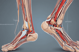
Talocrural Joint (images): Medial and lateral malleolus, and talus are key anatomical structures in images.
-
Medial Ligament (Deltoid Ligament): This ligament (aka deltoid ligament) is a key structure in the provided images.
-
Lateral Ligaments: This section discusses the critical anterior, calcaneofibular, and posterior talofibular ligaments.
-
Ankle Sprains: Grade 1 (stretched), Grade 2 (partial tear), and Grade 3 (ruptured) sprains are distinguished by the severity of ligament damage. Anterior talofibular ligament is the most common sprain.
-
Ankle Fracture: Treatment options such as ORIF (Open Reduction Internal Fixation) with plates and screws are mentioned. The image shows an ankle fracture with plates and screws.
-
Subtalar and Talocalcaneonavicular Joints: These are depicted in anatomical drawing images.
-
Talocalcaneonavicular Joint: Its stabilizing ligaments and facets are detailed.
-
Subtalar Joint: Includes interosseous talocalcaneal ligament.
-
Talonavicular & Calcaneocuboid Joints: Key structures for foot function, depicted in images.
-
Calcaneocuboid Joint: Fibularis longus tendon passes deep to long plantar ligament, supports the arch. Various plantar ligaments are detailed in relation to supporting the arch.
-
Intertarsal Joints: Articulations between adjacent tarsal bones.
-
Tarsometatarsal Joints: Joints of tarsal and metatarsal bones, depicted in images.
-
Intertarsal Ligaments: Capsule thickenings attaching to adjacent tarsal bones.
-
Metatarsophalangeal Joints (MTPs): The heads of metatarsal bones, bases of phalanges, deep transverse metatarsal ligaments, and plantar ligaments are shown in the accompanying visuals.
-
Interphalangeal Joints: Collateral and plantar ligaments (in images), metatarsophalangeal joints.
-
Dorsum of Foot Arteries are described: dorsalis pedis, deep plantar, and deep plantar arch (in imagery).
-
Dorsum of Foot Venous Arch includes the great and small saphenous veins (represented in diagrams).
-
Dorsum of Foot Extensor Retinacula are coverings of crural fascia that hold tendons against bones.
-
Subcutaneous Tendons in Foot: Locations and attachments for specific tendons are diagrammed.
-
Dorsum of Foot Cutaneous nerves (superficial fibular and deep fibular nerves.)
-
Plantar Foot: Cutaneous nerves, including medial plantar, lateral plantar nerves, and their relationships to major tendons.
-
Plantar Aponeurosis: An important anatomical structure (diagrammed) that provides support and flexibility to the foot
-
Plantar Muscles: Described in layers, highlighting origins, insertions, and actions of key muscles (detailed in diagrams).
-
Medial and Lateral Plantar Arteries: Paths and branches are illustrated, along with supplying the great toe and other foot regions.
-
Plantar Arteries Organization: Deep plantar arch is formed by the deep plantar artery.
-
Arch Support (Foot): The components of the arches, including ligaments and muscles are discussed.
-
Arches of Foot (images & text): These images show medial longitudinal, lateral longitudinal, and transverse arches, with ligamentous support structures detailed.
-
Foot and Ankle (images): Triple Arthrodesis refers to a surgical fusion of foot joints. Diagram and X-ray show the procedure.
-
Practice Question: A puncture wound to the lateral plantar surface of the foot (likely cutting the lateral plantar nerve) may cause loss of sensation on the plantar surface of the great toe, as indicated in the question.
Studying That Suits You
Use AI to generate personalized quizzes and flashcards to suit your learning preferences.