Podcast
Questions and Answers
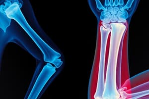
Which fat pad is typically visible on a lateral elbow projection?
Which fat pad is typically visible on a lateral elbow projection?
- Supinator fat stripe
- Anterior fat pad (correct)
- Distal fat stripe
- Posterior fat pad
In which position is the proximal radius and ulna completely superimposed?
In which position is the proximal radius and ulna completely superimposed?
- AP
- Lateral Oblique
- Medial Oblique (correct)
- Distal Oblique
What can the supinator fat stripe indicate?
What can the supinator fat stripe indicate?
- Inflammation of the bursae
- Infection of bone marrow
- Fractures that are easily seen
- Radial head or neck fractures not easily seen (correct)
Which condition is characterized by abnormally dense bone and often referred to as 'marble bone'?
Which condition is characterized by abnormally dense bone and often referred to as 'marble bone'?
What is the typical cause of osteomyelitis?
What is the typical cause of osteomyelitis?
Which projection best demonstrates the radial head and neck as well as the capitulum of the humerus?
Which projection best demonstrates the radial head and neck as well as the capitulum of the humerus?
What is the standard CR location for an AP elbow projection?
What is the standard CR location for an AP elbow projection?
How many degrees should the elbow be rotated for an external (lateral) oblique elbow projection?
How many degrees should the elbow be rotated for an external (lateral) oblique elbow projection?
What is the proper hand position for a medial (internal) AP oblique elbow projection?
What is the proper hand position for a medial (internal) AP oblique elbow projection?
Which Coyle method requires the hand to be pronated?
Which Coyle method requires the hand to be pronated?
Which clinical indication is appropriate for using the Axial Lateromedial Projection of the elbow?
Which clinical indication is appropriate for using the Axial Lateromedial Projection of the elbow?
What is the recommended angle for the central ray when performing the Axial Mediolateral Projection?
What is the recommended angle for the central ray when performing the Axial Mediolateral Projection?
What is the primary visual objective of the Axial Lateromedial Projection?
What is the primary visual objective of the Axial Lateromedial Projection?
In preparing for the Radial Head Laterals, how should the arm be positioned?
In preparing for the Radial Head Laterals, how should the arm be positioned?
What structure does the trochlea of the humerus directly articulate with?
What structure does the trochlea of the humerus directly articulate with?
Which evaluation criterion is key for the Axial Mediolateral Projection?
Which evaluation criterion is key for the Axial Mediolateral Projection?
Which fossa on the humerus receives the radial head?
Which fossa on the humerus receives the radial head?
What is an essential factor for the Axial Lateromedial Projection when positioning the elbow?
What is an essential factor for the Axial Lateromedial Projection when positioning the elbow?
What clinical indications warrant an AP projection of the elbow?
What clinical indications warrant an AP projection of the elbow?
In which scenario should a partial flexion AP projection be used?
In which scenario should a partial flexion AP projection be used?
What is the essential beam direction for an AP projection of the elbow?
What is the essential beam direction for an AP projection of the elbow?
How should the arm be positioned for a lateral oblique projection of the elbow?
How should the arm be positioned for a lateral oblique projection of the elbow?
Which anatomical structures are best visualized with a lateral projection of the elbow?
Which anatomical structures are best visualized with a lateral projection of the elbow?
What is the recommended Source to Image Distance (SID) for elbow projections?
What is the recommended Source to Image Distance (SID) for elbow projections?
When setting up for an elbow projection, how should the forearm and humerus be aligned?
When setting up for an elbow projection, how should the forearm and humerus be aligned?
What is the primary purpose of performing two projections during elbow radiography?
What is the primary purpose of performing two projections during elbow radiography?
Which part of the humerus articulates with the ulna?
Which part of the humerus articulates with the ulna?
What does the lateral epicondyle of the humerus represent?
What does the lateral epicondyle of the humerus represent?
What anatomical feature receives the coronoid process?
What anatomical feature receives the coronoid process?
In a true lateral elbow image, which arcs should be evaluated?
In a true lateral elbow image, which arcs should be evaluated?
Which depression of the distal humerus is located more laterally?
Which depression of the distal humerus is located more laterally?
What occurs when the arm is fully extended in relation to the olecranon fossa?
What occurs when the arm is fully extended in relation to the olecranon fossa?
What is the significance of the trochlear sulcus in elbow imaging?
What is the significance of the trochlear sulcus in elbow imaging?
Which projection of the elbow may be requested to better visualize the radial head?
Which projection of the elbow may be requested to better visualize the radial head?
Which part of the humerus is referred to as 'the little head'?
Which part of the humerus is referred to as 'the little head'?
What anatomical structure is located on the medial edge of the distal humerus?
What anatomical structure is located on the medial edge of the distal humerus?
Study Notes
Distal Humerus
- The expanded distal end of the humerus is called the humeral condyle.
- The medial condyle articulates with the ulna and is shaped like a pulley.
- The medial condyle contains a smooth depressed center called the trochlear sulcus.
- The lateral condyle, referred to as the capitulum, articulates with the radial head.
- The lateral epicondyle, a small projection located above the capitulum, is smaller than the medial epicondyle.
- The medial epicondyle, located on the medial edge of the distal humerus, is larger and more prominent than the lateral epicondyle.
- The coronoid fossa is an anterior depression that receives the coronoid process.
- The radial fossa, an anterior depression, is located lateral to the coronoid fossa and receives the radial head.
- The olecranon fossa, located posteriorly, receives the olecranon process when the arm is fully extended.
Elbow Joints
- The olecranon fossa is one of the structures that form the elbow joint.
- The proximal radioulnar joint is another joint that is located near the elbow.
Fat Pads
- The anterior fat pad is visible on the lateral elbow projection.
- Posterior fat pads are not typically visible on X-rays unless there is trauma or incorrect flexion.
- The supinator fat stripe may indicate a radial head or neck fracture.
Positioning Considerations
- The standard SID for elbow projections is 40 inches.
- Remove all artifacts or jewelry from the patient before radiographic procedures.
- Keep the shoulder and upper extremity on the same plane when positioning.
Routine Projections
- Common routine projections for the elbow include:
- AP
- Alternate AP – Partial Flexion
- Lateral (external)
- Medial (internal) Oblique
- Lateral Oblique
AP Projection
- Clinical Indications: Fractures, dislocations, osteomyelitis, and arthritis.
- The elbow should be fully extended with the hands supinated.
- The patient may need to lean laterally for a true AP projection.
- The epicondyles should be parallel to the IR.
- CR is perpendicular to the IR and directed at the mid-elbow joint (3/4 inch distal to the midpoint of the epicondyles).
Alternate AP – Partial Flexion
- Clinical Indications: Fractures, dislocations, osteomyelitis and arthritis.
- Used when the elbow cannot be fully extended.
- Two projections are needed:
- One with the forearm parallel to the IR.
- One with the humerus parallel to the IR.
- CR: Perpendicular to the IR, directed at the mid-elbow joint.
Lateral (External) Oblique Projection
- Clinical Indications: Fractures and dislocations, primarily those involving the radial head and neck.
- Best visualizes the radial head and neck of the radius and capitulum of the humerus.
- Arm should be fully extended and on the same plane as the shoulder.
- Hand should be supinated and rotated laterally approximately 45 degrees.
- CR is directed at the mid-elbow joint.
- Evaluation Criteria: The radial head, neck, and tuberosity should not be superimposed by the ulna.
Trauma Axial Laterals (Coyle Methods)
- Clinical Indications: Fractures and dislocations, particularly when the elbow cannot be fully extended for medial and lateral oblique projections.
- Two projections are used:
- Axial Lateromedial Projection (radial head best visualized)
- Axial Mediolateral Projection (Coronoid process best visualized)
Axial Lateromedial Projection (Radial Head)
- Clinical Indications: Fractures and dislocations of the radial head.
- Elbow should be flexed 90 degrees if possible.
- Hand should be pronated.
- CR angled 45 degrees toward the shoulder, centered at the radial head and mid-elbow joint.
- Evaluation Criteria: The joint space between the radial head and capitulum should be open, and the radial head, neck, and tuberosity should be free of superimposition.
Axial Mediolateral Projection (Coronoid Process)
- Clinical Indications: Fractures and dislocations involving the coronoid process.
- Elbow should be flexed to 80 degrees, hand pronated.
- CR angled 45 degrees from the shoulder, into the mid-elbow joint.
- Evaluation Criteria: The anterior portion of the coronoid process is elongated in profile, the joint space between the coronoid process and trochlea should be open, and the radial head and neck should be superimposed by the ulna.
Radial Head Laterals (Coyle Method)
- Clinical Indications: Occult fractures of the radial head or neck.
- Elbow flexed 90 degrees.
- Upper extremity and shoulder remain on the same plane.
- Four projections are obtained:
- Supinate hand and externally rotate as far as tolerated.
- Place hand in true lateral position.
- Pronate hand.
- Internally rotate hand as far as tolerated.
- CR perpendicular to the IR, directed at the radial head.
- Evaluation Criteria: The radial tuberosity should be visualized in different positions, depending on the rotation of the hand.
Common Pathologies
- Osteomyelitis: Infection of the bone or bone marrow, often caused by bacteria and associated with trauma or surgery.
- Bursitis: Inflammation of the bursae, fluid-filled sacs located around joints, resulting in pain and limited range of motion.
- Osteopetrosis: Rare genetic disorder characterized by abnormally dense bone, often referred to as "marble bone."
Studying That Suits You
Use AI to generate personalized quizzes and flashcards to suit your learning preferences.
Related Documents
Description
This quiz explores the detailed anatomy of the distal humerus and its role in the elbow joint. It covers features such as the humeral condyle, epicondyles, and various fossae that contribute to joint movement. Test your understanding of these critical anatomical structures!