Podcast
Questions and Answers
What is the normal color of gingiva?
What is the normal color of gingiva?
- Brown or black
- Pink or coral pink (correct)
- Yellowish
- Grayish white
What is the function of melanocytes in gingiva?
What is the function of melanocytes in gingiva?
- To attach the gingiva to the teeth and bones
- To produce toxins that incite inflammation
- To elevate the gingival surface
- To produce melanin that pigments the gingiva (correct)
What is the characteristic of the surface of attached gingiva?
What is the characteristic of the surface of attached gingiva?
- It is inflamed
- It appears stippled with shallow depressions (correct)
- It is freely mobile
- It is smooth and shiny
What is the primary function of the gingival sulcus?
What is the primary function of the gingival sulcus?
What is the average depth of the gingival sulcus in healthy individuals?
What is the average depth of the gingival sulcus in healthy individuals?
What is the significance of a gingival sulcus depth greater than 3 mm?
What is the significance of a gingival sulcus depth greater than 3 mm?
What is the main difference between the free gingiva and the attached gingiva?
What is the main difference between the free gingiva and the attached gingiva?
What is the function of the gingival sulcus?
What is the function of the gingival sulcus?
What is the significance of the gingival sulcus in relation to periodontal pockets?
What is the significance of the gingival sulcus in relation to periodontal pockets?
What is the origin of junctional epithelium?
What is the origin of junctional epithelium?
What is the characteristic of the epithelium in the attached gingiva?
What is the characteristic of the epithelium in the attached gingiva?
What is the shape of the interdental papilla of posterior teeth in a three-dimensional view?
What is the shape of the interdental papilla of posterior teeth in a three-dimensional view?
What is the region where the oral mucosa meets the surface of the tooth?
What is the region where the oral mucosa meets the surface of the tooth?
What is the percentage of gingiva that is nonkeratinized?
What is the percentage of gingiva that is nonkeratinized?
What type of cells are abundant in the lamina propria?
What type of cells are abundant in the lamina propria?
What is the function of the gingival ligament?
What is the function of the gingival ligament?
Flashcards are hidden until you start studying
Study Notes
Gingiva
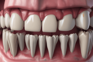
- The gingiva is the part of the firm oral masticatory mucosa surrounding the necks of the teeth and covering the alveolar processes of the jaws.
- Normal color of the gingiva is pink or coral pink, but it can have a grayish tinge.
- The gingiva can be pigmented brown or black due to melanin produced by melanocytes, specific cells found among the basal cells of the epithelium.
Clinical Parts of the Gingiva
- The gingiva has three clinical parts: free, attached, and interdental papilla.
Free Gingiva
- The free gingiva is freely mobile and not attached to the underlying structures.
- It has a shallow V-shaped notch called the free gingival groove, which divides the free gingiva from the attached gingiva.
- The gingival sulcus or crevice is a shallow groove between the free gingiva and the tooth surface, extending around the circumference of the tooth.
Attached Gingiva
- The attached gingiva is attached to the teeth and bones.
- Its surface appears stippled, with elevated portions of the epithelium and shallow depressions between them.
- Elongated rete pegs are seen, giving the surface a stippled appearance.
Dentogingival Junction
- The dentogingival junction is the region where the oral mucosa meets the surface of the tooth.
- Bacteria on the tooth surface can produce toxins that can cause inflammation and damage if they enter the mucosal tissues.
Gingival Sulcus
- In healthy individuals, the gingival sulcus is approximately 0.5 to 3 mm deep.
- Mild inflammation is present at an average depth of 1.8 mm.
- A depth greater than 3 mm is considered pathologic and represents periodontal disease.
Gingiva
- Part of the firm oral masticatory mucosa surrounding the necks of the teeth and covering the alveolar processes of the jaws
- Normally pink or coral pink in color, sometimes with a grayish tinge
- May be pigmented brown or black due to melanin produced by melanocytes in the basal cells of epithelium
Clinical Parts of Gingiva
- Free gingiva: freely mobile and not attached to underlying structures
- Has a free gingival groove (a shallow V-shaped notch)
- Has a gingival sulcus/crevice (a shallow groove between the free gingiva and tooth surface)
- Attached gingiva: attached to teeth and bones
- Has a stippled surface with elevated and depressed areas
- Has elongated rete pegs (stippling)
- Interdental papilla: part of the free gingiva that fills the space between teeth
- Pyramidal/triangular in shape on oral/vestibular aspect
- Has a central concave area (col) covered by thin nonkeratinized epithelium, more vulnerable to periodontal disease
Gingival Sulcus and Junctional Epithelium
- Gingival sulcus: a shallow groove between the free gingiva and tooth surface
- Average depth in healthy individuals is 0.5-3 mm
- Depth greater than 3 mm is considered pathologic and represents a periodontal pocket
- Junctional epithelium: epithelium attached to the tooth (enamel or sometimes cementum) surface
- Derived from the REE of the tooth germ
- Consists of cells aligned parallel to the tooth surface, increasing in thickness from apex to crown
- Attached to enamel by internal basal lamina and to connective tissue by external basal lamina
- Has hemidesmosomes in both basal laminas
Functional Epithelium and Lamina Propria
- Functional epithelium:
- 75% of population has parakeratinized epithelium, 15% has keratinized, and 10% has nonkeratinized epithelium
- Lamina propria:
- Consists of dense connective tissue, not highly vascularized
- Has macrophages, plasma cells, and lymphocytes
- Fibroblasts are abundant
- Gingival ligament: collagen fibers arranged in various groups to support the free gingiva, bind attached gingiva to alveolar bone and tooth, and link one tooth with another
Studying That Suits You
Use AI to generate personalized quizzes and flashcards to suit your learning preferences.