Podcast
Questions and Answers
What separates the gingival epithelium from the underlying connective tissue?
What separates the gingival epithelium from the underlying connective tissue?
- Basement membrane (correct)
- Lamina lucida
- Lamina densa
- Rete pegs
What is the main component of lamina lucida?
What is the main component of lamina lucida?
- Type IV collagen
- Mucopolysaccharides
- Keratin
- Glycoprotein laminin (correct)
What is the thickness of the oral epithelium?
What is the thickness of the oral epithelium?
- 0.2-0.3 mm (correct)
- 0.1-0.2 mm
- 0.3-0.4 mm
- 0.4-0.5 mm
What is the characteristic of sulcular epithelium?
What is the characteristic of sulcular epithelium?
What is the function of the junctional epithelium?
What is the function of the junctional epithelium?
What is the characteristic of junctional epithelium?
What is the characteristic of junctional epithelium?
What is the function of the basement membrane?
What is the function of the basement membrane?
What is the characteristic of oral epithelium?
What is the characteristic of oral epithelium?
What is the main function of keratinocytes in the oral epithelium?
What is the main function of keratinocytes in the oral epithelium?
What is the process by which keratinocytes undergo transformation?
What is the process by which keratinocytes undergo transformation?
What type of cells are Langerhans cells?
What type of cells are Langerhans cells?
Where are melanocytes located in the oral epithelium?
Where are melanocytes located in the oral epithelium?
What is the function of Merkel cells?
What is the function of Merkel cells?
What is the main difference between orthokeratinization and parakeratinization?
What is the main difference between orthokeratinization and parakeratinization?
What is the role of the epithelial cells in innate host defense?
What is the role of the epithelial cells in innate host defense?
What is the approximate turnover time for the gingival epithelium?
What is the approximate turnover time for the gingival epithelium?
What is the gingiva?
What is the gingiva?
What is the width of the gingiva?
What is the width of the gingiva?
What is the gingival sulcus?
What is the gingival sulcus?
What is the ideal depth of the gingival sulcus?
What is the ideal depth of the gingival sulcus?
What is the attached gingiva?
What is the attached gingiva?
What determines the shape of the interdental gingiva?
What determines the shape of the interdental gingiva?
What is the histological composition of the gingiva?
What is the histological composition of the gingiva?
What is the depth of a clinically normal gingival sulcus?
What is the depth of a clinically normal gingival sulcus?
What is the normal color of the gingiva in adults?
What is the normal color of the gingiva in adults?
What is the shape of the gingiva in relation to the tooth?
What is the shape of the gingiva in relation to the tooth?
What is the consistency of normal gingiva?
What is the consistency of normal gingiva?
What is the surface texture of normal gingiva?
What is the surface texture of normal gingiva?
At what age is stippling of gingiva usually seen?
At what age is stippling of gingiva usually seen?
What is the shape of the gingiva in the interdental area?
What is the shape of the gingiva in the interdental area?
What is one way epithelial cells may respond to bacteria?
What is one way epithelial cells may respond to bacteria?
What percentage of connective tissue is composed of collagen fibers?
What percentage of connective tissue is composed of collagen fibers?
What is the main function of the dentogingival group of gingival fibers?
What is the main function of the dentogingival group of gingival fibers?
What is the main component of the ground substance in connective tissue?
What is the main component of the ground substance in connective tissue?
What type of fibers provide tensile strength to the lamina propria?
What type of fibers provide tensile strength to the lamina propria?
What is the function of the circular group of gingival fibers?
What is the function of the circular group of gingival fibers?
What is one source of blood supply to the gingiva?
What is one source of blood supply to the gingiva?
What type of cells are the main cells of the connective tissue?
What type of cells are the main cells of the connective tissue?
Flashcards are hidden until you start studying
Study Notes
The Gingiva
- The gingiva is part of the oral mucosa, covering the alveolar processes of the jaws and surrounding the teeth, but not adherent to them.
- It extends to the mucogingival junction, with a varying width of 1-9 mm.
- The gingiva is divided anatomically into:
- Marginal gingiva
- Attached gingiva
- Interdental areas
Marginal Gingiva
- The free gingiva is a knife-edge part of the gingiva, located coronal or above the CEJ, surrounding the tooth in a turtleneck or collar-like manner.
- It forms the soft tissue wall of the gingival sulcus.
- The gingival sulcus is a shallow crevice or space around the tooth neck, bounded by the tooth surface and epithelium lining the free margin.
- The depth of the gingival sulcus is an important clinical parameter, ideally being 0 mm, but clinically normal sulcus is 2-3 mm.
Attached Gingiva
- It is firm, resilient, and extends from the free gingival groove to the mucogingival junction.
- The width of the attached gingiva is an important clinical parameter, differing in various areas of the mouth.
- The width is greatest in the incisor area (3.5-4.5 mm) and narrower in the posterior area (1.8-1.9 mm).
- The width increases with age and in supraerupted teeth.
Interdental Gingiva
- It is the part of the gingiva filling in the embrasure beneath the contact point.
- It is either pyramidal or can have a col shape, determined by tooth contact and the width of neighboring teeth.
- It is completely missing in case of diastema.
Microscopic Features
- The gingiva is composed of:
- Overlying stratified squamous epithelium (SSE)
- Core of connective tissue (lamina propria)
- A dividing line between the two, called the basement membrane
- The epithelium is joined to the underlying connective tissue by the basal lamina, which consists of lamina lucida and lamina densa.
- The basal lamina acts as a barrier to particulate matter, with a width of 300-400 A°.
Gingival Epithelium
- It consists of a continuous lining of SSE, with three different areas:
- The oral epithelium, extending from the gingival margin to the mucogingival junction.
- The sulcular epithelium, facing the tooth.
- The junctional epithelium, apical to the sulcular epithelium, where the gingiva is joined to the tooth.
- The principal cell type of the gingival epithelium is the keratinocyte, with other cells including Langerhans cells, Merkel cells, and melanocytes.
Gingival Epithelium Subtypes
- Oral epithelium: 0.2-0.3 mm in thickness, orthokeratinized or parakeratinized, with rete pegs.
- Sulcular epithelium: thin, non-keratinized, lacking granulosum and corneum strata, and normally having no Merkel cells.
- Junctional epithelium: 3-4 layers thick, composed of 2 strata, with a length ranging from 0.25-1.35 mm, and expressing K19.
Connective Tissue
- Mainly formed by collagen fibers (60%), fibroblasts, vessels, nerves, and matrix (35%).
- Consists of two layers:
- Papillary layer
- Reticular layer
- Has a cellular and extracellular compartment, with fibroblasts, mast cells, plasma cells, lymphocytes, and neutrophils, and a ground substance composed of proteoglycans, glycoprotein, and fibers.
Gingival Fibers
- Arranged in groups, including:
- Dentogingival group: originating from the cementum, extending in fan-like conformation toward the crest and outer marginal gingiva.
- Circular group: coursing through the connective tissue of marginal and interdental in a ring-like fashion.
- Transseptal group: located interproximally, forming horizontal bundles extending between the cementum of approximating teeth.
- Functions include:
- Bracing the marginal gingiva firmly against the tooth.
- Providing the rigidity necessary to withstand the forces of mastication.
- Uniting the free marginal gingiva with the cementum of the root and the adjacent attached gingiva.
Blood and Nerve Supply
- Blood supply is provided through three sources:
- Supraperiosteal arterioles
- Vessels of the periodontal ligament
- Arterioles that emerge from the crest of the interdental septa
- Nerve supply is provided by the fifth cranial nerve.
Clinical Features of Normal Gingiva
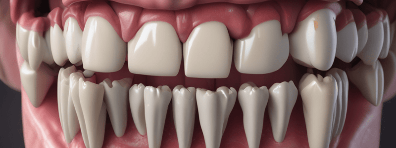
- Color: coral pink or salmon red, depending on the degree of vascularity, thickness of keratinization, and presence of pigmented cells.
- Shape: having a knife-edge with the tooth.
- Contour: normally following the contour of the tooth, but usually scalloped.
- Consistency: firm and resilient.
- Surface texture: orange-peel (stippled) appearance.
- Position: the position of the gingiva refers to the level at which the gingival margin is attached to the tooth.
Studying That Suits You
Use AI to generate personalized quizzes and flashcards to suit your learning preferences.