Podcast
Questions and Answers
What is the primary function of the gallbladder?
What is the primary function of the gallbladder?
- Filters blood from the portal vein
- Stores and concentrates bile (correct)
- Produces bile
- Secrets insulin into the bloodstream
Which statement about the portal vein is correct?
Which statement about the portal vein is correct?
- Supplies 75% of the liver's blood (correct)
- Is formed only by the splenic vein
- Drains blood from the liver into the inferior vena cava
- Is the main vessel draining the gallbladder
What condition is characterized by increased pressure in the portal venous system?
What condition is characterized by increased pressure in the portal venous system?
- Cholelithiasis
- Murphy's Sign
- Portal Hypertension (correct)
- Hepatomegaly
What anatomical feature connects the gallbladder to the biliary system?
What anatomical feature connects the gallbladder to the biliary system?
Which imaging technique is specifically used for diagnosing pathologies of the common bile duct?
Which imaging technique is specifically used for diagnosing pathologies of the common bile duct?
Which anatomic part of the gallbladder is defined as the main central portion?
Which anatomic part of the gallbladder is defined as the main central portion?
What is the role of the Sphincter of Oddi in relation to the biliary system?
What is the role of the Sphincter of Oddi in relation to the biliary system?
Which symptom is indicative of cholecystitis and often assessed using Murphy's Sign?
Which symptom is indicative of cholecystitis and often assessed using Murphy's Sign?
What is the approximate weight of the liver relative to body weight?
What is the approximate weight of the liver relative to body weight?
Which of the following structures separates the right and left lobes of the liver?
Which of the following structures separates the right and left lobes of the liver?
What is a primary function of the Kupffer cells in the liver?
What is a primary function of the Kupffer cells in the liver?
What is contained within the lesser omentum?
What is contained within the lesser omentum?
Which of the following statements about the anatomical lobes of the liver is true?
Which of the following statements about the anatomical lobes of the liver is true?
What percentage of the liver's blood supply is provided by the hepatic artery?
What percentage of the liver's blood supply is provided by the hepatic artery?
What is the primary role of the liver in metabolism?
What is the primary role of the liver in metabolism?
What is Morison's Pouch also known as?
What is Morison's Pouch also known as?
Which of the following structures demarcates the bare area of the liver?
Which of the following structures demarcates the bare area of the liver?
What percentage of the liver’s blood supply is provided by the portal vein?
What percentage of the liver’s blood supply is provided by the portal vein?
Which structure is located at the porta hepatis?
Which structure is located at the porta hepatis?
What is the primary function of the gallbladder?
What is the primary function of the gallbladder?
A patient presents with right upper quadrant pain and a positive Murphy's sign. What is the most likely diagnosis?
A patient presents with right upper quadrant pain and a positive Murphy's sign. What is the most likely diagnosis?
Porto-systemic anastomoses become clinically relevant in which condition?
Porto-systemic anastomoses become clinically relevant in which condition?
Which of the following is NOT a part of the common bile duct?
Which of the following is NOT a part of the common bile duct?
The round ligament (ligamentum teres) is the remnant of which fetal structure?
The round ligament (ligamentum teres) is the remnant of which fetal structure?
The liver is divided into eight functional segments based on which two factors?
The liver is divided into eight functional segments based on which two factors?
Where do the superficial lymphatics of the liver primarily drain?
Where do the superficial lymphatics of the liver primarily drain?
Which maneuver is used to control bleeding from the liver during surgery?
Which maneuver is used to control bleeding from the liver during surgery?
The portal vein is formed by the union of which two veins?
The portal vein is formed by the union of which two veins?
In portal hypertension, which site is most commonly associated with the formation of varices?
In portal hypertension, which site is most commonly associated with the formation of varices?
Which of the following is another common site where varices form due to portal hypertension?
Which of the following is another common site where varices form due to portal hypertension?
Why are porto-systemic anastomoses clinically relevant in portal hypertension?
Why are porto-systemic anastomoses clinically relevant in portal hypertension?
Which of the following surfaces of the liver is in contact with the diaphragm?
Which of the following surfaces of the liver is in contact with the diaphragm?
Which anatomical structure separates the right and left lobes of the liver anteriorly?
Which anatomical structure separates the right and left lobes of the liver anteriorly?
Which two vessels provide the liver’s blood supply?
Which two vessels provide the liver’s blood supply?
Where is the pancreas located in relation to the stomach?
Where is the pancreas located in relation to the stomach?
Which duct joins the common bile duct before draining into the duodenum at the major duodenal papilla?
Which duct joins the common bile duct before draining into the duodenum at the major duodenal papilla?
Acute pancreatitis is most commonly caused by which of the following?
Acute pancreatitis is most commonly caused by which of the following?
Lymph from the liver primarily drains into which group of nodes?
Lymph from the liver primarily drains into which group of nodes?
Where do the hepatic veins drain the blood from the liver?
Where do the hepatic veins drain the blood from the liver?
Which of the following is NOT a function of the pancreas?
Which of the following is NOT a function of the pancreas?
Which structure is responsible for transporting bile from the liver and gallbladder to the duodenum?
Which structure is responsible for transporting bile from the liver and gallbladder to the duodenum?
Which veins combine to form the portal vein?
Which veins combine to form the portal vein?
What is the consequence of increased pressure in the portal venous system (portal hypertension)?
What is the consequence of increased pressure in the portal venous system (portal hypertension)?
What is the role of the gallbladder in the digestive system?
What is the role of the gallbladder in the digestive system?
Which arteries supply blood to the pancreas?
Which arteries supply blood to the pancreas?
The pancreas performs both endocrine and exocrine functions. Which of the following is an exocrine function of the pancreas?
The pancreas performs both endocrine and exocrine functions. Which of the following is an exocrine function of the pancreas?
Where is the major duodenal papilla located, and what drains into it?
Where is the major duodenal papilla located, and what drains into it?
Which ligament attaches the liver to the anterior abdominal wall?
Which ligament attaches the liver to the anterior abdominal wall?
Liver segments are functionally independent units based on what criteria?
Liver segments are functionally independent units based on what criteria?
Which of the following is NOT a tributary of the hepatic portal vein?
Which of the following is NOT a tributary of the hepatic portal vein?
The gallbladder is located on the visceral surface of which liver lobe?
The gallbladder is located on the visceral surface of which liver lobe?
Study Notes
Liver: Overview
- Largest gland in the body, approximately 1500g, accounting for ~2% of body weight.
- Receives around 1500ml of blood per minute.
- Located in the right upper abdomen, from the 4th right intercostal space to the costal margin.
- Lies beneath ribs 7-11; can be palpated during inspiration.
Functions of the Liver
- Synthesis of proteins such as albumin and clotting factors.
- Metabolizes fats, carbohydrates, and proteins.
- Inactivates toxins, including drugs and alcohol.
- Produces bile for fat digestion and absorption.
- Stores glucose as glycogen and releases it when needed.
- Synthesizes cholesterol.
- Processes hemoglobin, converting heme to bilirubin.
- Regulates blood clotting mechanisms.
- Participates in immune responses via Kupffer cells that clear pathogens.
- Clears bilirubin from the breakdown of red blood cells.
Liver Anatomy
- Surface Anatomy:
- Diaphragmatic surface: Convex, facing anteriorly, superiorly, and posteriorly.
- Visceral surface: Concave, contacts gallbladder and peritoneum.
Peritoneal Recesses and Ligaments
- Recesses:
- Subphrenic recess: Between the diaphragm and anterior liver surface.
- Morison’s pouch: Between the liver and right kidney/suprarenal gland; contains peritoneal fluid.
- Ligaments:
- Falciform ligament: Attaches liver to anterior abdominal wall; divides liver into right and left lobes.
- Coronary ligament: Reflects peritoneum from diaphragm, marking the liver's bare area.
- Triangular ligaments: Right and left, located at edges of the coronary ligament.
- Lesser omentum: Houses the portal triad (bile duct, hepatic artery, portal vein) within the hepatoduodenal ligament.
- Round ligament (ligamentum teres): Fibrous remnant of umbilical vein, runs with the falciform ligament.
Liver Lobes and Segments
- Anatomical lobes:
- Right lobe: Larger and separated from the left lobe by the falciform ligament.
- Left lobe: Smaller, includes quadrate and caudate lobes.
- Functional segments: Eight segments defined by vascular and biliary drainage, operate independently.
Vasculature of the Liver
- Hepatic artery proper: Supplies 25% of blood to the liver.
- Portal vein: Provides 75% of blood, formed by superior mesenteric and splenic veins.
- Hepatic veins: Drain deoxygenated blood from liver to inferior vena cava (IVC).
Portal Venous System & Porto-Systemic Anastomoses
- Portal venous system: Drains blood from gastrointestinal tract and spleen into the liver.
- Porto-systemic anastomoses: Connections between portal system and systemic circulation, significant in portal hypertension (e.g., esophageal varices).
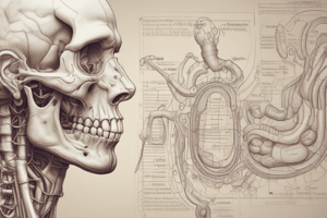
Biliary Tree and Gallbladder
- Gallbladder Anatomy:
- Situated in gallbladder fossa on the liver's visceral surface, near the quadrate lobe.
- Comprises three parts: fundus, body, and neck (which connects to cystic duct).
- Biliary System Components:
- Common bile duct (CBD): Formed by the cystic duct and common hepatic duct; has three segments: supraduodenal, retroduodenal, paraduodenal.
- Gallbladder Function: Stores and concentrates bile (50 mL capacity).
Major Duodenal Papilla
- Opening where common bile duct and pancreatic duct enter the duodenum, regulated by the Sphincter of Oddi.
Blood Supply to the Gallbladder
- Cystic artery: Usually branches from right hepatic artery.
- Venous drainage: Cystic vein drains into the portal vein.
Liver and Gallbladder: Clinical Correlations
- Liver Conditions:
- Portal hypertension: Increased pressure due to liver cirrhosis; leads to varices and ascites.
- Pringle’s maneuver: Surgical technique for controlling liver hemorrhage.
- Hepatomegaly: Liver enlargement caused by infections, storage disorders, or cirrhosis.
- Gallbladder Conditions:
- Cholelithiasis (gallstones): Formed from cholesterol; can block biliary tree.
- Murphy’s sign: Tenderness in right upper quadrant on inspiration, indicates cholecystitis.
- Diagnosis methods: Ultrasound, MRCP (Magnetic Resonance Cholangiopancreatography), ERCP (Endoscopic Retrograde Cholangiopancreatography).
Radiological Imaging
- Imaging techniques such as ultrasound, CT, and MRI visualize the liver, gallbladder, and bile ducts.
- Specialized imaging (MRCP, ERCP) aids in diagnosing common bile duct and gallbladder pathologies.
Studying That Suits You
Use AI to generate personalized quizzes and flashcards to suit your learning preferences.
Description
Explore the anatomy and functions of the liver, biliary tree, and gallbladder in this informative quiz. Learn about blood supply, location, and key functions such as protein synthesis and metabolism. Ideal for students studying human anatomy.