Understand the Problem
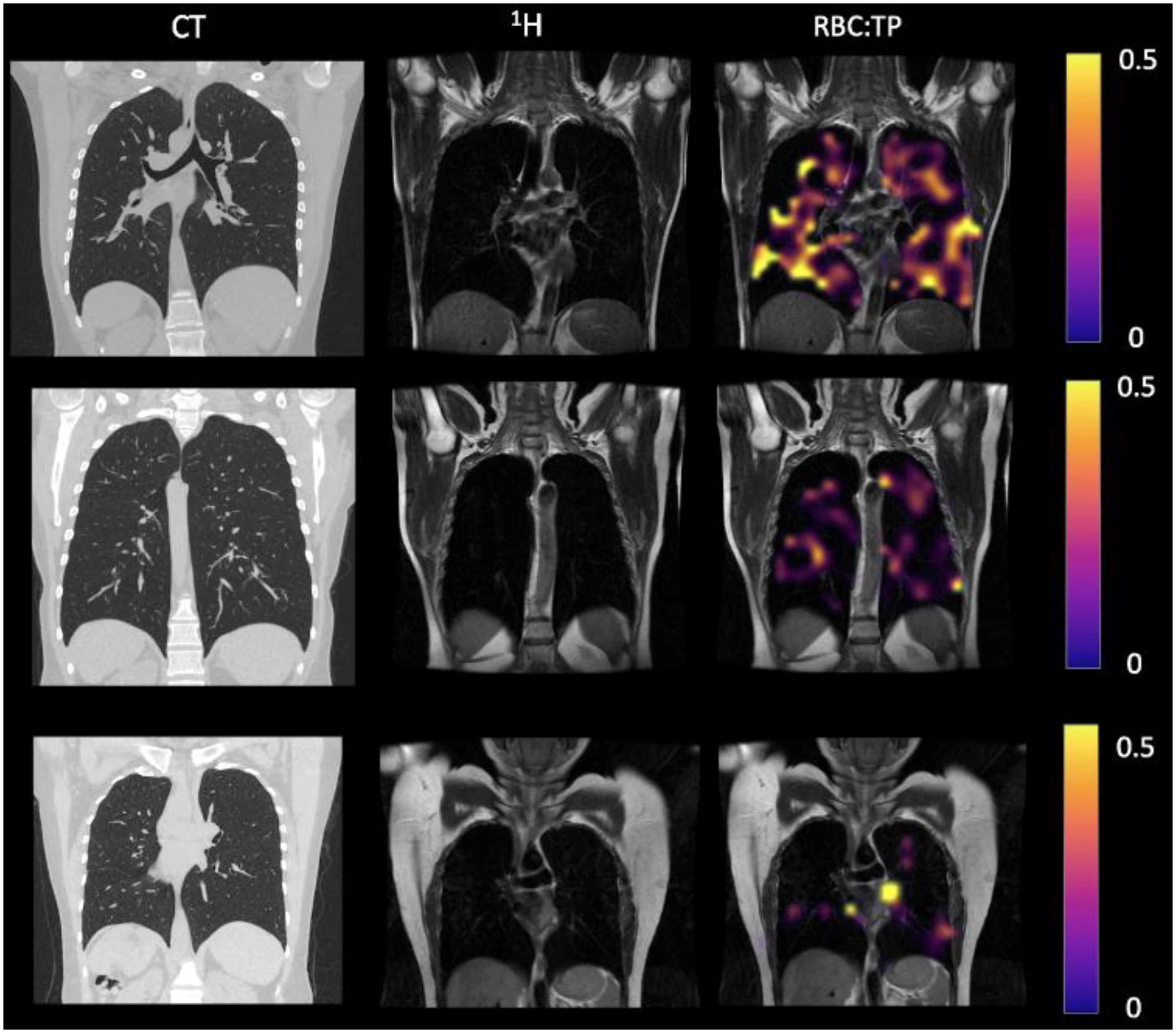
The image likely presents a comparison of different types of imaging techniques (CT, 1H imaging, and RBC:TP). It shows various lung scans, indicating different states or conditions of the lungs. The user may be interested in understanding the differences and applications of these imaging modalities.
Answer
CT and MRI (^1H and RBC:TP) lung scans comparison.
This image compares CT and MRI (^1H and RBC:TP) scans of the lungs. The CT scan shows detailed lung structure, while the ^1H MRI highlights soft tissue contrast and the RBC:TP image indicates functional imaging possibly related to blood or tracer distribution.
Answer for screen readers
This image compares CT and MRI (^1H and RBC:TP) scans of the lungs. The CT scan shows detailed lung structure, while the ^1H MRI highlights soft tissue contrast and the RBC:TP image indicates functional imaging possibly related to blood or tracer distribution.
More Information
CT scans are great for structural details, while MRI can highlight soft tissues and functionalities, like blood flow or tracer uptake in different areas.
AI-generated content may contain errors. Please verify critical information