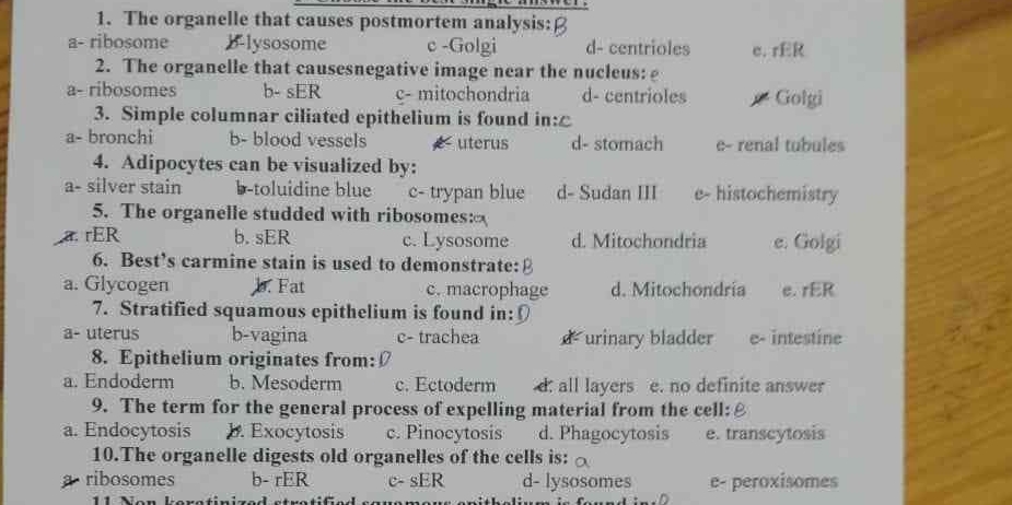
1. The organelle that causes postmortem analysis: 2. The organelle that causes negative image near the nucleus: 3. Simple columnar ciliated epithelium is found in: 4. Adipocytes ca... 1. The organelle that causes postmortem analysis: 2. The organelle that causes negative image near the nucleus: 3. Simple columnar ciliated epithelium is found in: 4. Adipocytes can be visualized by: 5. The organelle studded with ribosomes: 6. Best's carmine stain is used to demonstrate: 7. Stratified squamous epithelium is found in: 8. Epithelium originates from: 9. The term for the general process of expelling material from the cell: 10. The organelle digests organelles of the cell:

Understand the Problem
The question involves identifying and answering multiple-choice questions related to cell biology and histology. It covers topics like organelles, staining techniques, and types of epithelium. The high-level approach would involve recalling relevant biological concepts and selecting the appropriate answers based on knowledge of cell structure and function.
Answer
1. Lysosome; 2. Golgi; 3. Uterus; 4. Sudan III; 5. rER; 6. Fat; 7. Vagina; 8. All layers; 9. Exocytosis; 10. Lysosomes
- Lysosome; 2. Golgi; 3. Uterus; 4. Sudan III; 5. rER; 6. Fat; 7. Vagina; 8. All layers; 9. Exocytosis; 10. Lysosomes
Answer for screen readers
- Lysosome; 2. Golgi; 3. Uterus; 4. Sudan III; 5. rER; 6. Fat; 7. Vagina; 8. All layers; 9. Exocytosis; 10. Lysosomes
More Information
Simple columnar ciliated epithelium is commonly found lining reproductive tracts, aiding in movement and transportation. Best's carmine stain is used to demonstrate fat in tissues.
Tips
A common mistake is confusing organelle functions; for example, the Golgi apparatus is involved in packaging, not digestion.
Sources
AI-generated content may contain errors. Please verify critical information