Podcast
Questions and Answers
Which structure is located within the renal cortex?
Which structure is located within the renal cortex?
- Glomerulus (correct)
- Renal pyramid
- Loop of Henle
- Renal column
What histological feature distinguishes the proximal convoluted tubule (PCT) from the distal convoluted tubule (DCT)?
What histological feature distinguishes the proximal convoluted tubule (PCT) from the distal convoluted tubule (DCT)?
- Presence of prominent dark staining nuclei in the DCT.
- Presence of numerous microvilli creating a 'fuzzy' lumen in the PCT. (correct)
- Presence of light staining cytoplasm in the DCT.
- Presence of a clear, wide lumen in the PCT.
In the medulla of the kidney, which histological characteristic is associated with the thin descending limb of the nephron loop?
In the medulla of the kidney, which histological characteristic is associated with the thin descending limb of the nephron loop?
- Thin walled (correct)
- Light staining cytoplasm
- Prominent dark staining nuclei
- Fuzzy lumen
Which type of epithelium is characteristic of the ureter?
Which type of epithelium is characteristic of the ureter?
Identify the layers typically found in the ureter.
Identify the layers typically found in the ureter.
Which of the following is a key feature of the urinary bladder's histology?
Which of the following is a key feature of the urinary bladder's histology?
What type of epithelium is found in the female urethra?
What type of epithelium is found in the female urethra?
How does the muscularis layer in the urethra differ between males and females?
How does the muscularis layer in the urethra differ between males and females?
From which embryonic tissue does the urogenital system primarily derive?
From which embryonic tissue does the urogenital system primarily derive?
What are the functional components of the urogenital ridge in embryonic development?
What are the functional components of the urogenital ridge in embryonic development?
Which structures are formed by the nephrogenic cord during embryonic development?
Which structures are formed by the nephrogenic cord during embryonic development?
What is the role of the gonadal ridge during embryonic development?
What is the role of the gonadal ridge during embryonic development?
What is the fate of the pronephroi during kidney development?
What is the fate of the pronephroi during kidney development?
Which set of embryonic kidneys functions briefly and is characterized by glomeruli and mesonephric tubules?
Which set of embryonic kidneys functions briefly and is characterized by glomeruli and mesonephric tubules?
Which structure forms the permanent kidney?
Which structure forms the permanent kidney?
What two structures give rise to the permanent kidney?
What two structures give rise to the permanent kidney?
Which adult structure is derived from the ureteric bud?
Which adult structure is derived from the ureteric bud?
What structures does the metanephrogenic blastema give rise to?
What structures does the metanephrogenic blastema give rise to?
During kidney relocation, what critical process occurs as the kidneys ascend into the abdomen?
During kidney relocation, what critical process occurs as the kidneys ascend into the abdomen?
From which embryonic structure does the urinary bladder develop?
From which embryonic structure does the urinary bladder develop?
What happens to the allantois during the development of the urinary bladder?
What happens to the allantois during the development of the urinary bladder?
Which embryonic structure contributes to the formation of the male urethra, but not the female urethra?
Which embryonic structure contributes to the formation of the male urethra, but not the female urethra?
What is the origin of the epithelium lining the majority of the urethra?
What is the origin of the epithelium lining the majority of the urethra?
What is the role of the ureteric bud in kidney development?
What is the role of the ureteric bud in kidney development?
The relocation of the kidneys during development involves their relocation to the abdomen. During this process, what happens to their original blood supply?
The relocation of the kidneys during development involves their relocation to the abdomen. During this process, what happens to their original blood supply?
The pelvic part of the urogenital sinus contributes to the development of the urethra differently in males and females. What is the ultimate fate of this structure in each sex?
The pelvic part of the urogenital sinus contributes to the development of the urethra differently in males and females. What is the ultimate fate of this structure in each sex?
What event is critical for initiating the transformation of mesenchyme cells into nephrons during kidney development?
What event is critical for initiating the transformation of mesenchyme cells into nephrons during kidney development?
What process explains the separation of the urinary and genital systems during embryonic development?
What process explains the separation of the urinary and genital systems during embryonic development?
Flashcards
Renal/fibrous capsule
Renal/fibrous capsule
Fibrous outer layer of the kidney.
Kidney cortex
Kidney cortex
Contains renal corpuscles, proximal and distal convoluted tubules.
Kidney medulla
Kidney medulla
Contains the loop of Henle and collecting ducts.
Kidney general structures
Kidney general structures
Signup and view all the flashcards
Renal pyramid
Renal pyramid
Signup and view all the flashcards
Renal column
Renal column
Signup and view all the flashcards
Bowman's capsule
Bowman's capsule
Signup and view all the flashcards
Glomerulus
Glomerulus
Signup and view all the flashcards
Visceral layer
Visceral layer
Signup and view all the flashcards
Parietal layer
Parietal layer
Signup and view all the flashcards
Capsular space
Capsular space
Signup and view all the flashcards
Proximal convoluted tubule (PCT)
Proximal convoluted tubule (PCT)
Signup and view all the flashcards
Distal convoluted tubule (DCT)
Distal convoluted tubule (DCT)
Signup and view all the flashcards
Thick descending limb
Thick descending limb
Signup and view all the flashcards
Thin (ascending/descending) limb
Thin (ascending/descending) limb
Signup and view all the flashcards
Thick ascending limb
Thick ascending limb
Signup and view all the flashcards
Collecting duct (CD)
Collecting duct (CD)
Signup and view all the flashcards
Ureter
Ureter
Signup and view all the flashcards
Transitional epithelium (ureter)
Transitional epithelium (ureter)
Signup and view all the flashcards
Lamina propria
Lamina propria
Signup and view all the flashcards
Muscularis
Muscularis
Signup and view all the flashcards
Adventitia
Adventitia
Signup and view all the flashcards
Urinary bladder
Urinary bladder
Signup and view all the flashcards
Transitional epithelium (bladder)
Transitional epithelium (bladder)
Signup and view all the flashcards
Muscularis (bladder)
Muscularis (bladder)
Signup and view all the flashcards
Urethra
Urethra
Signup and view all the flashcards
Kidney development
Kidney development
Signup and view all the flashcards
Urogenital ridge
Urogenital ridge
Signup and view all the flashcards
Nephrogenic cord
Nephrogenic cord
Signup and view all the flashcards
Ureteric Bud
Ureteric Bud
Signup and view all the flashcards
Study Notes
- This lecture focuses on recalling how the body produces and expels filtrate; identifying gross and microscopic structures of the urinary system and tracing its pathway
Lecture Learning Objectives
- Visually identify the kidney, ureter, urinary bladder, and urethra
- Describe the urogenital ridge, including its location, embryonic tissue, and portions that form the urinary and reproductive systems
- Explain the three successive sets of kidneys that develop in the embryo, noting which parts remain
- Describe the ureteric bud and metanephrogenic blastema
- List the portions of the adult kidney formed by the ureteric bud and the metanephrogenic blastema
- Explain the relocation of the kidneys
- Describe the development of the urinary bladder and urethra, focusing on the portions of the urogenital sinus and embryonic tissue derivatives that develop each component
- Compare the development of the male and female urinary system
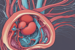
Kidney Review
- The kidney has a renal/fibrous capsule, cortex, and medulla
- The cortex contains specific structures
- The medulla has specific structures present in the pyramids of the medulla
Kidney General Structures
- Capsule
- Cortex
- Medulla
- Renal Pelvis
- Hilum
- Renal pyramid
- Renal column
Nephron Review
- Afferent arteriole
- Renal corpuscle component is the glomerulus
- The glomerular capsule has a visceral and parietal layer, plus a capsular space
- Proximal convoluted tubule
- Nephron loop with thick descending arm, thin arm and thick ascending arm
- Distal convoluted tubule has a macula densa
Renal Corpuscle (RC)
- Glomerulus contains a bundle of vessels within RC
- Glomerular capsule with visceral layer and parietal layer
- Capsular space is created by two layers of the glomerular capsule
Nephron Histological Characteristics - Cortex
- Proximal convoluted tubule (PCT) has a “fuzzy” lumen due to numerous microvilli
- Distal convoluted tubule with a clear lumen and very limited microvilli
Nephron Histological Characteristics - Medulla
- In the medulla, note these nephron loop characteristics:
- Thick descending limb has a fuzzy lumen and is part of the PCT
- Thin limb (ascending/descending) with thin walls
- Thick ascending limb has a clear lumen
- Collecting duct with prominent dark staining nuclei and light staining cytoplasm
- All three portions of the nephron loop can be in the same image because they are components of different nephrons
Kidney Cortex-Nephron
- Renal corpuscle with glomerulus
- Proximal convoluted tubule
- Distal convoluted tubule
- Glomerulus with a renal corpuscle
- Glomerular capsule with a visceral and parietal layer
- Capsular space
- Macula densa in the distal convoluted tubule
Kidney Medulla-Nephron
- Thick descending limb and thin limb
- Thick ascending limb
Kidney Medulla-Collecting Duct
- Collecting duct to compare to PCT and DCT
Ureter
- Look for transitional epithelium, thick lamina propria, two-layered muscularis (inner longitudinal layer and outer circular layer), and adventitia (A)
- Mucosa with transitional epithelium and lamina propria
- Muscularis includes an inner longitudinal layer and an outer circular layer
- Adventitia
Urinary Bladder
- Look for transitional epithelium and thin lamina propria
- Three layers of muscularis, including inner longitudinal, middle circular, and outer longitudinal layers
- Mucosa is followed by muscularis
Urethra
- Look for transitional epithelium (female) or stratified/squamous columnar epithelium (male)
- Also look for thick lamina propria and two layered muscularis, surrounded by additional tissue, such as:
- skeletal muscle in females
- erectile tissue in males
- Male: stratified columnar epithelium (spongy portion of male urethra) and lamina propria
- Female: transitional epithelium (upper most portion of female urethra) and lamina propria
Urinary Tract Embryology
- Closely linked to the genital system (urogenital system), derived from the intermediate mesoderm on the developing embryo's dorsal wall
- The urogenital ridge is a tissue ridge on either side of the primordial aorta
- The nephrogenic cord forms urinary system components
- The gonadal ridge forms reproductive systems
Development of the Kidneys
- Three sets of successive kidneys develop embryonically
- Pronephroi are rudimentary and nonfunctional
- Mesonephroi are well-developed and function briefly
- The metanephroi form the permanent kidney
Pronephroi
- Appear in the 4th week and are represented by a cluster of cells near the neck
- Connected to pronephric ducts that open into the cloaca
- Pronephroi degenerate with a remaining portion of pronephric ducts becoming mesonephric ducts
Mesonephroi
- Large, elongated excretory organs create urine between weeks 6-10, until the metaphrenos can function
- ~40 glomeruli and mesonephric tubules open into the mesonephric ducts (pronephric duct remnants)
- Mesonephroi degenerate around 3 months
- In males, mesonephroi and mesonephric duct remnants form portions of the adult reproductive system
Metanephroi
- Primordia of the permanent kidney.
- Begin to develop early in the 5th week and begin to function around the 9th week
- Urine continuously forms throughout fetal life, excreted into amniotic fluid
- Permanent kidneys form from the ureteric bud (outgrowth from the mesonephric duct) and the metanephrogenic blastema
Metanephrogenic Blastema
- The mass of cells surrounds the ureteric bud
- Some cells form nephrons (~2 million by 36 weeks)
- The ureteric bud penetrates metanephrogenic blastema.
- The root forms ureter
- Branches within blastema form the renal pelvis, calices, and collecting tubules
- Collecting tubules induce mesenchymal cells in metanephrogenic blastema to begin the formation of nephrons
Kidney Relocation
- Early in development, kidneys are close together in the pelvis
- Kidneys relocate in the abdomen. As the abdomen elongates, rotate medially 90 degrees
- Kidneys ascend cranially and move further apart, taking a vascular supply and maintain a connection with the ureter
- Eventually contact suprarenal/adrenal glands around the 9th week of development
Development of the Urinary Bladder and Urethra
- Formed from the urogenital sinus in the embryo's caudal part
- The urogenital sinus can be divided into three parts:
- Vesical: forms the bladder; continuous with the allantois (projects into the umbilical cord).
- The connection with the allantois degenerates and becomes the median umbilical ligament
- Pelvic: forms the proximal urethra portion (males) and the entire urethra (females)
- Phallic: forms the spongy urethra (males)
- The urinary bladder epithelium forms from endoderm of urogenital sinus
- Surrounding muscle and CT layers are derived from surrounding mesenchyme (mesoderm)
- The distal portion of the ureters forms the trigone
- Urogenital sinus separates from the developing GI tract/rectum
- The Connective tissue and smooth muscle that surround the urethra in both sexes are formed in mesenchyme (mesoderm) Epithelium of the Urethra
- Females have an epithelium of entire urethra, formed from urogenital sinus endoderm
- Males have an epithelium in most of the urethra that comes from urogenital sinus endoderm
- Epithelium in the glans penis (distal portion) is via external ectoderm
Studying That Suits You
Use AI to generate personalized quizzes and flashcards to suit your learning preferences.