Podcast
Questions and Answers
What is the primary function of the thoracic cage?
What is the primary function of the thoracic cage?
- To protect the upper abdominal organs
- To provide attachment for the muscles of the thorax
- To facilitate the movement of the shoulder girdle
- To surround and protect the heart, lungs, and adnexa (correct)
What is the name of the structure that separates the thorax from the abdomen?
What is the name of the structure that separates the thorax from the abdomen?
- Diaphragm (correct)
- Parietal pleura
- Thoracic wall
- Intercostal space
What is the name of the bony structure that forms the center of the thoracic cage?
What is the name of the bony structure that forms the center of the thoracic cage?
- Ribs
- Costal cartilages
- Thoracic vertebrae
- Sternum (correct)
What is the purpose of the parietal pleura?
What is the purpose of the parietal pleura?
What is the direction of the thoracic wall?
What is the direction of the thoracic wall?
What is the name of the space between the lungs and the thoracic walls?
What is the name of the space between the lungs and the thoracic walls?
What is the main function of the diaphragm?
What is the main function of the diaphragm?
What is the term for the region of the body between the neck and abdomen?
What is the term for the region of the body between the neck and abdomen?
What is the purpose of the visceral pleura?
What is the purpose of the visceral pleura?
What is the function of the parietal pleura?
What is the function of the parietal pleura?
What is the name of the upper part of the sternum?
What is the name of the upper part of the sternum?
What is the term for the handle-shaped bone?
What is the term for the handle-shaped bone?
What is the name of the joint where the manubrium articulates with the body of the sternum?
What is the name of the joint where the manubrium articulates with the body of the sternum?
What is the name of the cavity that contains the lungs and is lined by the pleura?
What is the name of the cavity that contains the lungs and is lined by the pleura?
Where is the manubrium located in relation to the thoracic vertebrae?
Where is the manubrium located in relation to the thoracic vertebrae?
What is the term for the thin membrane that covers the lungs?
What is the term for the thin membrane that covers the lungs?
What is the primary purpose of reviewing the basic anatomy of the thoracic wall?
What is the primary purpose of reviewing the basic anatomy of the thoracic wall?
What is the primary function of the thoracoabdominal muscles in normal respiration?
What is the primary function of the thoracoabdominal muscles in normal respiration?
What is the primary role of the major arteries and veins in the thoracic wall?
What is the primary role of the major arteries and veins in the thoracic wall?
What is the primary consequence of lesions of individual peripheral nerves in the thoracic wall?
What is the primary consequence of lesions of individual peripheral nerves in the thoracic wall?
What is the primary arrangement of the intercostal muscles and neurovascular elements in the intercostal space?
What is the primary arrangement of the intercostal muscles and neurovascular elements in the intercostal space?
What is the primary relationship between the lymphatic drainage of the thoracic wall and the axilla and breast?
What is the primary relationship between the lymphatic drainage of the thoracic wall and the axilla and breast?
What is the primary development of the thoracic wall?
What is the primary development of the thoracic wall?
What is the primary function of the diaphragm in normal respiration?
What is the primary function of the diaphragm in normal respiration?
What is the origin of the peripheral pleura and peritoneum covering the peripheral areas of the upper and lower surfaces of the diaphragm?
What is the origin of the peripheral pleura and peritoneum covering the peripheral areas of the upper and lower surfaces of the diaphragm?
What is the number of pairs of levatores costarum muscles?
What is the number of pairs of levatores costarum muscles?
What is the shape of the levatores costarum muscles?
What is the shape of the levatores costarum muscles?
What is the function of the serratus posterior superior muscle?
What is the function of the serratus posterior superior muscle?
What is the nerve supply of the thoracic wall?
What is the nerve supply of the thoracic wall?
What is the function of the levatores costarum muscles?
What is the function of the levatores costarum muscles?
What is the direction of the serratus posterior inferior muscle?
What is the direction of the serratus posterior inferior muscle?
What is the layer of muscles that the serratus posterior muscles belong to?
What is the layer of muscles that the serratus posterior muscles belong to?
Which nerve branch is equivalent to the lateral cutaneous branch of typical intercostal nerves?
Which nerve branch is equivalent to the lateral cutaneous branch of typical intercostal nerves?
What is the destination of the internal thoracic artery?
What is the destination of the internal thoracic artery?
What is the location of the internal thoracic artery in relation to the sternum?
What is the location of the internal thoracic artery in relation to the sternum?
Which arteries accompany the terminal branches of the corresponding intercostal nerves?
Which arteries accompany the terminal branches of the corresponding intercostal nerves?
Which vertebra does the lateral arcuate ligament extend from?
Which vertebra does the lateral arcuate ligament extend from?
What is the source of the internal thoracic artery?
What is the source of the internal thoracic artery?
What is the role of the pericardiacophrenic artery?
What is the role of the pericardiacophrenic artery?
What is the shape of the central tendon that the diaphragm inserts into?
What is the shape of the central tendon that the diaphragm inserts into?
What is the function of the muscle fibers of the right crus that surround the esophageal orifice?
What is the function of the muscle fibers of the right crus that surround the esophageal orifice?
Which nerve does not have an anterior cutaneous branch?
Which nerve does not have an anterior cutaneous branch?
Where is the pain referred in coronary artery disease?
Where is the pain referred in coronary artery disease?
What happens when the diaphragm is unable to rise due to air trapped in the respiratory tract?
What happens when the diaphragm is unable to rise due to air trapped in the respiratory tract?
What is the function of the diaphragm in assisting the contraction of the muscles of the anterolateral abdominal wall?
What is the function of the diaphragm in assisting the contraction of the muscles of the anterolateral abdominal wall?
What is the relationship between the superior surface of the central tendon and the inferior surface of the fibrous pericardium?
What is the relationship between the superior surface of the central tendon and the inferior surface of the fibrous pericardium?
What is the purpose of the median arcuate ligament?
What is the purpose of the median arcuate ligament?
What is the role of the diaphragm in a person taking a deep breath and holding it?
What is the role of the diaphragm in a person taking a deep breath and holding it?
What is the primary indication for intercostal nerve block?
What is the primary indication for intercostal nerve block?
What is the most common complication of intercostal nerve block?
What is the most common complication of intercostal nerve block?
Which nerve supplies the cutaneous innervation of the anterior chest wall above the level of the sternal angle?
Which nerve supplies the cutaneous innervation of the anterior chest wall above the level of the sternal angle?
At which point should the intercostal nerve be blocked to produce analgesia of the anterior and lateral thoracic and abdominal walls?
At which point should the intercostal nerve be blocked to produce analgesia of the anterior and lateral thoracic and abdominal walls?
Why is aspiration important before injecting anesthetic in intercostal nerve block?
Why is aspiration important before injecting anesthetic in intercostal nerve block?
What is the term for the pain in a dermatome that extends across the costal margin into the anterior abdominal wall?
What is the term for the pain in a dermatome that extends across the costal margin into the anterior abdominal wall?
Which nerve supplies the skin on the posterior surface of the chest wall?
Which nerve supplies the skin on the posterior surface of the chest wall?
What is the superior epigastric artery?
What is the superior epigastric artery?
What is the term for the condition caused by the reactivation of the latent varicella-zoster virus in a patient who has previously had chickenpox?
What is the term for the condition caused by the reactivation of the latent varicella-zoster virus in a patient who has previously had chickenpox?
What is the possible outcome of pneumothorax in intercostal nerve block?
What is the possible outcome of pneumothorax in intercostal nerve block?
Which nerves leave the thoracic wall and enter the anterior abdominal wall to supply dermatomes on the anterior abdominal wall?
Which nerves leave the thoracic wall and enter the anterior abdominal wall to supply dermatomes on the anterior abdominal wall?
What is the result of the reactivation of the latent varicella-zoster virus in the thorax?
What is the result of the reactivation of the latent varicella-zoster virus in the thorax?
Which structure is supplied by the intercostal nerve in addition to the skin?
Which structure is supplied by the intercostal nerve in addition to the skin?
What is the name of the plane that divides the mediastinum into superior and inferior parts?
What is the name of the plane that divides the mediastinum into superior and inferior parts?
What is the name of the structure that forms the boundary between the thoracic cavity and the vertebral column?
What is the name of the structure that forms the boundary between the thoracic cavity and the vertebral column?
What is the name of the membrane that covers each lung and passes from each lung at its root?
What is the name of the membrane that covers each lung and passes from each lung at its root?
What is the name of the cavities that contain the lungs and are lined by the pleura?
What is the name of the cavities that contain the lungs and are lined by the pleura?
What is the name of the joint where the manubrium articulates with the body of the sternum?
What is the name of the joint where the manubrium articulates with the body of the sternum?
What is the region of the mediastinum located above the sternal angle?
What is the region of the mediastinum located above the sternal angle?
What is the name of the structure that bifurcates?
What is the name of the structure that bifurcates?
What is the name of the cavity that contains the lungs and is lined by the pleura?
What is the name of the cavity that contains the lungs and is lined by the pleura?
What is the name of the part of the mediastinum that lies between the lungs?
What is the name of the part of the mediastinum that lies between the lungs?
What is the level of the mediastinum where the arch of the aorta and the descending thoracic aorta meet?
What is the level of the mediastinum where the arch of the aorta and the descending thoracic aorta meet?
What is the name of the membrane that covers the lungs?
What is the name of the membrane that covers the lungs?
What is the name of the membrane that forms two independent membranous sacs, one on each side of the thorax?
What is the name of the membrane that forms two independent membranous sacs, one on each side of the thorax?
What is the name of the lower part of the sternum?
What is the name of the lower part of the sternum?
What is the name of the space between the lungs and the thoracic walls?
What is the name of the space between the lungs and the thoracic walls?
What is the name of the subdivision of the mediastinum that lies below the sternal angle?
What is the name of the subdivision of the mediastinum that lies below the sternal angle?
What is the name of the part of the mediastinum that lies posterior to the pericardial sac?
What is the name of the part of the mediastinum that lies posterior to the pericardial sac?
What is the primary function of the fluid in the pleural cavity?
What is the primary function of the fluid in the pleural cavity?
What is the effect of increased production or impaired drainage of pleural fluid?
What is the effect of increased production or impaired drainage of pleural fluid?
What is the term for inflammation of the pleura?
What is the term for inflammation of the pleura?
What is the result of fibroblasts invading the exudate in pleurisy?
What is the result of fibroblasts invading the exudate in pleurisy?
What is the term for the accumulation of pus in the pleural cavity?
What is the term for the accumulation of pus in the pleural cavity?
What is the term for the accumulation of air in the pleural cavity?
What is the term for the accumulation of air in the pleural cavity?
What is the term for the fibers that surround the esophageal orifice?
What is the term for the fibers that surround the esophageal orifice?
What is the term for the nerves that supply the thoracic wall?
What is the term for the nerves that supply the thoracic wall?
What is the relationship between the right principal bronchus and the trachea?
What is the relationship between the right principal bronchus and the trachea?
What is the location of the arch of the aorta in relation to the trachea?
What is the location of the arch of the aorta in relation to the trachea?
Which structure is located posterior to the trachea?
Which structure is located posterior to the trachea?
What is the relationship between the left principal bronchus and the trachea?
What is the relationship between the left principal bronchus and the trachea?
What is the location of the azygos vein in relation to the trachea?
What is the location of the azygos vein in relation to the trachea?
What is the structure that bifurcates from the trachea?
What is the structure that bifurcates from the trachea?
What is the location of the stomach in relation to the trachea?
What is the location of the stomach in relation to the trachea?
What is the structure that forms the bifurcation of the trachea?
What is the structure that forms the bifurcation of the trachea?
What is the origin of the blood supply to the upper two thirds of the trachea?
What is the origin of the blood supply to the upper two thirds of the trachea?
What is the name of the structure that the bifurcation of the trachea forms?
What is the name of the structure that the bifurcation of the trachea forms?
What is the term for the smallest airways that terminate in one or more respiratory bronchioles?
What is the term for the smallest airways that terminate in one or more respiratory bronchioles?
What is the name of the nerve that is located near the apex of the heart and the cupola of the diaphragm?
What is the name of the nerve that is located near the apex of the heart and the cupola of the diaphragm?
What is the term for the process by which the bronchi divide into smaller airways?
What is the term for the process by which the bronchi divide into smaller airways?
What is the name of the structure that is formed by the bifurcation of the trachea and the opening into the right main bronchus?
What is the name of the structure that is formed by the bifurcation of the trachea and the opening into the right main bronchus?
What is the origin of the blood supply to the bronchi?
What is the origin of the blood supply to the bronchi?
What is the term for the airways that are formed by the dichotomous division of the bronchi?
What is the term for the airways that are formed by the dichotomous division of the bronchi?
What is the orientation of the horizontal fissure in the right lung?
What is the orientation of the horizontal fissure in the right lung?
What is the characteristic of the middle lobe in both lungs?
What is the characteristic of the middle lobe in both lungs?
What is the function of the bronchopulmonary segments in the lungs?
What is the function of the bronchopulmonary segments in the lungs?
Which vessels run in the connective tissue between adjacent bronchopulmonary segments?
Which vessels run in the connective tissue between adjacent bronchopulmonary segments?
What is the relationship between the upper and lower lobes in both lungs?
What is the relationship between the upper and lower lobes in both lungs?
What is the course of the oblique fissure in the right lung?
What is the course of the oblique fissure in the right lung?
What is the characteristic of the left lung in terms of fissures?
What is the characteristic of the left lung in terms of fissures?
What is the purpose of the bronchopulmonary segments in terms of lymphatic vessels and autonomic nerve supply?
What is the purpose of the bronchopulmonary segments in terms of lymphatic vessels and autonomic nerve supply?
What is the main characteristic of the cartilage plates in the smaller bronchi?
What is the main characteristic of the cartilage plates in the smaller bronchi?
What is the location of the pulmonary veins in relation to the bronchopulmonary segments?
What is the location of the pulmonary veins in relation to the bronchopulmonary segments?
What is the characteristic of the terminal bronchiole?
What is the characteristic of the terminal bronchiole?
What is the component of the bronchopulmonary segment that is centrally located?
What is the component of the bronchopulmonary segment that is centrally located?
What is the structure that separates the bronchopulmonary segments?
What is the structure that separates the bronchopulmonary segments?
What is the characteristic of the bronchi as they become smaller?
What is the characteristic of the bronchi as they become smaller?
What is the relationship between the bronchioles and the bronchi?
What is the relationship between the bronchioles and the bronchi?
What is the component of the lung that is surrounded by connective tissue?
What is the component of the lung that is surrounded by connective tissue?
Which of the following nerves is seen on the posterior surface of the heart?
Which of the following nerves is seen on the posterior surface of the heart?
Which artery arises from the arch of the aorta?
Which artery arises from the arch of the aorta?
What is the name of the vein that drains into the coronary sinus?
What is the name of the vein that drains into the coronary sinus?
What is the name of the groove that separates the atria from the ventricles?
What is the name of the groove that separates the atria from the ventricles?
Which of the following arteries supplies the circumflex branch?
Which of the following arteries supplies the circumflex branch?
What is the name of the structure that forms the base of the heart?
What is the name of the structure that forms the base of the heart?
Which of the following veins drains into the right atrium?
Which of the following veins drains into the right atrium?
What is the name of the artery that arises from the arch of the aorta and supplies the head and neck?
What is the name of the artery that arises from the arch of the aorta and supplies the head and neck?
What is the effect of postganglionic sympathetic fibers on the heart?
What is the effect of postganglionic sympathetic fibers on the heart?
What is the function of the parasympathetic nerves in the heart?
What is the function of the parasympathetic nerves in the heart?
What is the purpose of the slight delay in the passage of the impulse from the atria to the ventricles?
What is the purpose of the slight delay in the passage of the impulse from the atria to the ventricles?
What is the role of the sinoatrial node?
What is the role of the sinoatrial node?
What is the function of the atrioventricular node?
What is the function of the atrioventricular node?
What is the distribution of the vagal cardiac nerve branches?
What is the distribution of the vagal cardiac nerve branches?
Where is the pain referred in coronary artery disease?
Where is the pain referred in coronary artery disease?
What is the nerve that is equivalent to the lateral cutaneous branch of typical intercostal nerves?
What is the nerve that is equivalent to the lateral cutaneous branch of typical intercostal nerves?
What is the primary function of the fibrous skeleton of the heart?
What is the primary function of the fibrous skeleton of the heart?
What is the main difference between the left and right ventricular walls?
What is the main difference between the left and right ventricular walls?
What is the purpose of the fibrous rings around the atrioventricular orifices?
What is the purpose of the fibrous rings around the atrioventricular orifices?
What is the effect of the fibrous skeleton on the electrical impulses of myocardial contractions?
What is the effect of the fibrous skeleton on the electrical impulses of myocardial contractions?
What is the result of afferent fibers running with the sympathetic nerves being stimulated?
What is the result of afferent fibers running with the sympathetic nerves being stimulated?
What is the shape of the left ventricle in cross-section?
What is the shape of the left ventricle in cross-section?
What is the difference between the intraventricular blood pressure of the left and right ventricles?
What is the difference between the intraventricular blood pressure of the left and right ventricles?
What is the primary function of the sympathetic supply to the heart?
What is the primary function of the sympathetic supply to the heart?
What is the purpose of the trabeculae carneae and papillary muscles in the ventricles?
What is the purpose of the trabeculae carneae and papillary muscles in the ventricles?
Which part of the autonomic nervous system forms the extrinsic nerve supply to the heart?
Which part of the autonomic nervous system forms the extrinsic nerve supply to the heart?
What is the origin of the sympathetic nerve supply to the heart?
What is the origin of the sympathetic nerve supply to the heart?
What is the function of the conducting system of the heart?
What is the function of the conducting system of the heart?
Where do the sympathetic cervical and thoracic cardiac nerves enter the heart?
Where do the sympathetic cervical and thoracic cardiac nerves enter the heart?
What is the normal heart rate of a resting adult?
What is the normal heart rate of a resting adult?
What is the intrinsic ability of cardiac muscle cells?
What is the intrinsic ability of cardiac muscle cells?
What is the role of the extrinsic nerve supply in the heart?
What is the role of the extrinsic nerve supply in the heart?
What is the likely cause of pain originating in the heart during acute myocardial ischemia?
What is the likely cause of pain originating in the heart during acute myocardial ischemia?
Which spinal nerves are involved in the transmission of afferent pain fibers from the heart?
Which spinal nerves are involved in the transmission of afferent pain fibers from the heart?
What is the reason why painful acute esophagitis can mimic the pain of myocardial infarction?
What is the reason why painful acute esophagitis can mimic the pain of myocardial infarction?
Which nerve communicates with the skin areas supplied by the upper four intercostal nerves?
Which nerve communicates with the skin areas supplied by the upper four intercostal nerves?
In which areas is the pain referred to in myocardial infarction involving the inferior wall or diaphragmatic surface of the heart?
In which areas is the pain referred to in myocardial infarction involving the inferior wall or diaphragmatic surface of the heart?
What is the nature of the pain originating in the heart during acute myocardial ischemia?
What is the nature of the pain originating in the heart during acute myocardial ischemia?
Which nerve fibers are responsible for transmitting pain signals from the heart to the central nervous system?
Which nerve fibers are responsible for transmitting pain signals from the heart to the central nervous system?
In which dermatomes is the pain referred to in myocardial infarction involving the inferior wall or diaphragmatic surface of the heart?
In which dermatomes is the pain referred to in myocardial infarction involving the inferior wall or diaphragmatic surface of the heart?
What happens to the atrioventricular valves just after the start of systole?
What happens to the atrioventricular valves just after the start of systole?
What is the purpose of the papillary muscles?
What is the purpose of the papillary muscles?
What is the significance of the atrioventricular bundle and its branches?
What is the significance of the atrioventricular bundle and its branches?
What is the name of the first heart sound?
What is the name of the first heart sound?
What is the function of the semilunar valves?
What is the function of the semilunar valves?
What causes the second heart sound?
What causes the second heart sound?
What happens to the tricuspid and bicuspid valves during ventricular relaxation?
What happens to the tricuspid and bicuspid valves during ventricular relaxation?
What is the significance of the cardiac impulse along the atrioventricular bundle and its terminal branches?
What is the significance of the cardiac impulse along the atrioventricular bundle and its terminal branches?
What is the primary factor that causes the heart tube to bend?
What is the primary factor that causes the heart tube to bend?
What is the result of the narrowing of the passage between the atrium and ventricle?
What is the result of the narrowing of the passage between the atrium and ventricle?
What is the shape of the heart tube after the bend becomes U-shaped?
What is the shape of the heart tube after the bend becomes U-shaped?
What happens to the atrium during development?
What happens to the atrium during development?
What is the process by which the atrioventricular canal widens transversely?
What is the process by which the atrioventricular canal widens transversely?
What is the result of the heart tube migrating from the neck region?
What is the result of the heart tube migrating from the neck region?
What is the purpose of the atrioventricular canal?
What is the purpose of the atrioventricular canal?
What is the significance of the venous and arterial ends being fixed by the pericardium?
What is the significance of the venous and arterial ends being fixed by the pericardium?
What is the characteristic radiographic finding associated with collateral circulation in aortic coarctation?
What is the characteristic radiographic finding associated with collateral circulation in aortic coarctation?
What is the remnant of the ductus arteriosus in the fetus?
What is the remnant of the ductus arteriosus in the fetus?
What is the normal fate of the ductus arteriosus after birth?
What is the normal fate of the ductus arteriosus after birth?
What is the consequence of a persistent patent ductus arteriosus?
What is the consequence of a persistent patent ductus arteriosus?
At which level of the thoracic vertebra does the descending thoracic aorta pass behind the diaphragm?
At which level of the thoracic vertebra does the descending thoracic aorta pass behind the diaphragm?
What is the site of attachment of the ligamentum arteriosum in relation to the aorta?
What is the site of attachment of the ligamentum arteriosum in relation to the aorta?
What is the consequence of a patent ductus arteriosus?
What is the consequence of a patent ductus arteriosus?
What is the consequence of aortic coarctation on the aortic wall?
What is the consequence of aortic coarctation on the aortic wall?
What is the structure that hooks around the lower border of the ductus arteriosus?
What is the structure that hooks around the lower border of the ductus arteriosus?
What is the name of the condition that results from an unusual quantity of ductus arteriosus muscle tissue in the wall of the aorta?
What is the name of the condition that results from an unusual quantity of ductus arteriosus muscle tissue in the wall of the aorta?
What is the purpose of the collateral circulation in aortic coarctation?
What is the purpose of the collateral circulation in aortic coarctation?
Where does the descending thoracic aorta begin?
Where does the descending thoracic aorta begin?
What is the characteristic clinical sign of aortic coarctation?
What is the characteristic clinical sign of aortic coarctation?
What is the result of the failure of the ductus arteriosus to close?
What is the result of the failure of the ductus arteriosus to close?
What is the direction of the descending thoracic aorta?
What is the direction of the descending thoracic aorta?
What is the surgical procedure necessary to correct a patent ductus arteriosus?
What is the surgical procedure necessary to correct a patent ductus arteriosus?
What is the location of the azygos vein as it ascends through the diaphragm?
What is the location of the azygos vein as it ascends through the diaphragm?
What is the primary source of blood drainage into the inferior vena cava?
What is the primary source of blood drainage into the inferior vena cava?
What is the origin of the azygos vein?
What is the origin of the azygos vein?
What is the destination of the azygos vein?
What is the destination of the azygos vein?
What is the location of the brachiocephalic veins in the thorax?
What is the location of the brachiocephalic veins in the thorax?
What is the purpose of ligating a patent ductus arteriosus?
What is the purpose of ligating a patent ductus arteriosus?
What is the relationship between the azygos vein and the right lung?
What is the relationship between the azygos vein and the right lung?
What is the function of the tributaries of the azygos vein?
What is the function of the tributaries of the azygos vein?
What is the location of the thoracic duct at the root of the neck?
What is the location of the thoracic duct at the root of the neck?
What is the path of the vagus nerve in the thorax?
What is the path of the vagus nerve in the thorax?
Where does the thoracic duct cross the median plane?
Where does the thoracic duct cross the median plane?
What is the destination of the thoracic duct?
What is the destination of the thoracic duct?
What is the location of the thoracic duct in relation to the aorta?
What is the location of the thoracic duct in relation to the aorta?
What is the direction of the thoracic duct in the neck?
What is the direction of the thoracic duct in the neck?
What is the relationship between the thoracic duct and the esophagus?
What is the relationship between the thoracic duct and the esophagus?
What is the origin of the lymph that the thoracic duct conveys?
What is the origin of the lymph that the thoracic duct conveys?
Where does the vagus nerve pass through after leaving the pulmonary plexus?
Where does the vagus nerve pass through after leaving the pulmonary plexus?
What type of fibers do the phrenic nerves possess?
What type of fibers do the phrenic nerves possess?
What is the function of the efferent fibers in the phrenic nerves?
What is the function of the efferent fibers in the phrenic nerves?
Where do the terminal branches of the phrenic nerves pierce?
Where do the terminal branches of the phrenic nerves pierce?
What is the location of the thoracic sympathetic trunk?
What is the location of the thoracic sympathetic trunk?
What is the function of the afferent fibers in the phrenic nerves?
What is the function of the afferent fibers in the phrenic nerves?
What is the course of the vagus nerve in the thorax?
What is the course of the vagus nerve in the thorax?
What is the number of ganglia in the thoracic sympathetic trunk?
What is the number of ganglia in the thoracic sympathetic trunk?
What is the origin of the phrenic nerves?
What is the origin of the phrenic nerves?
Which structure separates the right phrenic nerve from the right atrium?
Which structure separates the right phrenic nerve from the right atrium?
What is the destination of the right phrenic nerve?
What is the destination of the right phrenic nerve?
What is the course of the left phrenic nerve in the thorax?
What is the course of the left phrenic nerve in the thorax?
What is the origin of the vagus nerves?
What is the origin of the vagus nerves?
What is the function of the vagus nerves?
What is the function of the vagus nerves?
What is the destination of the cardiac branches of the vagus nerves?
What is the destination of the cardiac branches of the vagus nerves?
What is the course of the right phrenic nerve in the thorax?
What is the course of the right phrenic nerve in the thorax?
What is the main purpose of performing preganglionic sympathectomy of the second and third thoracic ganglia?
What is the main purpose of performing preganglionic sympathectomy of the second and third thoracic ganglia?
What is the name of the nerve that arises from ganglia 5 to 9?
What is the name of the nerve that arises from ganglia 5 to 9?
What is the common symptom that may be caused by disease in the thoracic and abdominal walls or viscera?
What is the common symptom that may be caused by disease in the thoracic and abdominal walls or viscera?
Where do the three abdominal splanchnic nerves enter the abdomen?
Where do the three abdominal splanchnic nerves enter the abdomen?
What is the effect of preganglionic sympathectomy on the arterioles?
What is the effect of preganglionic sympathectomy on the arterioles?
What is the function of the gray rami communicantes?
What is the function of the gray rami communicantes?
What is the result of diaphragm paralysis on one side?
What is the result of diaphragm paralysis on one side?
What is the relationship between the severity of chest pain and the seriousness of the underlying cause?
What is the relationship between the severity of chest pain and the seriousness of the underlying cause?
What is the purpose of reviewing the anatomy of the sympathetic trunk?
What is the purpose of reviewing the anatomy of the sympathetic trunk?
What is the origin of the thoracic splanchnic nerves?
What is the origin of the thoracic splanchnic nerves?
What is the function of the abdominal splanchnic nerves?
What is the function of the abdominal splanchnic nerves?
What is the location of the thoracic part of the sympathetic trunk?
What is the location of the thoracic part of the sympathetic trunk?
What is the consequence of malignant tumors in the mediastinum?
What is the consequence of malignant tumors in the mediastinum?
What is the purpose of crushing or sectioning the phrenic nerve in the neck?
What is the purpose of crushing or sectioning the phrenic nerve in the neck?
What is the destination of the postganglionic fibers carried by the thoracic splanchnic nerves?
What is the destination of the postganglionic fibers carried by the thoracic splanchnic nerves?
What is the result of the diaphragm being unable to rise due to air trapped in the respiratory tract?
What is the result of the diaphragm being unable to rise due to air trapped in the respiratory tract?
From where do the deep lymph vessels of the posterior parts of the intercostal spaces drain?
From where do the deep lymph vessels of the posterior parts of the intercostal spaces drain?
What is the primary destination of the lymph vessels in the thorax?
What is the primary destination of the lymph vessels in the thorax?
Where do the upper ends of the thoracic lymph vessels typically collect drainage from?
Where do the upper ends of the thoracic lymph vessels typically collect drainage from?
What is the origin of the thoracic duct?
What is the origin of the thoracic duct?
What is the primary function of the lymph vessels of the skin of the anterior thoracic wall?
What is the primary function of the lymph vessels of the skin of the anterior thoracic wall?
What is the effect of disease and enlargement of the nodes in the mediastinum?
What is the effect of disease and enlargement of the nodes in the mediastinum?
Where do the two pulmonary veins from each lung carry oxygenated blood to?
Where do the two pulmonary veins from each lung carry oxygenated blood to?
What is the primary region of the body that the thoracic lymph vessels collect lymph from?
What is the primary region of the body that the thoracic lymph vessels collect lymph from?
Where do the phrenic nerves arise from?
Where do the phrenic nerves arise from?
What is the path of the right phrenic nerve in the thorax?
What is the path of the right phrenic nerve in the thorax?
Where do the vagus nerves carry preganglionic parasympathetic fibers into?
Where do the vagus nerves carry preganglionic parasympathetic fibers into?
What is the destination of the terminal branches of the phrenic nerve?
What is the destination of the terminal branches of the phrenic nerve?
Where does the left phrenic nerve descend in the thorax?
Where does the left phrenic nerve descend in the thorax?
What is the path of the right vagus nerve in the thorax?
What is the path of the right vagus nerve in the thorax?
What is the function of the cardiac branches of the vagus nerves?
What is the function of the cardiac branches of the vagus nerves?
Where do the terminal branches of the phrenic nerve pass through?
Where do the terminal branches of the phrenic nerve pass through?
What is the location of the thoracic duct at the root of the neck?
What is the location of the thoracic duct at the root of the neck?
Where does the thoracic duct ascend through?
Where does the thoracic duct ascend through?
What is the path of the left vagus nerve in the thorax?
What is the path of the left vagus nerve in the thorax?
What is the point of entry of the thoracic duct into the neck?
What is the point of entry of the thoracic duct into the neck?
What is the course of the left vagus nerve after crossing the left side of the aortic arch?
What is the course of the left vagus nerve after crossing the left side of the aortic arch?
Where does the thoracic duct cross the median plane?
Where does the thoracic duct cross the median plane?
What is the path of the thoracic duct in the thorax?
What is the path of the thoracic duct in the thorax?
What is the relationship between the thoracic duct and the lymph from the lower limbs, pelvic cavity, abdominal cavity, left side of the thorax, and left side of the head, neck, and arm?
What is the relationship between the thoracic duct and the lymph from the lower limbs, pelvic cavity, abdominal cavity, left side of the thorax, and left side of the head, neck, and arm?
What type of fibers do the gray rami communicantes carry?
What type of fibers do the gray rami communicantes carry?
What is the effect of surgical crushing or sectioning of the phrenic nerve in the neck?
What is the effect of surgical crushing or sectioning of the phrenic nerve in the neck?
What is the origin of the thoracic splanchnic (visceral) branches?
What is the origin of the thoracic splanchnic (visceral) branches?
What is the destination of the postganglionic fibers carried by the thoracic splanchnic (visceral) branches?
What is the destination of the postganglionic fibers carried by the thoracic splanchnic (visceral) branches?
What is the origin of the abdominal splanchnic (visceral) nerves?
What is the origin of the abdominal splanchnic (visceral) nerves?
What type of fibers do the abdominal splanchnic (visceral) nerves mainly carry?
What type of fibers do the abdominal splanchnic (visceral) nerves mainly carry?
What is the effect of diaphragm paralysis on the lung?
What is the effect of diaphragm paralysis on the lung?
What was the historical use of phrenic nerve paralysis in the treatment of tuberculosis?
What was the historical use of phrenic nerve paralysis in the treatment of tuberculosis?
Which nerve arises from ganglia 10 and 11?
Which nerve arises from ganglia 10 and 11?
What is the purpose of preganglionic sympathectomy of the second and third thoracic ganglia?
What is the purpose of preganglionic sympathectomy of the second and third thoracic ganglia?
Where do the abdominal splanchnic nerves enter the abdomen?
Where do the abdominal splanchnic nerves enter the abdomen?
What is the location of the thoracic part of the sympathetic trunk?
What is the location of the thoracic part of the sympathetic trunk?
What is the consequence of chest pain?
What is the consequence of chest pain?
What is the relationship between the severity of chest pain and the seriousness of the cause?
What is the relationship between the severity of chest pain and the seriousness of the cause?
Where does the thoracic part of the sympathetic trunk continue above and below?
Where does the thoracic part of the sympathetic trunk continue above and below?
What is the function of the preganglionic sympathectomy in the treatment of Raynaud disease?
What is the function of the preganglionic sympathectomy in the treatment of Raynaud disease?
Flashcards
Thoracic cage
Thoracic cage
The bony structure surrounding and protecting the heart and lungs.
Sternum
Sternum
The flat bone in the midline of the anterior chest wall, consisting of the manubrium, body, and xiphoid process.
Visceral pleura
Visceral pleura
Thin membrane that covers the lungs, continuing as the parietal pleura on the chest wall.
Mediastinum
Mediastinum
Signup and view all the flashcards
Diaphragm
Diaphragm
Signup and view all the flashcards
Pleural membrane
Pleural membrane
Signup and view all the flashcards
Trachea
Trachea
Signup and view all the flashcards
Bronchial tree
Bronchial tree
Signup and view all the flashcards
Bronchopulmonary segment
Bronchopulmonary segment
Signup and view all the flashcards
Bronchioles
Bronchioles
Signup and view all the flashcards
Left ventricle
Left ventricle
Signup and view all the flashcards
Conducting system of the heart
Conducting system of the heart
Signup and view all the flashcards
Cardiac pain
Cardiac pain
Signup and view all the flashcards
Systole
Systole
Signup and view all the flashcards
Diastole
Diastole
Signup and view all the flashcards
Thoracic aorta
Thoracic aorta
Signup and view all the flashcards
Aortic aneurysm
Aortic aneurysm
Signup and view all the flashcards
Coarctation of the aorta
Coarctation of the aorta
Signup and view all the flashcards
Patent ductus arteriosus
Patent ductus arteriosus
Signup and view all the flashcards
Azygos vein
Azygos vein
Signup and view all the flashcards
Thoracic duct
Thoracic duct
Signup and view all the flashcards
Vagus nerve
Vagus nerve
Signup and view all the flashcards
Phrenic nerve
Phrenic nerve
Signup and view all the flashcards
Sympathetic trunk
Sympathetic trunk
Signup and view all the flashcards
Anterior mediastinal nodes
Anterior mediastinal nodes
Signup and view all the flashcards
Posterior mediastinal nodes
Posterior mediastinal nodes
Signup and view all the flashcards
Pulmonary veins
Pulmonary veins
Signup and view all the flashcards
Mediastinal lymph nodes
Mediastinal lymph nodes
Signup and view all the flashcards
Right phrenic nerve
Right phrenic nerve
Signup and view all the flashcards
Left phrenic nerve
Left phrenic nerve
Signup and view all the flashcards
Study Notes
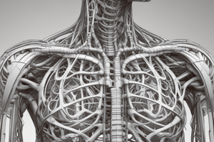
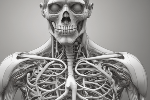
Thoracic Wall
- The thoracic wall is formed by the thoracic part of the vertebral column posteriorly, ribs and intercostal spaces laterally on either side, and the sternum and costal cartilages anteriorly.
- The thoracic cage protects the lungs and heart and provides attachment for the muscles of the thorax, upper extremity, abdomen, and back.
Thoracic Cage
- The thoracic cage is an osseocartilaginous, cage-like unit that surrounds and protects the heart, lungs, and adnexa.
- It also covers all or parts of certain upper abdominal organs (e.g., liver, stomach, spleen, kidneys).
Sternum
- The sternum is an elongate, flat bone that lies in the midline of the anterior chest wall.
- It consists of three parts: manubrium, body, and xiphoid process.
- The manubrium is the upper part of the sternum, articulating with the body of the sternum at the manubriosternal joint, and with the clavicles and the first costal cartilage and the upper part of the second costal cartilage on each side.
Pleurae and Lungs
- The lungs are covered by a thin membrane called the visceral pleura.
- The visceral pleura passes from each lung at its root (i.e., where the main air passages and blood vessels enter) to the inner surface of the chest wall, where it is called the parietal pleura.
- Two membranous sacs called the pleural cavities are formed, one on each side of the thorax, between the lungs and the thoracic walls.
Muscles of the Thorax
- The diaphragm is the most important muscle used in inspiration, and its contraction assists the contraction of the muscles of the anterolateral abdominal wall in raising the intra-abdominal pressure for micturition, defecation, and parturition.
- The diaphragm inserts into a central tendon, which is shaped like three leaves, and is partially fused with the inferior surface of the fibrous pericardium.
- The right crus of the diaphragm passes up to the left and surrounds the esophageal orifice in a sling-like loop, acting as a sphincter and possibly assisting in the prevention of regurgitation of the stomach contents into the thoracic part of the esophagus.
- The sensory innervation of the peripheral parts of the diaphragm is from the lower six thoracic nerves.
Levatores Costarum
- The levatores costarum muscles comprise 12 pairs, each triangular in shape and inserting into the rib below its origin.
- They elevate the ribs, but their role in respiration is questionable, and they may serve as proprioceptive devices.
Serratus Posterior Muscles
- The serratus posterior superior and inferior muscles are thin, flat, and comprise the intermediate layer of muscles of the back.
- They are supplied by adjacent intercostal nerves and may have proprioceptive functions rather than motor actions.
Nerves
- The intercostal nerves supply the entire thoracic wall, and the first intercostal nerve joins the brachial plexus by a large branch.
- The second intercostal nerve joins the medial cutaneous nerve of the arm by a large branch named the intercosto-brachial nerve.
- The intercostal nerves supply the skin of the chest wall and the parietal pleura covering the outer and inner surfaces of each intercostal space.
Internal Thoracic Artery
- The internal thoracic artery supplies the anterior wall of the body from the clavicle to the umbilicus.
- It is a branch of the first part of the subclavian artery in the neck and descends vertically on the pleura behind the costal cartilages.
Clinical Notes
- Skin innervation of the chest wall and referred pain: the supraclavicular nerves (C3 and 4) provide the cutaneous innervation of the anterior chest wall above the level of the sternal angle.
- Below this level, the anterior and lateral cutaneous branches of the intercostal nerves supply oblique bands of skin in regular sequence.
- Herpes zoster is a relatively common condition caused by the reactivation of the latent varicella-zoster virus in a patient who has previously had chickenpox.
- Intercostal nerve block is indicated for repair of lacerations of the thoracic and abdominal walls, for relief of pain in rib fractures, and to allow pain-free respiratory movements.
Thoracic Cavity
- The thoracic cavity can be divided into a median partition called the mediastinum and the laterally placed pleurae and lungs.
- The mediastinum is divided into superior and inferior mediastinum by an imaginary plane passing from the sternal angle anteriorly to the lower border of the body of the fourth thoracic vertebra posteriorly.
- This plane marks several key structures, including:
- The joint between the manubrium and body of the sternum
- The second costosternal joint
- The demarcation between the ascending aorta and the arch of the aorta
- The demarcation between the arch of the aorta and the descending thoracic aorta
- The bifurcation of the trachea
- The level of the left primary bronchus
Pleural Cavities
- Each lung is covered by a thin, serous pleural membrane that passes from the lung at its root and continues onto the inner surface of the thoracic wall.
- This arrangement forms two independent membranous sacs called the pleural cavities, one on each side of the thorax, between the lungs and the thoracic walls.
Mediastinum
- The mediastinum is the area between the sternum, the two pleural cavities, and the vertebral column.
- The mediastinum is divided into superior, middle, and inferior mediastinum.
- The superior mediastinum contains the trachea, esophagus, and thoracic duct.
- The middle mediastinum contains the heart and pericardium.
- The inferior mediastinum contains the diaphragm and abdominal organs.
Pleural Fluid
- The pleural cavity normally contains 5 to 10 mL of clear fluid, which lubricates the apposing surfaces of the visceral and parietal pleurae during respiratory movements.
- Hydrostatic and osmotic pressures stimulate the formation of the fluid.
- Any condition that increases the production of the fluid or impairs the drainage of the fluid results in abnormal accumulation of fluid, called a pleural effusion.
Trachea
- The trachea is a tube that connects the larynx to the bronchi.
- The trachea bifurcates into the right and left principal bronchi.
- The right principal bronchus is wider and is a more direct continuation of the trachea than the left.
- The trachea is supplied by the inferior thyroid arteries and the bronchial arteries.
Bronchial Tree
- The bronchi divide dichotomously, eventually giving rise to several million terminal bronchioles that terminate in one or more respiratory bronchioles.
- The bronchial tree is supplied by the bronchial arteries.
- The bronchi are lined by ciliated epithelium and contain mucous glands.
Lung Lobes and Fissures
- Right lung has 3 lobes: upper (superior), middle, and lower (inferior)
- Right lung has 2 fissures: horizontal and oblique, which intersect in the midaxillary line
- Horizontal fissure runs along the fourth costal cartilage
- Oblique fissure runs at a T2 (posterior) to T6 (anterior) angulation
- Left lung has 2 lobes: upper (superior) and lower (inferior)
- Left lung has 1 oblique fissure, similar to the right lung, but no horizontal fissure
Bronchopulmonary Segments
- A bronchopulmonary segment is a structurally and functionally independent unit of a lung lobe
- Each segment is bounded by connective tissue walls
- Each segment has its own lymphatic vessels, autonomic nerve supply, and branch of the pulmonary artery
- Segmental bronchus divides repeatedly within a bronchopulmonary segment
- Pulmonary veins run in the connective tissue between adjacent bronchopulmonary segments
Bronchioles
- Bronchi become smaller and more irregular as they divide
- Cartilage plates become smaller and fewer in number
- Bronchioles eventually divide and give rise to terminal bronchioles
- Terminal bronchioles are surrounded by connective tissue
Heart Structure and Blood Vessels
- The left ventricle walls are three times thicker than those of the right ventricle.
- The left intraventricular blood pressure is six times higher than that inside the right ventricle.
- In cross-section, the left ventricle is circular, while the right ventricle is crescentic due to the bulging of the ventricular septum.
- The left ventricle has well-developed trabeculae carneae and two large papillary muscles, but no moderator band.
Heart Nerve Supply
- The heart receives both sympathetic and parasympathetic fibers from the autonomic nervous system.
- The sympathetic supply originates in upper thoracic spinal cord segments and routes through the cervical and upper thoracic sympathetic chain ganglia.
- The parasympathetic supply comes from the vagus nerves.
- Activation of sympathetic nerves results in cardiac acceleration, increased force of contraction, and dilation of the coronary arteries.
- Activation of parasympathetic nerves results in a reduction in the rate and force of contraction of the heart and constriction of the coronary arteries.
Conducting System of the Heart
- The conducting system is a network of specialized cardiac muscle cells designed to generate rhythmic cardiac impulses.
- The normal heart contracts rhythmically at about 70 to 90 beats per minute in the resting adult.
- The contractile process originates spontaneously in the conducting system and the impulse travels to different regions of the heart.
- The functions of the conducting system are to initiate excitation of the atria, delay excitation of the ventricles, and spread excitation across the ventricles.
Cardiac Pain
- Cardiac pain is caused by oxygen deficiency and the accumulation of metabolites, which stimulate the sensory nerve endings in the myocardium.
- Afferent nerve fibers ascend to the central nervous system through the cardiac branches of the sympathetic trunk and enter the spinal cord through the posterior roots of the upper four thoracic nerves.
- The pain is not felt in the heart but is referred to the skin areas supplied by the upper four intercostal nerves and by the intercostobrachial nerve (T2).
- The intercostobrachial nerve communicates with the medial cutaneous nerve of the arm and is distributed to the skin on the medial side of the upper part of the arm.
Cardiac Cycle
- Ventricular contraction (shortening and emptying) occurs during systole, accompanied by the first heart sound ("lub") and closure of the atrioventricular (cuspid) valves
- Ventricular relaxation (elongation and filling) occurs during diastole, accompanied by the second heart sound ("dub") and closure of the semilunar valves
- Aortic and pulmonary valves open during systole and close during diastole
- Papillary muscles contract and take up the slack of the chordae tendineae during systole
- The atrioventricular bundle and its branches, including the Purkinje fibers, ensure synchronized myocardial contraction throughout the ventricles
Heart Development
- The heart tube elongates and bends, forming a U-shaped and then a compound S shape, with the atrium posterior to the ventricle
- The atrium and ventricle become separated by the atrioventricular canal
- The single primitive atrium divides into two separate right and left atria
Thoracic Aorta
- The descending thoracic aorta lies in the posterior mediastinum and begins at the level of the fourth thoracic vertebra
- It runs downward and forward, inclining medially to reach the anterior surface of the vertebral column
- At the level of the 12th thoracic vertebra, it passes behind the diaphragm through the aortic opening
Aneurysm and Coarctation of the Aorta
- Aneurysm is a gross dilatation of the aorta, which may show as a pulsatile swelling in the suprasternal notch
- Coarctation of the aorta is a congenital narrowing of the aorta, often just proximal, opposite, or distal to the site of attachment of the ligamentum arteriosum
- Collateral circulation develops to compensate for the diminished volume of blood reaching the lower part of the body, resulting in dilatation of the internal thoracic, subclavian, and posterior intercostal arteries
Patent Ductus Arteriosus
- The ductus arteriosus represents the distal portion of the sixth left aortic arch and connects the left pulmonary artery to the beginning of the descending aorta
- Failure of the ductus arteriosus to close may occur as an isolated congenital abnormality or in association with congenital heart disease
- A patent ductus arteriosus results in high-pressure aortic blood passing into the pulmonary artery, producing pulmonary hypertension and hypertrophy of the right ventricle
Large Thoracic Veins
- The azygos vein originates from the union of the right ascending lumbar vein and the right subcostal vein in the abdomen
- It ascends through the aortic opening in the diaphragm on the right side of the aorta to empty into the posterior surface of the superior vena cava
- The azygos vein has numerous tributaries, including the eight lower right intercostal veins, the right superior intercostal vein, and the numerous mediastinal veins
Thoracic Duct and Lymphatic System
- The thoracic duct receives lymph from the lower limbs, pelvic cavity, abdominal cavity, left side of the thorax, and left side of the head, neck, and arm.
- It conveys all lymph from these areas into adjacent large veins.
- The thoracic duct ascends through the aortic opening of the diaphragm on the right side of the descending aorta.
Vagus Nerves
- The vagus nerves carry preganglionic parasympathetic fibers into the thoracic and abdominal cavities.
- They give off cardiac branches in the neck that descend into the chest.
- These branches contribute to the pulmonary, esophageal, and cardiac plexuses, supplying the lungs, esophagus, and heart.
Phrenic Nerves
- The phrenic nerves arise in the neck from the anterior rami of the third, fourth, and fifth cervical nerves.
- They descend in the thorax, passing in front of the root of the lung and assists in the formation of the pulmonary plexus.
- The phrenic nerves possess both efferent and afferent fibers.
- Efferent fibers provide the sole motor supply to the diaphragm.
- Afferent fibers carry sensation from the peritoneum covering the central region of the undersurface of the diaphragm, the pleura covering the central region of the upper surface of the diaphragm, and the pericardium and mediastinal parietal pleura.
Thoracic Sympathetic Trunk
- The thoracic part of the sympathetic trunk is continuous above with the cervical and below with the lumbar parts of the sympathetic trunk.
- It is the most laterally placed structure in the mediastinum and runs downward on the heads of the ribs.
- The thoracic sympathetic trunk has 12 (often only 11) segmentally arranged ganglia, each with a white and gray ramus communicans passing to the corresponding spinal nerve.
Branches of Thoracic Sympathetic Trunk
- White rami communicantes connect individual thoracic spinal nerves with the sympathetic chain, conveying preganglionic fibers from the spinal nerves into the sympathetic chain.
- Gray rami communicantes connect sympathetic chain ganglia to their matching thoracic spinal nerves, carrying postganglionic fibers to blood vessels, sweat glands, and arrector pili muscles of the skin of the body wall and limbs.
- Thoracic splanchnic (visceral) branches arise from the first four or five thoracic chain ganglia, carrying postganglionic fibers to the pulmonary, cardiac, and esophageal plexuses and on to the lungs, heart, aorta, and esophagus.
- Abdominal splanchnic (visceral) nerves arise from the lower eight thoracic chain ganglia, carrying mainly preganglionic fibers to the abdominal viscera.
Lymphatic System in the Thorax
- Lymph vessels in the thorax collect lymph from virtually all parts of the body below the neck and upper limbs.
- The lymph vessels in the posterior parts of the intercostal spaces drain backward to the posterior intercostal nodes lying near the heads of the ribs.
- From there, the lymph enters the thoracic duct.
- The thoracic duct begins below in the abdomen as a dilated sac, the cisterna chyli, and ascends through the aortic opening of the diaphragm.
- It gradually crosses the median plane behind the esophagus and reaches the left border of the esophagus at the level of the lower border of the body of the fourth thoracic vertebra (sternal angle).
- The thoracic duct receives the left jugular, subclavian, and bronchomediastinal lymph trunks at the root of the neck, although they may drain directly into the adjacent large veins.
- The thoracic duct conveys all lymph from the lower limbs, pelvic cavity, abdominal cavity, left side of the thorax, and left side of the head, neck, and arm.
Thoracic Wall
- The lymph vessels of the skin of the anterior thoracic wall drain to the anterior mediastinal nodes.
- The lymph vessels of the skin of the posterior thoracic wall drain to the posterior mediastinal nodes.
Pulmonary Veins
- Two pulmonary veins leave each lung, carrying oxygenated blood to the left atrium of the heart.
Mediastinum
- The mediastinum contains nodes that drain lymph from mediastinal structures and empty into the bronchomediastinal trunks and thoracic duct.
- Disease and enlargement of these nodes may exert pressure on important neighboring mediastinal structures, such as the trachea and superior vena cava.
Vagus Nerves
- The vagus nerves carry preganglionic parasympathetic fibers (and other components) into the thoracic and abdominal cavities.
- Both give off cardiac branches in the neck that descend into the chest and contribute to the pulmonary, esophageal, and cardiac plexuses.
- The left vagus nerve descends in the thorax between the left common carotid and left subclavian arteries.
- The right vagus nerve descends in the thorax along the right side of the right brachiocephalic vein and the superior vena cava.
Phrenic Nerves
- The phrenic nerves arise in the neck from the anterior rami of the third, fourth, and fifth cervical nerves.
- The right phrenic nerve descends in the thorax along the right side of the right brachiocephalic vein and the superior vena cava.
- The left phrenic nerve descends in the thorax along the left side of the left subclavian artery.
- The phrenic nerves pass through the diaphragm to supply the central part of the peritoneum on its abdominal aspect.
Sympathetic Trunks
- The thoracic part of the sympathetic trunk is continuous above with the cervical and below with the lumbar parts of the sympathetic trunk.
- It is the most laterally placed structure in the mediastinum and runs downward on the heads of the ribs.
- The sympathetic trunk leaves the thorax on the side of the body of the 12th thoracic vertebra by passing behind the medial arcuate ligament of the diaphragm.
Clinical Notes
- Diaphragm paralysis can occur due to pressure from malignant tumors in the mediastinum or surgical crushing or sectioning of the phrenic nerve in the neck.
- Sympathetic trunk surgery can be performed to treat conditions such as Raynaud disease.
- Chest pain can be a symptom of many conditions, including disease in the thoracic and abdominal walls or in many different thoracic and abdominal viscera.
Studying That Suits You
Use AI to generate personalized quizzes and flashcards to suit your learning preferences.