Podcast
Questions and Answers
Which bones are primarily involved in forming the thoracic cage?
Which bones are primarily involved in forming the thoracic cage?
- Radius, Ulna, Carpals
- Clavicle, Scapula, Sternum
- Pelvis, Femur, Tibia
- Vertebral column, Ribs, Sternum (correct)
What type of ribs are classified as 'floating ribs'?
What type of ribs are classified as 'floating ribs'?
- Ribs that attach indirectly to the sternum
- Ribs that attach directly to the sternum
- Ribs that connect to the xiphoid process
- Ribs with no anterior attachment to the sternum (correct)
Which aspect differentiates typical thoracic vertebrae from atypical ones?
Which aspect differentiates typical thoracic vertebrae from atypical ones?
- Number of transverse processes
- Presence of a circular vertebral foramen
- Presence of a heart-shaped body
- Presence of costal facets on the vertebral body (correct)
What is a significant clinical aspect associated with the sternum?
What is a significant clinical aspect associated with the sternum?
Which part of the rib anatomy is recognized for supporting subclavian vessels?
Which part of the rib anatomy is recognized for supporting subclavian vessels?
In the anatomy of thoracic vertebrae, which feature is unique to them compared to others?
In the anatomy of thoracic vertebrae, which feature is unique to them compared to others?
Which characteristic applies specifically to the 11th and 12th ribs?
Which characteristic applies specifically to the 11th and 12th ribs?
How can you identify an atypical thoracic vertebra?
How can you identify an atypical thoracic vertebra?
At which rib does the inferior margin of the parietal pleura begin in the midclavicular line?
At which rib does the inferior margin of the parietal pleura begin in the midclavicular line?
Which of the following accurately describes the characteristics of the right bronchus?
Which of the following accurately describes the characteristics of the right bronchus?
What type of epithelium lines the distal passages known as alveolar ducts and alveoli?
What type of epithelium lines the distal passages known as alveolar ducts and alveoli?
Which structures are embedded in elastic connective tissue within the lung?
Which structures are embedded in elastic connective tissue within the lung?
What is the primary function of the cells found within the lung tissue for defense against microbes?
What is the primary function of the cells found within the lung tissue for defense against microbes?
What shape best describes the thorax?
What shape best describes the thorax?
Which structure is NOT found passing through the inlet of the thorax?
Which structure is NOT found passing through the inlet of the thorax?
Which of the following muscles are associated with the inlet of the thorax?
Which of the following muscles are associated with the inlet of the thorax?
Which boundary is not part of the thoracic inlet?
Which boundary is not part of the thoracic inlet?
What defines the shape and position of the diaphragm?
What defines the shape and position of the diaphragm?
Which of the following structures is primarily located at the thoracic outlet?
Which of the following structures is primarily located at the thoracic outlet?
What aspect of the diaphragm is occupied by tendons?
What aspect of the diaphragm is occupied by tendons?
Which vessels pass through the inlet of the thorax?
Which vessels pass through the inlet of the thorax?
How are the cupolae of the diaphragm characterized?
How are the cupolae of the diaphragm characterized?
Which of the following structures is not associated with the boundaries of the thoracic outlet?
Which of the following structures is not associated with the boundaries of the thoracic outlet?
Which surface of the lung maintains a concave shape?
Which surface of the lung maintains a concave shape?
What anatomical structure enters and exits the lungs at the hilum?
What anatomical structure enters and exits the lungs at the hilum?
How many lobes does the right lung have compared to the left lung?
How many lobes does the right lung have compared to the left lung?
What is located in the mediastinum?
What is located in the mediastinum?
What type of pleura directly covers the lungs?
What type of pleura directly covers the lungs?
Where is the apex of the lung located?
Where is the apex of the lung located?
What is the primary role of the pleural cavity?
What is the primary role of the pleural cavity?
Which thoracic vertebrae level is associated with the lung hilum?
Which thoracic vertebrae level is associated with the lung hilum?
Which structure is not part of the lung root?
Which structure is not part of the lung root?
What is the costal surface of the lung related to?
What is the costal surface of the lung related to?
Which part of the diaphragm originates from the deep surfaces of the lower six ribs and their costal cartilages?
Which part of the diaphragm originates from the deep surfaces of the lower six ribs and their costal cartilages?
What structure passes through the vena caval opening of the diaphragm?
What structure passes through the vena caval opening of the diaphragm?
Where is the esophageal opening of the diaphragm located?
Where is the esophageal opening of the diaphragm located?
What is the function of the muscle fibers of the right crus surrounding the esophageal orifice?
What is the function of the muscle fibers of the right crus surrounding the esophageal orifice?
Which of the following openings is not classified as a major opening in the diaphragm?
Which of the following openings is not classified as a major opening in the diaphragm?
Which major blood vessel passes through the aortic opening of the diaphragm?
Which major blood vessel passes through the aortic opening of the diaphragm?
Which structure is located at the space of LARREY in the diaphragm?
Which structure is located at the space of LARREY in the diaphragm?
Which nerve pierces the left cupola of the diaphragm?
Which nerve pierces the left cupola of the diaphragm?
Which lumbar vertebrae contribute to the vertebral part of the diaphragm?
Which lumbar vertebrae contribute to the vertebral part of the diaphragm?
What is transmitted through the minor openings of the diaphragm?
What is transmitted through the minor openings of the diaphragm?
Flashcards are hidden until you start studying
Study Notes
Thoracic Cage
- The thoracic cage is the bony framework of the chest and is formed by the vertebral column posteriorly, ribs and intercostal spaces laterally, and the sternum and costal cartilages anteriorly.
- The thoracic cage serves as a protective barrier for the heart, lungs, and other internal organs.
Bones Forming the Thoracic Cage
- The sternum comprises three parts: manubrium, body, and xiphoid process.
- The manubrium articulates with the clavicle and the first and second ribs.
- The sternal angle is a landmark that marks the articulation of the manubrium and body of the sternum, as well as the level of the second rib.
- There are 12 pairs of ribs.
- 7 true ribs directly attach to the sternum.
- 5 false ribs attach indirectly or not at all to the sternum.
- 2 of the 5 false ribs are called floating ribs and do not have any ventral attachment.
- Typical ribs are numbered 3-9 and consist of a head, neck, tubercle, angle, shaft, and subcostal groove.
- Atypical ribs (1st, 2nd, and 10th-12th) have unique features:
- The 1st rib is shorter, flatter, wider, and supports the subclavian vessels.
- The 2nd rib is also long and flat.
- The 10th-12th ribs only articulate with vertebrae.
- The 11th and 12th rib don't articulate with the transverse processes posteriorly or the sternum anteriorly.
- Thoracic vertebrae have specific characteristics:
- They have transverse costal facets and costal facets on the vertebral body.
- Their spinous processes are long and point inferiorly.
- The superior articular facets face dorsally/posteriorly, while the inferior articular facets face ventrally/anteriorly.
- The vertebral foramen is circular and the body is heart-shaped.
Apertures of the Thoracic Cage
- The thoracic cage, formed by osseo-cartilaginous structures, is elastic and crucial for respiration.
- It has two main openings: the inlet and outlet.
- The inlet of the thorax, also known as the superior aperture, is narrow, kidney-shaped, and bounded by the manubrium sterni anteriorly, T1 body posteriorly, and the first rib and cartilage laterally.
- Structures traversing through the inlet include important viscera like the trachea, esophagus, lung apices, and remnants of the thymus.
- Large vessels passing through the inlet include the brachiocephalic artery, left common carotid artery, left subclavian artery, and right and left brachiocephalic veins.
- The inlet also accommodates muscles such as the sternohyoid, sternothyroid, and longus colli, as well as nerves like the right and left phrenic, vagus, sympathetic trunks, and first thoracic nerves.
Diaphragm
- The diaphragm is a dome-shaped musculo-aponeurotic partition located between the thorax and abdomen.
- Its superior surface faces the thorax and is convex, while its central part is depressed, forming summits called cupolae.
- The right cupolae is generally higher due to the presence of the liver.
- The diaphragm's peripheral part is muscular (striated) and its central part is tendinous, occupied by the central tendon.
- It originates from the posterior surface of the xiphoid process (sternal part), deep surfaces of the lower six ribs and their costal cartilages (costal part), and the upper three lumbar vertebrae (vertebral/lumbar part).
- Fibers of the right crus surrounding the esophageal orifice act as a sphincter, potentially preventing the regurgitation of stomach contents into the thoracic esophagus.
Openings in the Diaphragm
- The diaphragm has major and minor openings.
- The three major openings allow for the passage of vital structures:
- The vena caval opening (T8 vertebra) allows passage of the inferior vena cava and branches of the right phrenic nerve.
- The esophageal opening (T10 vertebra) allows passage of the esophagus, the right and left vagus nerves, the esophageal branches of the left gastric vessels, and lymph vessels.
- The aortic opening (anterior to T12 vertebra) allows passage of the aorta, thoracic duct, and azygos vein.
- The minor openings include:
- The space of Larrey: located between the sternal origin and 7th costal cartilage, transmitting superior epigastric vessels, few lymphatics of the liver. When this opening is enlarged, it becomes "foramen of Morgagni."
- Other minor openings include the passage of the greater and lesser splanchnic nerves through the right and left crus, the left phrenic nerve piercing through the left cupola, and the sympathetic trunk and least splanchnic nerve passing medial to the arcuate ligament.
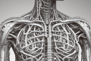
Lungs
- The lungs are the essential organs of respiration, located in the thoracic cavity on either side of the mediastinum.
- Each lung has an apex, base, costal surface, and medial surface.
- The apex is located above the border of the first rib and is close to the blood vessels and nerves in the root of the neck.
- The base of the lung is related to the thoracic surface of the diaphragm.
- The costal surface of the lung is convex and related to the costal cartilage, ribs, and intercostal muscles.
- The medial surface of the lung is concave and has a hilum (depression or slit-like opening) at the level of the 5th, 6th, and 7th thoracic vertebrae.
- The lung root, composed of the primary bronchus, pulmonary artery, two pulmonary veins, bronchial arteries and veins, lymphatic vessels, and nerves, enters and leaves the hilum.
Mediastinum
- The mediastinum is the area between the lungs, containing vital structures such as the heart, major blood vessels (great vessels), trachea, right and left bronchi, esophagus, lymph nodes, lymph vessels, and nerves.
Pleura and Pleural Cavity
- The parietal pleura lines the thoracic cavity, while the visceral pleura covers the lung surface.
- The pleural cavity, a potential space between the two pleurae, contains a small amount of serous fluid.
- The two layers are continuous at the hilum.
Interior of the Lung
- The lung interior consists of bronchi, smaller air passages (bronchioles), alveoli, connective tissue, blood vessels, lymph vessels, and nerves, all embedded in elastic connective tissue.
Pulmonary Blood Supply
- The lungs receive blood from both the pulmonary and bronchial circulations:
- The pulmonary arteries deliver deoxygenated blood from the right ventricle for oxygenation.
- The bronchial arteries supply oxygenated blood to the lung tissue itself.
Bronchi and Bronchioles
- The trachea divides into the right and left bronchi at the level of the 5th thoracic vertebra.
- The right bronchus is wider, shorter, and more vertical than the left, making it more susceptible to obstruction by inhaled foreign bodies.
- It further divides into three lobar bronchi within the lung.
- The left bronchus is longer, narrower, and more oblique, and divides into two branches.
Blood Supply, Venous Drainage, Nerve Supply of the Bronchi and Bronchioles
- The bronchial arteries supply blood to the bronchi, while the right and left bronchial veins drain it.
- The right side drains into the azygos vein, while the left drains into the superior intercostal vein.
- The vagus nerve provides parasympathetic innervation to the bronchi and bronchioles, causing bronchoconstriction.
- The sympathetic nervous system causes bronchodilation.
Respiratory Bronchioles and Alveoli
- The lung is further divided into lobules by fine sheets of connective tissue.
- As airway divides and becomes smaller, the wall becomes thinner along with gradual decrease in smooth muscle and connective tissue.
- Alveolar ducts and alveoli have a single layer of squamous epithelium surrounded by dense capillaries, forming the respiratory membrane.
- Septal cells (type II pneumocytes), located between squamous epithelial cells, produce surfactant, which reduces surface tension and allows for proper lung expansion.
Nerve Supply of the Alveoli
- Parasympathetic stimulation from the vagus nerve causes bronchoconstriction, while sympathetic stimulation causes bronchodilation.
Functions of the Alveoli
- Alveoli provide a large surface area for gas exchange.
- They are also involved in defense against microbes, with plasma cells, macrophages, and lymphocytes acting in the distal air passages.
Studying That Suits You
Use AI to generate personalized quizzes and flashcards to suit your learning preferences.