Podcast
Questions and Answers
What type of joints does each rib form?
What type of joints does each rib form?
- Two costovertebral joints
- One costotransverse joint
- One costovertebral joint
- Two joints: one costovertebral and one costotransverse (correct)
What is the function of the conchae in the nasal cavity?
What is the function of the conchae in the nasal cavity?
- To increase surface area and warm the air (correct)
- To produce mucus to moisturize the air
- To detect odors in the air
- To filter out large particles from the air
What is the name of the flap of cartilage that prevents food and drink from entering the trachea?
What is the name of the flap of cartilage that prevents food and drink from entering the trachea?
- Epiglottis (correct)
- Laryngeal cartilage
- Glottis
- Uvula
Which part of the pharynx is posterior to the oral cavity and inferior to the level of the soft palate?
Which part of the pharynx is posterior to the oral cavity and inferior to the level of the soft palate?
What is the name of the tube that air travels through to reach the lungs?
What is the name of the tube that air travels through to reach the lungs?
What is the purpose of the laryngeal cartilage?
What is the purpose of the laryngeal cartilage?
What is the name of the system of airways that allow passage of air into the lungs?
What is the name of the system of airways that allow passage of air into the lungs?
What is the boundary between the oral cavity and the nasal cavity?
What is the boundary between the oral cavity and the nasal cavity?
What happens to the air after it passes through the larynx?
What happens to the air after it passes through the larynx?
Why do we sometimes choke on food or drink?
Why do we sometimes choke on food or drink?
What is the purpose of the thoracic cage?
What is the purpose of the thoracic cage?
Which of the following structures is NOT part of the sternum?
Which of the following structures is NOT part of the sternum?
Where is the sternal angle located?
Where is the sternal angle located?
Which ribs articulate with the body of the sternum?
Which ribs articulate with the body of the sternum?
What is the name of the depression on the superior aspect of the manubrium?
What is the name of the depression on the superior aspect of the manubrium?
Which of the following structures articulates with the medial ends of the clavicles?
Which of the following structures articulates with the medial ends of the clavicles?
What is the level of the T10 vertebrae?
What is the level of the T10 vertebrae?
What is the function of the costal cartilages?
What is the function of the costal cartilages?
Which nerve innervates the diaphragm?
Which nerve innervates the diaphragm?
What are the three areas of attachment of the diaphragm?
What are the three areas of attachment of the diaphragm?
What is the effect of diaphragm contraction on the thoracic cavity?
What is the effect of diaphragm contraction on the thoracic cavity?
Which of the following structures passes through the diaphragm at the T8 level?
Which of the following structures passes through the diaphragm at the T8 level?
What is the function of the external intercostal muscles?
What is the function of the external intercostal muscles?
Which muscle is NOT an accessory muscle of inhalation?
Which muscle is NOT an accessory muscle of inhalation?
What is the arrangement of the neurovascular bundle in the intercostal spaces?
What is the arrangement of the neurovascular bundle in the intercostal spaces?
What is the name of the movement of the ribs during inspiration?
What is the name of the movement of the ribs during inspiration?
What is the function of the internal intercostal muscles?
What is the function of the internal intercostal muscles?
What is the central tendon of the diaphragm?
What is the central tendon of the diaphragm?
What is the primary function of the trachealis muscle?
What is the primary function of the trachealis muscle?
Why are the tracheal rings shaped like 'C' rather than complete circles?
Why are the tracheal rings shaped like 'C' rather than complete circles?
How many secondary bronchi are found in the left lung?
How many secondary bronchi are found in the left lung?
What is the composition of surfactant secreted by Type 2 pneumocytes?
What is the composition of surfactant secreted by Type 2 pneumocytes?
What characteristic distinguishes the right bronchus from the left bronchus?
What characteristic distinguishes the right bronchus from the left bronchus?
What type of epithelium lines the majority of the respiratory tree from the nasal cavity to the bronchi?
What type of epithelium lines the majority of the respiratory tree from the nasal cavity to the bronchi?
What happens to inhaled foreign objects in the bronchi?
What happens to inhaled foreign objects in the bronchi?
What is the primary role of Type 1 pneumocytes in the alveoli?
What is the primary role of Type 1 pneumocytes in the alveoli?
What does widening or distortion of the carina typically indicate?
What does widening or distortion of the carina typically indicate?
Which structure is directly responsible for trapping pathogens in the respiratory tract?
Which structure is directly responsible for trapping pathogens in the respiratory tract?
Which ribs are classified as true ribs?
Which ribs are classified as true ribs?
What distinguishes atypical ribs from typical ribs?
What distinguishes atypical ribs from typical ribs?
How do ribs 8-10 articulate with the sternum?
How do ribs 8-10 articulate with the sternum?
What is the role of the costal groove found on typical ribs?
What is the role of the costal groove found on typical ribs?
Which of the following statements about rib anatomy is correct?
Which of the following statements about rib anatomy is correct?
What is the primary function of the rib cage?
What is the primary function of the rib cage?
Which rib is characterized by a single facet for articulation?
Which rib is characterized by a single facet for articulation?
How many costal cartilages connect with the sternum?
How many costal cartilages connect with the sternum?
What structural feature do all thoracic vertebrae possess regarding ribs?
What structural feature do all thoracic vertebrae possess regarding ribs?
What characterizes the left lung compared to the right lung?
What characterizes the left lung compared to the right lung?
Which structure is located at the hilum of the lungs?
Which structure is located at the hilum of the lungs?
How many bronchopulmonary segments are found in the right lung?
How many bronchopulmonary segments are found in the right lung?
What is the primary function of pulmonary arteries?
What is the primary function of pulmonary arteries?
Where do bronchial veins on the left side drain into?
Where do bronchial veins on the left side drain into?
What is the main role of the diaphragm in respiration?
What is the main role of the diaphragm in respiration?
Which fissure is found only in the right lung?
Which fissure is found only in the right lung?
What does the term 'costal surface' refer to?
What does the term 'costal surface' refer to?
Which of the following structures is involved in the drainage of the right bronchial veins?
Which of the following structures is involved in the drainage of the right bronchial veins?
What is the purpose of bronchoscopy?
What is the purpose of bronchoscopy?
At what gestational age does surfactant production typically begin in a developing fetus?
At what gestational age does surfactant production typically begin in a developing fetus?
What is the primary function of the lungs?
What is the primary function of the lungs?
Which cell type primarily promotes mucus motility in the larger airways?
Which cell type primarily promotes mucus motility in the larger airways?
What distinguishes the visceral pleura from the parietal pleura?
What distinguishes the visceral pleura from the parietal pleura?
What is the consequence of being born prematurely with insufficient surfactant?
What is the consequence of being born prematurely with insufficient surfactant?
Which of the following describes the histology of the proximal conducting zone airways?
Which of the following describes the histology of the proximal conducting zone airways?
How many lobes does the right lung have?
How many lobes does the right lung have?
What structure separates the thoracic cavity from the abdominal cavity?
What structure separates the thoracic cavity from the abdominal cavity?
What is the preferred term for the space between the parietal pleura and visceral pleura?
What is the preferred term for the space between the parietal pleura and visceral pleura?
What type of epithelium is commonly found in the bronchioles?
What type of epithelium is commonly found in the bronchioles?
Study Notes
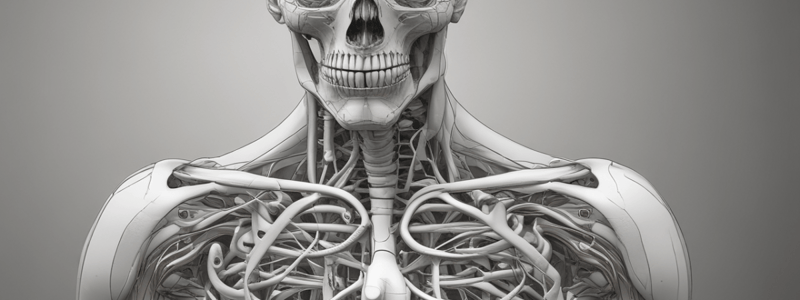
The Thoracic Cage (Rib Cage)
- The thoracic cage is made up of:
- Sternum (breastbone)
- 12 pairs of ribs
- Thoracic vertebrae
- Its purpose is to provide structure and protect thoracic contents (heart and lungs)
- The thoracic cage has the ability to change its volume of space during inhalation/exhalation to allow lungs to expand
The Sternum
- The sternum (breastbone) consists of:
- Manubrium
- Body
- Xiphoid process
- Sternal angle (angle of Louis) is located at the level of T4 vertebrae
- The superior aspect of the manubrium is concave, producing a depression called the jugular notch
Ribs
- There are 12 pairs of ribs, classified as:
- True ribs (1-7): articulate directly with the sternum
- False ribs (8-10): do not articulate directly with the sternum
- Floating ribs (11 and 12): have no anterior connection with other ribs or the sternum
- Typical ribs have a head, neck, and body (shaft)
- Atypical ribs (1, 2, 10, 11, and 12) have variations on this structure
Costovertebral Joints
- The ribs articulate posteriorly with the thoracic vertebrae
- Each thoracic vertebra has two demi-facets on either side of its vertebral body
- The demi-facets articulate with the heads of two different ribs
- Each rib forms two joints: costovertebral joint and costotransverse joint
The Respiratory System
- The respiratory system can be divided into upper and lower parts:
- Upper: nose, nasal passages, paranasal sinuses, pharynx, and larynx above the vocal cords
- Lower: larynx below vocal cords, trachea, bronchi, bronchioles, and lungs
- The nasal cavity:
- Air enters the nose via the nasal cavity
- Conchae increase surface area and spin the air to warm and moisten it
- The oral cavity:
- Acts as an air inlet in addition to the nasal cavity
- Lies inferior to the nasal cavity, bordered superiorly by the hard palate and soft palate
- The pharynx:
- Can be divided into three parts: nasopharynx, oropharynx, and laryngopharynx
- Serves as a common passageway for air and food
The Tracheobronchial Tree
- The trachea, bronchi, and bronchioles form the tracheobronchial tree
- The trachea bifurcates into the left and right primary bronchi
- The bronchi have characteristic hyaline cartilage rings
- The left and right primary bronchi divide further into secondary bronchi, and then tertiary bronchi
The Bronchi
- Each tertiary bronchus gives rise to many terminal bronchioles
- Terminal bronchioles give rise to respiratory bronchioles
- Each respiratory bronchiole ends in an acinus of clustered alveoli
Epithelial Types Recap
- Pseudostratified columnar ciliated epithelium lines the trachea and bronchi
- Simple columnar to simple cuboidal epithelium lines the bronchioles
- Simple squamous epithelium lines the alveoli
The Alveoli
-
The alveolar wall has a different structure than the bronchioles
-
Type 1 pneumocytes are responsible for gas exchange
-
Type 2 pneumocytes secrete surfactant, which decreases surface tension and helps keep alveoli open
-
Surfactant production begins in the developing fetus at around 24-28 weeks gestation### Descending Aorta
-
The descending aorta transports blood to the thorax, abdomen, pelvis, and lower limbs.
-
It is divided into two parts: thoracic aorta and abdominal aorta.
-
The thoracic aorta gives rise to bronchial arteries, which supply the trachea and bronchial tree.
-
The abdominal aorta begins at the diaphragm and extends to the common iliac arteries.
Vasculature
- The trachea and bronchial tree receive blood supply from the thoracic aorta through bronchial arteries.
- Bronchial veins drain into the accessory hemiazygos vein on the left and the azygos vein on the right, which ultimately drain into the Superior Vena Cava (SVC).
Lymphatics
- The inferior tracheobronchial lymph nodes can become enlarged and distort the carina, indicating pathology.
- Bronchoscopy involves traveling down the trachea to enter a main bronchus, and cancer cells can metastasize to the inferior tracheobronchial lymph nodes.
Diaphragm
- The diaphragm is the most important muscle of respiration, separating the thoracic and abdominal cavities.
- It is comprised of two domes and is peripherally muscular, with a central tendon.
- The diaphragm is innervated by the phrenic nerve (C3, C4, C5) and has three areas of attachment: costal cartilages, lumbar vertebrae, and the xiphoid process of the sternum.
- The parts of the diaphragm that arise from the vertebrae are known as the right and left crura.
Movement of Diaphragm
- Contraction of the diaphragm results in a downward movement of the central tendon, increasing the vertical size of the thoracic cavity during inhalation.
- Relaxation of the diaphragm allows it to return to its resting position, decreasing the vertical size of the thoracic cavity during exhalation.
- The diaphragm's movement is often described as a "bucket handle" movement, with the diaphragm increasing thoracic volume in three planes during inspiration.
Openings in the Diaphragm
- There are three major openings in the diaphragm that allow the oesophagus, inferior vena cava, and aorta to pass from the thoracic to the abdominal cavity.
- These openings occur at specific levels: IVC at T8, oesophagus at T10, and aorta at T12.
Intercostal Spaces
- The intercostal spaces are numbered according to the rib superior to them and contain intercostal muscles that help move the ribs during breathing.
- There are three layers of intercostal muscles: external, internal, and innermost.
Intercostal Muscles
- External intercostal muscles are the most superficial layer, running from the inferior border of one rib to the superior border of the rib below, and contract to elevate the ribs.
- Internal intercostal muscles are the middle layer, running from the superior border of one rib to the inferior border of the rib above, and contract to draw adjacent ribs together.
- Innermost intercostal muscles are the deepest layer, separated from the other two layers by the neurovascular bundle, and have the same attachment points and action as internal intercostal muscles.
Neurovascular Supply to Intercostal Spaces
- Each intercostal space has a separate neurovascular bundle that runs along the inferior aspect of the rib in the costal groove.
- The neurovascular bundle contains the intercostal vein, intercostal artery, and intercostal nerve, always in that arrangement (superior to inferior).
Accessory Muscles of Respiration
- Accessory muscles of inhalation include the sternocleidomastoid muscle, pectoralis minor muscle, and scalene muscles.
- Accessory muscles of exhalation include the external oblique muscle, internal oblique muscle, transversus abdominis muscle, and rectus abdominis muscle.
Studying That Suits You
Use AI to generate personalized quizzes and flashcards to suit your learning preferences.
Description
Learn about the thoracic cage, also known as the rib cage, and its components. Understand its purpose and how it functions during inhalation and exhalation.


