Podcast
Questions and Answers
How does adjusting the diaphragm tension of a stethoscope bell affect its performance?
How does adjusting the diaphragm tension of a stethoscope bell affect its performance?
- Increasing tension allows for the detection of higher frequency sounds. (correct)
- Decreasing tension allows for the detection of higher frequency sounds.
- Increasing tension allows for the detection of lower frequency sounds.
- Adjusting tension has no impact on the frequency range detected.
What is the fundamental principle behind how a piezoelectric transducer generates ultrasound waves?
What is the fundamental principle behind how a piezoelectric transducer generates ultrasound waves?
- Radio waves are converted into sound waves.
- Applying mechanical pressure generates an electrical current.
- A magnetic field causes the crystal to vibrate.
- An electrical potential difference causes the crystal to expand and contract. (correct)
In medical ultrasound imaging, what is the primary reason for using a water-based gel between the transducer and the patient's skin?
In medical ultrasound imaging, what is the primary reason for using a water-based gel between the transducer and the patient's skin?
- To lubricate the skin for easier transducer movement.
- To sterilize the skin and prevent infection.
- To cool the transducer during operation.
- To improve acoustic impedance matching and eliminate air. (correct)
How does the frequency range of ultrasound used in medical applications compare to the range of human hearing?
How does the frequency range of ultrasound used in medical applications compare to the range of human hearing?
Which of the following is the correct order of energy conversion in ultrasound imaging, starting from the device and ending with image production?
Which of the following is the correct order of energy conversion in ultrasound imaging, starting from the device and ending with image production?
A sound wave travels through different mediums. Which of the following correctly orders the mediums from fastest to slowest sound propagation?
A sound wave travels through different mediums. Which of the following correctly orders the mediums from fastest to slowest sound propagation?
If the period of a sound wave is doubled, what happens to its frequency?
If the period of a sound wave is doubled, what happens to its frequency?
Which of the following is NOT a commonly observed symptom associated with exposure to intense infrasonic noise?
Which of the following is NOT a commonly observed symptom associated with exposure to intense infrasonic noise?
A sound wave has a frequency of 2 kHz and a wavelength of 0.17 meters. What is the velocity of the sound wave?
A sound wave has a frequency of 2 kHz and a wavelength of 0.17 meters. What is the velocity of the sound wave?
The seismocardiogram, which measures micro-vibrations produced by heart contraction, utilizes signals in what frequency range?
The seismocardiogram, which measures micro-vibrations produced by heart contraction, utilizes signals in what frequency range?
Which of the following characteristics is NOT a general property of sound?
Which of the following characteristics is NOT a general property of sound?
Why can infrasound travel long distances with minimal power loss?
Why can infrasound travel long distances with minimal power loss?
What is a major advantage of using ultrasound for medical imaging compared to X-rays, particularly in fetal imaging?
What is a major advantage of using ultrasound for medical imaging compared to X-rays, particularly in fetal imaging?
What is the range of frequencies that the average human ear can typically detect?
What is the range of frequencies that the average human ear can typically detect?
Sound intensity is defined as the energy carried by a wave per unit area per unit time. Which of the following parameters is NOT required to calculate sound intensity?
Sound intensity is defined as the energy carried by a wave per unit area per unit time. Which of the following parameters is NOT required to calculate sound intensity?
Which of the following scenarios would most likely involve the use of infrasound?
Which of the following scenarios would most likely involve the use of infrasound?
The human ear perceives sound in terms of loudness and pitch. What physical property of sound primarily determines its loudness?
The human ear perceives sound in terms of loudness and pitch. What physical property of sound primarily determines its loudness?
A sound wave is applied perpendicularly to the interface between two media with different acoustic impedances, Z1 and Z2. If Z2 is significantly larger than Z1, what would you expect?
A sound wave is applied perpendicularly to the interface between two media with different acoustic impedances, Z1 and Z2. If Z2 is significantly larger than Z1, what would you expect?
What happens to the wavelength of a sound wave if the frequency increases while the velocity of the wave remains constant?
What happens to the wavelength of a sound wave if the frequency increases while the velocity of the wave remains constant?
In the context of sound reflection and transmission between two media, what condition results in complete transmission of a sound wave from one medium to another?
In the context of sound reflection and transmission between two media, what condition results in complete transmission of a sound wave from one medium to another?
How does the bell of a stethoscope serve as an impedance matcher between the body and the air in the tubing?
How does the bell of a stethoscope serve as an impedance matcher between the body and the air in the tubing?
Which of the following best explains why a thick liquid is applied between the ultrasound transducer and the patient's skin?
Which of the following best explains why a thick liquid is applied between the ultrasound transducer and the patient's skin?
An ultrasound wave travels from one tissue to another with a significantly different sound velocity. What is the most likely consequence of this transition?
An ultrasound wave travels from one tissue to another with a significantly different sound velocity. What is the most likely consequence of this transition?
To minimize refraction artifacts in ultrasound imaging, how should the ultrasound transducer be oriented relative to the interface between two media?
To minimize refraction artifacts in ultrasound imaging, how should the ultrasound transducer be oriented relative to the interface between two media?
What two processes contribute to the attenuation of an ultrasound beam as it propagates through tissue?
What two processes contribute to the attenuation of an ultrasound beam as it propagates through tissue?
In ultrasound imaging, what is the primary difference between reflection from a smooth surface compared to a rough surface?
In ultrasound imaging, what is the primary difference between reflection from a smooth surface compared to a rough surface?
In A-mode ultrasound, what type of information is primarily obtained?
In A-mode ultrasound, what type of information is primarily obtained?
When an ultrasound wave encounters a boundary between two tissues with different acoustic impedances (Z), what phenomenon occurs?
When an ultrasound wave encounters a boundary between two tissues with different acoustic impedances (Z), what phenomenon occurs?
What determines the quality of ultrasound imaging?
What determines the quality of ultrasound imaging?
In A-mode ultrasound, the depth of an interface between different tissues is determined by what factor?
In A-mode ultrasound, the depth of an interface between different tissues is determined by what factor?
What is the significance of comparing echoes from the left and right sides of the head in echoencephalography?
What is the significance of comparing echoes from the left and right sides of the head in echoencephalography?
In ophthalmology, A-scan ultrasound uses high frequencies (up to 20 MHz). What is the primary reason for using those high frequencies?
In ophthalmology, A-scan ultrasound uses high frequencies (up to 20 MHz). What is the primary reason for using those high frequencies?
In B-mode ultrasound, what principle is used to create 2D images of the body?
In B-mode ultrasound, what principle is used to create 2D images of the body?
M-mode ultrasound combines features of A-mode and B-mode to study motion of the heart. What is held stationary during M-mode imaging, similar to A-mode?
M-mode ultrasound combines features of A-mode and B-mode to study motion of the heart. What is held stationary during M-mode imaging, similar to A-mode?
If an adult patient's echoencephalography shows a shift of greater than 3 mm in the midline structures, what does this indicate?
If an adult patient's echoencephalography shows a shift of greater than 3 mm in the midline structures, what does this indicate?
Which ultrasound mode is most suitable for visualizing the motion of heart valves and obtaining diagnostic information about their function?
Which ultrasound mode is most suitable for visualizing the motion of heart valves and obtaining diagnostic information about their function?
What additional element does D-mode ultrasound add to 3D ultrasound images?
What additional element does D-mode ultrasound add to 3D ultrasound images?
Flashcards
Sound Wave
Sound Wave
Energy traveling away from a sound source, transferring energy without matter.
Sound
Sound
Mechanical disturbance propagating through an elastic medium.
Sound in Air
Sound in Air
Local increase (compression) or decrease (rarefaction) of pressure relative to atmospheric pressure.
Frequency (f)
Frequency (f)
Signup and view all the flashcards
Wavelength (λ)
Wavelength (λ)
Signup and view all the flashcards
Sonic Spectrum
Sonic Spectrum
Signup and view all the flashcards
Infrasound
Infrasound
Signup and view all the flashcards
Human Hearing Range
Human Hearing Range
Signup and view all the flashcards
Natural Frequency (Fres)
Natural Frequency (Fres)
Signup and view all the flashcards
SONAR (Medical)
SONAR (Medical)
Signup and view all the flashcards
Transducer
Transducer
Signup and view all the flashcards
Piezoelectric Principle
Piezoelectric Principle
Signup and view all the flashcards
Focal Zone
Focal Zone
Signup and view all the flashcards
Infrasonic Noise
Infrasonic Noise
Signup and view all the flashcards
Seismocardiogram
Seismocardiogram
Signup and view all the flashcards
Ultrasound
Ultrasound
Signup and view all the flashcards
Intensity of a Sound Wave (I)
Intensity of a Sound Wave (I)
Signup and view all the flashcards
Acoustic Impedance (Z)
Acoustic Impedance (Z)
Signup and view all the flashcards
Loudness (Volume)
Loudness (Volume)
Signup and view all the flashcards
Pitch
Pitch
Signup and view all the flashcards
Stethoscope
Stethoscope
Signup and view all the flashcards
Refraction (Ultrasound)
Refraction (Ultrasound)
Signup and view all the flashcards
Ultrasound Reflection
Ultrasound Reflection
Signup and view all the flashcards
Attenuation (Ultrasound)
Attenuation (Ultrasound)
Signup and view all the flashcards
Quality of Ultrasound Imaging
Quality of Ultrasound Imaging
Signup and view all the flashcards
Spatial Resolution
Spatial Resolution
Signup and view all the flashcards
A-Mode (1D)
A-Mode (1D)
Signup and view all the flashcards
A-Mode Ultrasound
A-Mode Ultrasound
Signup and view all the flashcards
Depth Calculation in A-Mode
Depth Calculation in A-Mode
Signup and view all the flashcards
Echoencephalography
Echoencephalography
Signup and view all the flashcards
A-Scan in Ophthalmology
A-Scan in Ophthalmology
Signup and view all the flashcards
B-Mode Ultrasound
B-Mode Ultrasound
Signup and view all the flashcards
M-Mode Ultrasound
M-Mode Ultrasound
Signup and view all the flashcards
D-Mode Ultrasound
D-Mode Ultrasound
Signup and view all the flashcards
Abnormal brain shift
Abnormal brain shift
Signup and view all the flashcards
Study Notes
- The lecture covers sound in medicine, discussing sound wave characteristics, reflection, transmission, intensity, and medical applications.
- A key focus is on ultrasound (US) in medicine, covering generation, image production, imaging modes, and physiological effects.
General Properties of Sound
- Sound is a disturbance pattern caused by energy traveling from a source, transferring energy without transferring matter.
- Sound is a mechanical disturbance propagating through an elastic material medium at a specific velocity.
- In air, sound is defined by local increases aka compressions or decreases aka rarefactions in pressure relative to atmospheric pressure.
- Sound travels fastest in solids, slower in liquids, and slowest in gases.
- The speed of sound is given by a formula including frequency and wavelength
Wave Properties
- Frequency is the number of rarefactions and compressions per unit time, denoted as f=1/T, where T is time.
- Wavelength is the distance between successive compression and rarefaction.
Sonic Spectrum
- The sonic spectrum is classified by frequency into infrasound, audible sound, and ultrasound.
- The human ear hears sounds roughly from 20 Hz to 20 kHz.
- Infrasound refers to frequencies below 20Hz, often from natural phenomena.
- Intense infrasonic noise produces symptoms like respiratory issues, aural pain, fear, visual hallucinations and chills.
- Infrasound is used in the study of heart function via seismocardiograms, measuring micro-vibrations.
- Ultrasound has a frequency range above 20kHz and has many clinical applications often providing more information than X-rays, and is safer for fetuses.
Intensity and Loudness
- The intensity 'I' of a sound wave is the energy carried per unit area per unit time, expressed in W/m².
- Acoustic impedance 'Z' is a property of the medium.
- The absolute sound intensity cannot be measured directly and is compared to a reference intensity (I₀).
- Intensity is compared to a reference intensity (I₀).
- The audible sound intensity ranges from 10⁻¹² W/m² (hearing threshold) to 1 W/m² (pain threshold).
- Two characteristics of sound include loudness, which depends on intensity, and pitch, which is determined by frequency.
Reflection and Transmission
- Sound waves applied perpendicularly to the interface between two media with differing acoustic impedances result in reflection and transmission.
- The ratio of reflected or transmitted waves to incident waves can be measured.
- A large difference in impedance 'Z' leads to a high reflection ratio.
- A sign change indicates a phase change in the reflected wave.
- A large impedance mismatch results in high reflection and low transmission.
Medical Applications
Percussion
- Percussion involves striking a body surface to detect underlying structures
- Types of sounds from percussion include resonant, hyper-resonant, and dull.
Stethoscope
- Stethoscopes amplify body sounds from the heart, lungs, or other body sites.
- Modern stethoscopes consist of a bell, a thin diaphragm, tubing, and earpieces.
- The frequency of sounds must resonate in the bell membrane.
- To selectively pick up certain frequency ranges the appropriate bell size and diaphragm tension must be chosen.
Ultrasound (US)
- Diagnostic ultrasound uses frequencies from 20kHz to 1GHz.
- SONAR (Sound Navigation and Ranging) is a device using US waves to generate images of soft tissue.
- Transducers convert electrical energy to ultrasound energy and vice versa, and vary by frequency and footprint.
Ultrasound Generation
- Ultrasound signals are generated and detected by a sensor using the piezoelectric principle.
- The piezoelectric principle involves using crystals that vibrate in response to alternating current (AC) voltage, producing ultrasound waves.
- In clinical applications, applying an electric potential difference to a piezoelectric crystal causes it to expand or contract, creating compression and rarefaction regions.
Basic Principles of SONAR
- In medical diagnosis, US pulses transmit into the body using water or jelly paste to eliminate air for impedance matching.
- Backed echoes are detected as weak signals, amplified, and shown on an oscilloscope.
US Image Production
- Three concepts affect US image production: focal zone, acoustic impedance, and refraction.
- For the best US image, the object should be at the focal zone (near field).
- When an ultrasound wave goes through different tissues a fraction of the sound wave is backscattered towards the transducer while the rest is transmitted.
Acoustic Impendance
- The liquid aka jelly between the transducer and the patients skin helps keep away any air bubbles and allows the passage of ultrasound waves.
- Small changes in impedance result in low reflection and high transmission while large changes results in high reflection and low transmission.
- Refraction involves changes in direction, and minimizing it requires transducers positioned perpendicular to the interface between two media.
Quality of Ultrasound Imaging
- Ultrasound imaging quality depends on the interaction these interactions includes spatial resolution, attenuation and reflection, and transmission.
- Spatial resolution is defined by the wavelength of sound.
Attenuation
- Attenuation of ultrasound beams propagating through tissue, resulting in decreased intensity due to absorption and scattering.
- The material/tissue is relevant to the images.
- Higher frequency attenuates better in the tissue resulting in a better image.
- Higher attenuation and decreased penetration gives a better resolution.
Image Quality
- Is determined by a compromise between good resolution and deep penetration.
- The choice of the ultrasound is determined by a compromise between good resolution and deep penetration.
- Smooth vs Rough surfaces matter for images.
- Perpendicular reflection relates to echo while non-perpendicular reflection relates to intensity in the signal.
Types of US Image Modes
A-Mode (1D)
- It is used to obtain diagnostic information about the depth of structure (image with 1-dimention).
- It sends US waves into the body and measures the required to receive the reflected sound.
- The depth of the inteface recorded is proportional to the time it takes for the echo to return.
A-Mode applications
- Used to diagnostic brain tumors and in ophthalmology and biometry.
- For ophthalmology the frequency is upto 20 MHz.
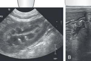
B-Mode (2D)
- B-Mode provides 2D images of the body.
- It's used to view internal structure of the body and changes over time of the liver, breast, heart, and fetus.
M-Mode (2D + motion)
- M-Mode is used to study motion such as of the heart and heart valves.
D-Mode (3D + motion; or 4D)
- D-Mode take images with 3-dimensions and adds the process of time.
Physiological Effects
- Various physiological and chemical effects occur when ultrasonic waves pass through the body, and they can cause physiological effects.
- The magnitude of physiological effects depends on amplitude and frequency.
- Different effects with varying usage.
- Low intensity US (~ 0.01 W/cm²) relate to no harmful effects and diagnostic work.
- Continues US (~1 W/cm²) relates to a deep heating effect and increased temperature.
- Continues US (1-10 W/cm²) relates to movements and pressure regions in tissues aka micro-massages.
- Continues US (~ 35 W/cm²) relates to destroying tissue as in the rupture of the DNA.
- Continues and focused US (~ 103 W/cm²) relates to selectively destroying deep tissue by using a focused beam.
Studying That Suits You
Use AI to generate personalized quizzes and flashcards to suit your learning preferences.