Podcast
Questions and Answers
کیفیت بدنه آن از T1 تا T12 به چه شکلی است؟
کیفیت بدنه آن از T1 تا T12 به چه شکلی است؟
- giảm تدریجی
- نوسان
- افزایش تدریجی (correct)
- ثابت
چه کدام از فضا در قفسه سینه قرار دارد؟
چه کدام از فضا در قفسه سینه قرار دارد؟
- شریان های بزرگ
- همه موارد فوق (correct)
- ریه ها
- قلب
چه تعداد جفت از دنده ها در قفسه سینه حضور دارند؟
چه تعداد جفت از دنده ها در قفسه سینه حضور دارند؟
- 13
- 10
- 12 (correct)
- 7
کدام قسمت از قفسه سینه شامل ریه ها است؟
کدام قسمت از قفسه سینه شامل ریه ها است؟
کدام قسمت از قلب در قفسه سینه قرار دارد؟
کدام قسمت از قلب در قفسه سینه قرار دارد؟
چه تعداد اتاق از قلب وجود دارد؟
چه تعداد اتاق از قلب وجود دارد؟
Flashcards are hidden until you start studying
Study Notes
Thoracic Vertebrae
- 12 vertebrae (T1-T12) that form the middle section of the spine
- Characterized by:
- Heart-shaped body
- Long spinous process
- Costal facets for rib articulation
- Gradual increase in size from T1 to T12
- Intervertebral discs separate each vertebra, allowing for flexibility and shock absorption
Thoracic Cavity
- The space within the thorax that contains the lungs, heart, and major blood vessels
- Boundaries:
- Superiorly: apex of the thorax (T1 vertebra)
- Inferiorly: diaphragm
- Anteriorly: sternum and costal cartilages
- Posteriorly: thoracic vertebrae
- Laterally: ribs and intercostal spaces
- Divided into three compartments:
- Right pleural cavity (contains right lung)
- Left pleural cavity (contains left lung)
- Mediastinum (contains heart, trachea, esophagus, and major blood vessels)
Ribcage Structure
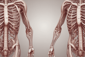
- 12 pairs of ribs (true and false ribs) that form the bony framework of the thorax
- True ribs (1-7): attach directly to the sternum via costal cartilages
- False ribs (8-10): attach to the 7th rib's costal cartilage
- Floating ribs (11-12): do not attach to the sternum
- Ribs 1-7: increasing in length and curvature from top to bottom
- Intercostal spaces: areas between adjacent ribs that contain intercostal muscles and nerves
Heart
- Located in the mediastinum, between the lungs
- Divided into four chambers:
- Right atrium (receives deoxygenated blood)
- Right ventricle (pumps blood to the lungs)
- Left atrium (receives oxygenated blood)
- Left ventricle (pumps blood to the rest of the body)
- Separated by the septum, a thin wall of tissue
- Valves regulate blood flow between chambers and into vessels
Mediastinum
- The region between the lungs that contains the heart, trachea, esophagus, and major blood vessels
- Divided into:
- Anterior mediastinum (in front of the heart)
- Middle mediastinum (around the heart)
- Posterior mediastinum (behind the heart)
- Contains lymph nodes, nerves, and fat tissue
- Important structures in the mediastinum include:
- Trachea and bronchi
- Esophagus
- Aorta and its branches
- Pulmonary arteries and veins
- Phrenic nerve and vagus nerve
Studying That Suits You
Use AI to generate personalized quizzes and flashcards to suit your learning preferences.


