Podcast
Questions and Answers
Which structure forms the base of the rib cage?
Which structure forms the base of the rib cage?
- Intercostal muscles
- Sternum (correct)
- Costal cartilages
- Vertebral column
What is the function of the alveoli in the lungs?
What is the function of the alveoli in the lungs?
- To provide structural support
- To produce mucus
- To facilitate gas exchange (correct)
- To regulate blood pressure
Which organ continually moves blood throughout the body?
Which organ continually moves blood throughout the body?
- Diaphragm
- Heart (correct)
- Lungs
- Kidneys
How do floating ribs differ from true ribs?
How do floating ribs differ from true ribs?
What is the main function of the diaphragm?
What is the main function of the diaphragm?
Which organ does the pericardium enclose?
Which organ does the pericardium enclose?
What is the function of the diaphragm?
What is the function of the diaphragm?
Which part of the thorax contains bronchioles and alveoli?
Which part of the thorax contains bronchioles and alveoli?
During exhalation, what happens to the diaphragm?
During exhalation, what happens to the diaphragm?
What is the main function of the mediastinum in the body?
What is the main function of the mediastinum in the body?
What causes downward pressure on organs in the abdominal cavity?
What causes downward pressure on organs in the abdominal cavity?
Which structure runs vertically downward in the posterior portion of the mediastinum?
Which structure runs vertically downward in the posterior portion of the mediastinum?
Flashcards are hidden until you start studying
Study Notes
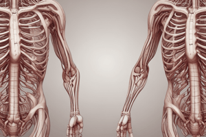
The human body's torso region is called the thorax. This area includes several vital organs such as the rib cage, lungs, heart, and the diaphragm, which work together to enable breathing and support our internal system. Here's a more detailed look into each component of the thorax:
Rib Cage
The rib cage is formed by the rib bones and cartilages between them. It protects the lungs, heart, spinal cord, and other important structures within the chest cavity from injury. The sternum (breastbone) forms its base, while the vertebral column supports it above. The true ribs connect directly to the spine via their costal cartilages, false ribs attach indirectly via intercostals muscles, floating ribs have no connection with the thoracic wall and only function as attachment points for various abdominal muscles.
Lungs
Lungs are two large air sacs located near the bottom of the thorax. They facilitate gas exchange - taking in oxygen and expelling carbon dioxide through the process of respiration. Each lung has three layers: The outer layer contains bronchioles, small blood vessels, nerves, lymphatic vessels, and other structures; the middle layer is filled mostly with thin walls called alveoli where oxygen diffuses across from the trachea to reach your bloodstream; the inner core is composed mainly of elastic fibers to keep the lungs expanded during exhalation.
Heart
The heart acts like a pump inside the thorax, continually moving blood throughout the rest of our bodies. It pumps oxygenated blood towards the lungs so they can extract more oxygen before sending it back out again for circulation elsewhere. Blood returns to the right side when low on oxygen due to partial saturation, while returning fully oxygenated on left because there are higher concentrations of O2 here too.
Diaphragm
This essential muscle separates the chest cavity containing the organs, from the abdomen below. When we breathe deeply, this dome-like muscle contracts causing downward pressure against organs contained within the abdominal cavity, thereby raising the diaphragm upwards into the thorax increasing capacity allowing us to take deeper breaths. Conversely, during exhalation, gravity pulls everything back down into place forming an efficient system.
Mediastinum
This refers to all spaces occupied by any type of tissue lying between the pleural cavities containing lungs. In general terms though, most think of it simply as 'the space between the lungs.' Some key landmarks within the mediastinum include the pericardium enclosing the heart, esophagus traversing its center horizontally just behind and slightly above heart level, trachea running vertically downward in its posterior portion, and superior vena cava draining into the right atrium high upon top of the heart. Also note that various nerve trunks pass through areas around these structures.
Studying That Suits You
Use AI to generate personalized quizzes and flashcards to suit your learning preferences.


