Podcast
Questions and Answers
What is the primary function of the urethra?
What is the primary function of the urethra?
- Regulate blood pressure
- Transport oxygen to the bloodstream
- Aid in digestion
- Drain urine from the bladder and convey it out of the body (correct)
The internal urethral sphincter is a voluntary muscle.
The internal urethral sphincter is a voluntary muscle.
False (B)
What are the three named regions of the male urethra?
What are the three named regions of the male urethra?
Prostatic urethra, membranous urethra, spongy urethra
What is one of the primary functions of the kidneys?
What is one of the primary functions of the kidneys?
The left kidney is located lower than the right kidney due to the presence of the liver.
The left kidney is located lower than the right kidney due to the presence of the liver.
The female urethra is tightly bound to the anterior _______ wall.
The female urethra is tightly bound to the anterior _______ wall.
What organ is responsible for temporarily storing urine?
What organ is responsible for temporarily storing urine?
Match the following parts of the urethra with their descriptions:
Match the following parts of the urethra with their descriptions:
The kidneys are located in a ______ position in the superior lumbar region.
The kidneys are located in a ______ position in the superior lumbar region.
Match the following components related to kidney structure with their functions:
Match the following components related to kidney structure with their functions:
Which layer of tissue provides a fibrous capsule around the kidney?
Which layer of tissue provides a fibrous capsule around the kidney?
The medulla of the kidney consists of granular tissue.
The medulla of the kidney consists of granular tissue.
What structure transports urine from the kidneys to the bladder?
What structure transports urine from the kidneys to the bladder?
What is the main function of the proximal convoluted tubule (PCT)?
What is the main function of the proximal convoluted tubule (PCT)?
The distal convoluted tubule (DCT) contains microvilli.
The distal convoluted tubule (DCT) contains microvilli.
What are filtration slits?
What are filtration slits?
What is the primary function of peritubular capillary beds?
What is the primary function of peritubular capillary beds?
The two types of nephrons are cortical nephrons and ______ nephrons.
The two types of nephrons are cortical nephrons and ______ nephrons.
Match the parts of the renal tubule with their characteristics:
Match the parts of the renal tubule with their characteristics:
Vasa recta are associated with superficial nephrons.
Vasa recta are associated with superficial nephrons.
What occurs in the glomerulus?
What occurs in the glomerulus?
What are the three major processes involved in urine formation?
What are the three major processes involved in urine formation?
The ________ is a triangular area outlined by the openings for the ureters and the urethra.
The ________ is a triangular area outlined by the openings for the ureters and the urethra.
Cortical nephrons make up the majority of nephrons in the kidneys.
Cortical nephrons make up the majority of nephrons in the kidneys.
Match the components of the urinary system with their characteristics:
Match the components of the urinary system with their characteristics:
What mechanism causes high blood pressure in the glomerulus?
What mechanism causes high blood pressure in the glomerulus?
Which layer is NOT found in the wall of the ureters?
Which layer is NOT found in the wall of the ureters?
Each glomerulus is fed by an ______ arteriole.
Each glomerulus is fed by an ______ arteriole.
Which segment of the Loop of Henle has simple squamous cells?
Which segment of the Loop of Henle has simple squamous cells?
The bladder wall is composed of four distinct layers.
The bladder wall is composed of four distinct layers.
What structure actively propels urine to the bladder?
What structure actively propels urine to the bladder?
The smooth, collapsible, muscular sac that temporarily stores urine is called the ________.
The smooth, collapsible, muscular sac that temporarily stores urine is called the ________.
What type of capillaries arise from efferent arterioles?
What type of capillaries arise from efferent arterioles?
What is the primary function of the renal pelvis?
What is the primary function of the renal pelvis?
The glomerulus is a type of renal tubule.
The glomerulus is a type of renal tubule.
What is the purpose of the major calyces?
What is the purpose of the major calyces?
The _____ is the union of the glomerulus and the Bowman’s capsule.
The _____ is the union of the glomerulus and the Bowman’s capsule.
Which layer of the glomerular capsule consists of modified, branching epithelial podocytes?
Which layer of the glomerular capsule consists of modified, branching epithelial podocytes?
Approximately one-fourth of the cardiac output flows through the kidneys each minute.
Approximately one-fourth of the cardiac output flows through the kidneys each minute.
What is the role of the fenestrated endothelium in the glomerulus?
What is the role of the fenestrated endothelium in the glomerulus?
The renal columns are __________ extensions of cortical tissue.
The renal columns are __________ extensions of cortical tissue.
Match the following components of the nephron with their functions:
Match the following components of the nephron with their functions:
What type of support does the renal plexus provide to the kidneys?
What type of support does the renal plexus provide to the kidneys?
What is the primary function of the kidneys?
What is the primary function of the kidneys?
The left kidney is positioned higher than the right kidney.
The left kidney is positioned higher than the right kidney.
What is the role of the renal capsule?
What is the role of the renal capsule?
The kidneys are located in a __________ position in the body.
The kidneys are located in a __________ position in the body.
Match the following urinary system organs with their functions:
Match the following urinary system organs with their functions:
Which tissue layer surrounds and attaches the kidneys?
Which tissue layer surrounds and attaches the kidneys?
The renal hilus is a convex surface on the medial aspect of the kidney.
The renal hilus is a convex surface on the medial aspect of the kidney.
What are the pyramids in the medulla of the kidney composed of?
What are the pyramids in the medulla of the kidney composed of?
What separates the renal pyramids in the kidney?
What separates the renal pyramids in the kidney?
The renal pelvis is a funnel-shaped structure located medial to the hilus within the renal sinus.
The renal pelvis is a funnel-shaped structure located medial to the hilus within the renal sinus.
What are the structural and functional units of the kidney that form urine?
What are the structural and functional units of the kidney that form urine?
The _________ is the combination of the glomerulus and Bowman’s capsule.
The _________ is the combination of the glomerulus and Bowman’s capsule.
Match the following kidney structures with their descriptions:
Match the following kidney structures with their descriptions:
What type of blood vessels provide supply to the kidneys?
What type of blood vessels provide supply to the kidneys?
The glomerular endothelium allows protein-rich filtrate to pass from the blood into the glomerular capsule.
The glomerular endothelium allows protein-rich filtrate to pass from the blood into the glomerular capsule.
What vessel type arises from the efferent arterioles in the nephron?
What vessel type arises from the efferent arterioles in the nephron?
The visceral layer of the glomerular capsule consists of modified, branching epithelial __________.
The visceral layer of the glomerular capsule consists of modified, branching epithelial __________.
Approximately how much of the systemic cardiac output flows through the kidneys each minute?
Approximately how much of the systemic cardiac output flows through the kidneys each minute?
What is the proximal convoluted tubule (PCT) composed of?
What is the proximal convoluted tubule (PCT) composed of?
The Loop of Henle is primarily involved in the reabsorption of water.
The Loop of Henle is primarily involved in the reabsorption of water.
What two types of nephrons are found in the kidney?
What two types of nephrons are found in the kidney?
The openings between the foot processes that allow filtrate to pass into the capsular space are called __________.
The openings between the foot processes that allow filtrate to pass into the capsular space are called __________.
Match the following components of the nephron with their functions:
Match the following components of the nephron with their functions:
What type of urethral sphincter is responsible for involuntarily keeping the urethra closed?
What type of urethral sphincter is responsible for involuntarily keeping the urethra closed?
The external urethral sphincter is a voluntary muscle surrounding the urethra.
The external urethral sphincter is a voluntary muscle surrounding the urethra.
How does the structure of afferent and efferent arterioles contribute to the high blood pressure in the glomerulus?
How does the structure of afferent and efferent arterioles contribute to the high blood pressure in the glomerulus?
Cortical nephrons are located at the cortex-medulla junction.
Cortical nephrons are located at the cortex-medulla junction.
What is the function of the urethra?
What is the function of the urethra?
The male urethra has three named regions: prostatic urethra, membranous urethra, and ________.
The male urethra has three named regions: prostatic urethra, membranous urethra, and ________.
What is the primary function of juxtamedullary nephrons?
What is the primary function of juxtamedullary nephrons?
Match the following urethra types with their characteristics:
Match the following urethra types with their characteristics:
The tubular structure that actively reabsorbs water and solutes is the __________.
The tubular structure that actively reabsorbs water and solutes is the __________.
Match the features with the nephron types:
Match the features with the nephron types:
What are the three major processes involved in urine formation?
What are the three major processes involved in urine formation?
Ureters have a single-layered wall.
Ureters have a single-layered wall.
What is the triangular area outlined by the openings for the ureters and the urethra in the bladder called?
What is the triangular area outlined by the openings for the ureters and the urethra in the bladder called?
The ________ are long, straight efferent arterioles of juxtamedullary nephrons.
The ________ are long, straight efferent arterioles of juxtamedullary nephrons.
Match the following layers of the urinary bladder wall with their descriptions:
Match the following layers of the urinary bladder wall with their descriptions:
What is the primary function of peritubular capillary beds?
What is the primary function of peritubular capillary beds?
The walls of the ureters are made solely of stratified squamous epithelium.
The walls of the ureters are made solely of stratified squamous epithelium.
What allows the bladder to collapse when it is empty?
What allows the bladder to collapse when it is empty?
Ureters actively propel urine to the bladder via response to smooth muscle ________.
Ureters actively propel urine to the bladder via response to smooth muscle ________.
What type of capillaries are peritubular beds?
What type of capillaries are peritubular beds?
Flashcards
Kidney Function
Kidney Function
Kidneys filter blood, removing toxins, wastes, and excess ions, regulating blood volume and composition, and maintaining water and electrolyte balance.
Urinary Bladder
Urinary Bladder
A temporary storage reservoir for urine.
Ureters
Ureters
Tubular structures that transport urine from the kidneys to the bladder.
Urethra
Urethra
Signup and view all the flashcards
Kidney Location
Kidney Location
Signup and view all the flashcards
Renal Capsule
Renal Capsule
Signup and view all the flashcards
Adipose Capsule
Adipose Capsule
Signup and view all the flashcards
Renal Fascia
Renal Fascia
Signup and view all the flashcards
Renal Hilus
Renal Hilus
Signup and view all the flashcards
Kidney Cortex
Kidney Cortex
Signup and view all the flashcards
Kidney Medulla
Kidney Medulla
Signup and view all the flashcards
Renal columns
Renal columns
Signup and view all the flashcards
Renal lobe
Renal lobe
Signup and view all the flashcards
Renal pelvis
Renal pelvis
Signup and view all the flashcards
Major calyces
Major calyces
Signup and view all the flashcards
Nephron
Nephron
Signup and view all the flashcards
Glomerulus
Glomerulus
Signup and view all the flashcards
Glomerular (Bowman's) capsule
Glomerular (Bowman's) capsule
Signup and view all the flashcards
Renal corpuscle
Renal corpuscle
Signup and view all the flashcards
Glomerular endothelium
Glomerular endothelium
Signup and view all the flashcards
Podocytes
Podocytes
Signup and view all the flashcards
Filtration slits
Filtration slits
Signup and view all the flashcards
Proximal convoluted tubule (PCT)
Proximal convoluted tubule (PCT)
Signup and view all the flashcards
Loop of Henle
Loop of Henle
Signup and view all the flashcards
Distal convoluted tubule (DCT)
Distal convoluted tubule (DCT)
Signup and view all the flashcards
Cortical nephrons
Cortical nephrons
Signup and view all the flashcards
Juxtamedullary nephrons
Juxtamedullary nephrons
Signup and view all the flashcards
Glomerulus
Glomerulus
Signup and view all the flashcards
Peritubular capillaries
Peritubular capillaries
Signup and view all the flashcards
Afferent arteriole
Afferent arteriole
Signup and view all the flashcards
Efferent arteriole
Efferent arteriole
Signup and view all the flashcards
Urethra function
Urethra function
Signup and view all the flashcards
Urethral sphincters
Urethral sphincters
Signup and view all the flashcards
Internal sphincter
Internal sphincter
Signup and view all the flashcards
External sphincter
External sphincter
Signup and view all the flashcards
Female urethra location
Female urethra location
Signup and view all the flashcards
Male urethra regions
Male urethra regions
Signup and view all the flashcards
Peritubular capillaries
Peritubular capillaries
Signup and view all the flashcards
Vasa recta
Vasa recta
Signup and view all the flashcards
Glomerular filtration
Glomerular filtration
Signup and view all the flashcards
Tubular reabsorption
Tubular reabsorption
Signup and view all the flashcards
Secretion
Secretion
Signup and view all the flashcards
Ureters
Ureters
Signup and view all the flashcards
Ureter wall layers
Ureter wall layers
Signup and view all the flashcards
Urinary Bladder
Urinary Bladder
Signup and view all the flashcards
Trigone
Trigone
Signup and view all the flashcards
Bladder wall layers
Bladder wall layers
Signup and view all the flashcards
Kidney Function
Kidney Function
Signup and view all the flashcards
Urinary Bladder
Urinary Bladder
Signup and view all the flashcards
Ureters
Ureters
Signup and view all the flashcards
Urethra
Urethra
Signup and view all the flashcards
Kidney Location
Kidney Location
Signup and view all the flashcards
Renal Capsule
Renal Capsule
Signup and view all the flashcards
Adipose Capsule
Adipose Capsule
Signup and view all the flashcards
Renal Fascia
Renal Fascia
Signup and view all the flashcards
Renal Hilus
Renal Hilus
Signup and view all the flashcards
Kidney Cortex
Kidney Cortex
Signup and view all the flashcards
Kidney Medulla
Kidney Medulla
Signup and view all the flashcards
Renal columns
Renal columns
Signup and view all the flashcards
Renal lobe
Renal lobe
Signup and view all the flashcards
Renal pelvis
Renal pelvis
Signup and view all the flashcards
Major calyces
Major calyces
Signup and view all the flashcards
Nephron
Nephron
Signup and view all the flashcards
Glomerulus
Glomerulus
Signup and view all the flashcards
Glomerular (Bowman's) capsule
Glomerular (Bowman's) capsule
Signup and view all the flashcards
Renal corpuscle
Renal corpuscle
Signup and view all the flashcards
Glomerular endothelium
Glomerular endothelium
Signup and view all the flashcards
Filtration slits
Filtration slits
Signup and view all the flashcards
Proximal convoluted tubule (PCT)
Proximal convoluted tubule (PCT)
Signup and view all the flashcards
Loop of Henle
Loop of Henle
Signup and view all the flashcards
Distal convoluted tubule (DCT)
Distal convoluted tubule (DCT)
Signup and view all the flashcards
Cortical nephrons
Cortical nephrons
Signup and view all the flashcards
Juxtamedullary nephrons
Juxtamedullary nephrons
Signup and view all the flashcards
Glomerulus
Glomerulus
Signup and view all the flashcards
Peritubular capillaries
Peritubular capillaries
Signup and view all the flashcards
Afferent arteriole
Afferent arteriole
Signup and view all the flashcards
Efferent arteriole
Efferent arteriole
Signup and view all the flashcards
Peritubular capillaries
Peritubular capillaries
Signup and view all the flashcards
Urethra Function
Urethra Function
Signup and view all the flashcards
Vasa recta
Vasa recta
Signup and view all the flashcards
Glomerular filtration
Glomerular filtration
Signup and view all the flashcards
Urethral Sphincters
Urethral Sphincters
Signup and view all the flashcards
Internal Urethral Sphincter
Internal Urethral Sphincter
Signup and view all the flashcards
Tubular reabsorption
Tubular reabsorption
Signup and view all the flashcards
External Urethral Sphincter
External Urethral Sphincter
Signup and view all the flashcards
Secretion
Secretion
Signup and view all the flashcards
Ureters
Ureters
Signup and view all the flashcards
Female Urethra Location
Female Urethra Location
Signup and view all the flashcards
Ureter wall layers
Ureter wall layers
Signup and view all the flashcards
Male Urethra Regions
Male Urethra Regions
Signup and view all the flashcards
Urinary Bladder
Urinary Bladder
Signup and view all the flashcards
Trigone
Trigone
Signup and view all the flashcards
Bladder wall layers
Bladder wall layers
Signup and view all the flashcards
Kidney Function
Kidney Function
Signup and view all the flashcards
Study Notes
The Urinary System
- The kidneys filter 200 liters of blood daily, removing toxins, metabolic waste, and excess ions.
- They regulate blood volume and chemical composition.
- The kidneys maintain the proper balance of water, salts, acids, and bases.
Other Urinary System Organs
- The urinary bladder is a temporary reservoir for urine.
- Paired ureters transport urine from the kidneys to the bladder.
- The urethra carries urine from the bladder out of the body.
Urinary System Organs (Anatomy)
- The kidneys are bean-shaped and lie in a retroperitoneal position in the superior lumbar region, extending from the twelfth thoracic to the third lumbar vertebra.
- The right kidney sits lower than the left due to the liver.
- The lateral surface is convex, and the medial surface is concave with a vertical cleft: the renal hilus. This cleft leads to the renal sinus.
Kidney Support
- A fibrous capsule prevents kidney infection.
- Adipose capsule cushions the kidneys and helps attach them to the body wall.
- Dense fibrous connective tissue (renal fascia) anchors the kidneys.
Kidney Internal Anatomy
- A frontal section reveals three regions.
- Cortex: light colored, granular superficial region.
- Medulla: cone-shaped medullary (renal) pyramids.
- Pyramids: composed of parallel bundles of collecting tubules.
- Renal columns: inward extensions of cortical tissue that separate the pyramids.
- A medullary pyramid along with its surrounding capsule forms a lobe.
- Renal pelvis: a flat, funnel shaped tube lateral to the hilus within the renal sinus.
- Major calyces: large branches of the renal pelvis. They collect urine from papillae and empty it into the pelvis. Urine then flows to the bladder via the ureters.
Blood and Nerve Supply
- Approximately one-fourth (1200 mL) of systemic cardiac output flows through the kidneys each minute.
- Arterial and venous flow follows similar paths.
- Nerve supply is via the renal plexus.
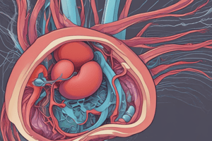
The Nephron
- Nephrons are the structural and functional units that form urine.
- They consist of: -Glomerulus: a tuft of capillaries associated with a renal tubule. -Glomerular (Bowman's) capsule: a blind, cup-shaped end of a renal tubule that completely surrounds the glomerulus.
- Renal corpuscle: glomerulus and its Bowman's capsule.
- Glomerular endothelium: fenestrated epithelium that permits solute-rich, virtually protein-free filtrate to pass from blood to the glomerular capsule.
Anatomy of the Glomerular Capsule
- External parietal layer: structural.
- Visceral layer: modified, branching epithelial podocytes.
- Podocyte extensions: terminate in foot processes.
- Filtration slits: openings between foot processes. Filtrate passes through them into the capsular space.
Renal Tubule
- Proximal convoluted tubule (PCT): cuboidal cells with microvilli and mitochondria.
- Reabsorbs water and solutes from filtrate and secretes substances into filtrate.
- Loop of Henle: hairpin-shaped loop of the renal tubule. -Proximal part: similar to proximal convoluted tubule. Followed by a thin segment (simple squamous cells) and a thick segment (cuboidal to columnar cells).
- Distal convoluted tubule (DCT): cuboidal cells without microvilli. Functions primarily in secretion, not reabsorption.
Connecting Tubules
- Distal portion of the DCT is connected to the collecting ducts.
Nephrons (Types)
- Cortical nephrons: account for 85% of nephrons and are located in the cortex.
- Juxtamedullary nephrons: located at the cortex-medulla junction. Have loops of Henle that deeply invade the medulla and extensive thin segments; they concentrate urine.
Capillary Beds
- Every nephron has two capillary beds:
- Glomerulus
- Peritubular capillaries.
- Each glomerulus:
- Fed by an afferent arteriole
- Drained by an efferent arteriole.
- Blood pressure in the glomerulus is high.
- Arterioles are high resistance vessels.
- Afferent arterioles have larger diameters than efferent arterioles.
- Fluids and solutes are forced out of the blood through the entire glomerular length.
- Peritubular beds: low-pressure, porous capillaries for absorption.
- Arise from efferent arterioles
- Empty into the renal venous system.
- Vasa recta: long, straight efferent arterioles of juxtamedullary nephrons.
Filtration Membrane
- Three layers:
- Glomerular capillary endothelium (fenestrated)
- Basement membrane
- Podocyte foot processes (filtration slits).
Mechanisms of Urine Formation
- Urine formation and blood composition adjustment involves three major processes:
- Glomerular filtration
- Tubular reabsorption
- Secretion.
Ureters
- Slender tubes conveying urine from the kidneys to the bladder.
- Trilayered wall:
- Transitional epithelial mucosa
- Smooth muscle muscularis
- Fibrous connective tissue adventitia.
- Ureters actively propel urine to the bladder by smooth muscle stretch.
Urinary Bladder
- Smooth, collapsible, muscular sac that temporarily stores urine.
- Trigone: triangular area outlined by ureters and urethra openings. Clinically important due to infections.
- Bladder wall has three layers:
- Transitional epithelial mucosa
- Thick muscular layer
- Fibrous adventitia.
- Distensible and collapses when empty.
Urethra
- Muscular tube that drains urine from the bladder and conveys it out of the body.
- Sphincters regulate urine flow:
- Internal urethral sphincter: involuntary, at the bladder-urethra junction.
- External urethral sphincter: voluntary, surrounding the urethra as it passes through the urogenital diaphragm.
- Female urethra: tightly bound to the anterior vaginal wall. External opening is anterior to the vaginal opening and posterior to the clitoris.
- Male urethra: three named regions.
- Prostatic urethra: within the prostate gland
- Membranous urethra: through the urogenital diaphragm
- Spongy (penile) urethra: through the penis, opening at the external urethral orifice.
Studying That Suits You
Use AI to generate personalized quizzes and flashcards to suit your learning preferences.