Podcast
Questions and Answers
What are the three primary functions of the lymphatic system?
What are the three primary functions of the lymphatic system?
- Return fluid called lymph to the circulatory system. 2. Transport dietary fats. 3. Help the immune system.
Describe the route that lymph takes through the lymphatic system, starting from the capillaries.
Describe the route that lymph takes through the lymphatic system, starting from the capillaries.
Lymph capillaries pick up interstitial fluid, which becomes lymph. Lymph then moves through lymph vessels, passing through lymph nodes, and eventually connect to the venous system via lymphatic ducts.
What are the primary and secondary organs of the lymphatic system, and what is the main function of each?
What are the primary and secondary organs of the lymphatic system, and what is the main function of each?
Primary organs are the bone marrow and thymus, where lymphocytes mature. Secondary organs include lymph nodes, the spleen, tonsils, and mucous membranes, where immune responses occur.
Explain how the right lymphatic duct and the thoracic duct contribute to the overall function of the lymphatic system.
Explain how the right lymphatic duct and the thoracic duct contribute to the overall function of the lymphatic system.
Describe the role of chylomicrons in the lymphatic system and their ultimate destination.
Describe the role of chylomicrons in the lymphatic system and their ultimate destination.
In what ways does the lymphatic system 'filter' fluids in the body?
In what ways does the lymphatic system 'filter' fluids in the body?
How do lymphatic capillaries differ from blood capillaries in terms of structure and function?
How do lymphatic capillaries differ from blood capillaries in terms of structure and function?
Discuss the arrangement of overlapping simple squamous epithelium in lymphatic capillaries and its functional significance.
Discuss the arrangement of overlapping simple squamous epithelium in lymphatic capillaries and its functional significance.
What mechanisms facilitate the movement of lymph fluid through lymphatic vessels?
What mechanisms facilitate the movement of lymph fluid through lymphatic vessels?
Describe the structure of a lymph node, including its connective tissue covering and internal organization.
Describe the structure of a lymph node, including its connective tissue covering and internal organization.
What types of cells are found within lymph nodes, and what roles do they play in immune function?
What types of cells are found within lymph nodes, and what roles do they play in immune function?
What are the two primary lymphatic organs, and how do T and B lymphocytes relate to them?
What are the two primary lymphatic organs, and how do T and B lymphocytes relate to them?
Discuss the role of reticular cells in lymphatic tissue and how they contribute to immune function.
Discuss the role of reticular cells in lymphatic tissue and how they contribute to immune function.
What is MALT, and where is it typically located in the body?
What is MALT, and where is it typically located in the body?
Describe the structure of the spleen and how its different regions (red pulp and white pulp) contribute to its overall function.
Describe the structure of the spleen and how its different regions (red pulp and white pulp) contribute to its overall function.
What is the difference between afferent and efferent lymphatic vessels in relation to lymph nodes?
What is the difference between afferent and efferent lymphatic vessels in relation to lymph nodes?
How does the location of lymph nodes in areas like the groin, armpits, neck, chest, and abdomen contribute to their function in the body?
How does the location of lymph nodes in areas like the groin, armpits, neck, chest, and abdomen contribute to their function in the body?
Why is it important that lymphatic capillaries are not found in the central nervous system and bone marrow?
Why is it important that lymphatic capillaries are not found in the central nervous system and bone marrow?
Describe how skeletal muscle contraction aids in lymph movement, and explain the role of valves in this process.
Describe how skeletal muscle contraction aids in lymph movement, and explain the role of valves in this process.
Explain how changes in pressure within the thoracic cavity during inspiration affect lymph fluid movement into vessels.
Explain how changes in pressure within the thoracic cavity during inspiration affect lymph fluid movement into vessels.
Flashcards
Lymphatic System
Lymphatic System
A separate circulatory system that returns fluid to circulation, transports fats, and aids the immune system.
Lymph
Lymph
Fluid derived from blood and tissues that circulates through lymphatic vessels.
Lymph Nodes
Lymph Nodes
Small, hollow structures that filter lymph to remove debris and pathogens.
Right Lymphatic Duct
Right Lymphatic Duct
Signup and view all the flashcards
Thoracic Duct
Thoracic Duct
Signup and view all the flashcards
Lymphatic Capillaries
Lymphatic Capillaries
Signup and view all the flashcards
Lacteals
Lacteals
Signup and view all the flashcards
Chylomicrons
Chylomicrons
Signup and view all the flashcards
Primary Lymphatic Organs
Primary Lymphatic Organs
Signup and view all the flashcards
Secondary Lymphatic Organs
Secondary Lymphatic Organs
Signup and view all the flashcards
Lymphocytes
Lymphocytes
Signup and view all the flashcards
Spleen
Spleen
Signup and view all the flashcards
MALT
MALT
Signup and view all the flashcards
Afferent Vessels
Afferent Vessels
Signup and view all the flashcards
Efferent Vessels
Efferent Vessels
Signup and view all the flashcards
Study Notes
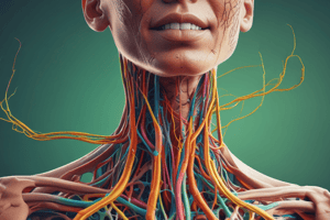
- The lymphatic system is a separate circulatory system with three main functions.
- Returning fluid (lymph) to circulation.
- Transporting dietary fats.
- Aiding the immune system.
- The lymphatic system includes:
- Vessels.
- Lymph nodes.
- Lymph nodes serve as the "filters of the lymph."
- Primary lymphatic organs:
- Bone marrow.
- Thymus.
- Lymphocytes mature here.
- Secondary lymphatic organs:
- Lymph nodes.
- Spleen.
- Tonsils.
- Certain tissues.
- Various mucous membranes.
- The lymphatic system connects to the venous system at the subclavian veins via:
- Right lymphatic duct (drains the right side of the head, neck, and trunk).
- Thoracic duct (drains the remainder of the body).
- Lymphatic capillaries:
- Pick up interstitial fluid lost by the circulatory system and returns it to venous circulation as lymph.
- Transport dietary fats from the gastrointestinal system; lacteals are lymphatic structures in villi of the small intestine where fats are absorbed through chylomicrons and transported to the venous circulation.
- The lymphatic system contains a large part of the immune system where lymph nodes help destroy pathogens.
Lymphatic Capillaries
- Distributed throughout the interstitium.
- Absent in the central nervous system, bone marrow, and tissues lacking blood flow (epidermis, cartilage).
- Allow one-way fluid flow into the capillary.
- Consist of overlapping simple squamous epithelium forming one-way valves.
- This arrangement increases the ease of permeability and fluid movement toward the venous circulation.
- Form larger structures called lymphatic vessels that are similar to veins with three layers:
- Inner endothelium.
- Middle smooth muscle layer.
- Outer layer of thin fibrous connective tissue.
Lymphatic Vessels
- Contains valves to allow the one way flow of blood.
- Smooth muscle contraction helps in blood movement.
- Some wall cells generate action potentials promoting muscle contraction.
- Lymph fluid moves through skeletal muscle contraction.
- Pressure is applied externally causing the vessels to constrict.
- Valves only allow the one way flow.
- Lymph fluid:
- Moves into vessels in the thoracic cavity because of dilation.
- Dilation results in decreased thoracic cavity pressure during inspiration.
- Fluid moves toward the area of lower pressure.
- Asymmetrical lymph drainage:
- Right: right lymphatic duct drains the right side of the head, neck, and trunk.
- Left: the thoracic duct drains the remainder of the body.
Lymph Nodes
- Located throughout the lymphatic system, connected by lymphatic vessels.
- Small, oval structures generally not felt unless enlarged or calcified.
- Remove pathogens from lymph.
- Distributed throughout the body, concentrated in cervical, axillary, inguinal, popliteal, and mammary glands.
- Lymph node structure:
- Dense connective tissue covering.
- Trabeculated internal structure.
- Reticular connective tissue forming an interconnected web.
- Afferent vessels (entry).
- Efferent vessels (exit).
- Lymph node layers:
- Outer cortex: contains open areas called sinuses.
- Inner medulla: contains medullary cords (branching lymphatic tissue) and medullary sinuses.
- White blood cells in lymph nodes:
- Macrophages in the sinuses phagocytize bacteria and debris.
- Lymphocytes in the germinal centers activate and move into the bloodstream.
Lymphatic Organs
- Spleen and thymus are lymphatic organs.
- Contain lymphatic tissue (primarily white blood cells like macrophages, lymphocytes, and reticular cells that produce reticular fibers).
- T and B lymphocytes are produced in bone marrow and carried to the lymphatic system.
- Activation causes them to divide and attach pathogens.
- Lymphatic tissue:
- Usually in a lymph node or organ.
- May be in the mucous membranes of the digestive, urinary, respiratory, and reproductive systems (MALT).
- The spleen is located in the left upper quadrant of the abdominal area and is about the size of an adult fist:
- Structure: Outer connective tissue capsule and trabeculated inner part with red and white pulp; contains venous sinuses.
- White pulp: lymphatic tissue with arteries.
- Red pulp: contains white and red blood cells (associated with veins).
- The splenic artery and vein enter/exit at the hilum.
- Blood flows into the spleen :
- Through the trabeculated network.
- Cells here can destroy pathogens and trigger the immune system.
- Also serves as a blood reservoir.
Studying That Suits You
Use AI to generate personalized quizzes and flashcards to suit your learning preferences.