Podcast
Questions and Answers
What primary function does the respiratory system serve in the human body?
What primary function does the respiratory system serve in the human body?
- To circulate nutrients throughout the body.
- To regulate body temperature through sweating.
- To filter waste products from the blood.
- To exchange oxygen and carbon dioxide between the blood and the air. (correct)
Which structural division of the respiratory system includes the nose, nasal cavity, sinuses and pharynx?
Which structural division of the respiratory system includes the nose, nasal cavity, sinuses and pharynx?
- The pulmonary division.
- The lower respiratory tract.
- The thoracic cavity.
- The upper respiratory tract. (correct)
What is the main role of the nasal cavity's lining of highly vascular ciliated columnar epithelium?
What is the main role of the nasal cavity's lining of highly vascular ciliated columnar epithelium?
- To warm, moisten, and filter incoming air. (correct)
- To secrete digestive enzymes.
- To provide a protective barrier against pathogens.
- To facilitate the sense of smell.
The posterior nares connect the nasal cavity to which structure?
The posterior nares connect the nasal cavity to which structure?
How do the paranasal sinuses contribute to the function of the respiratory system?
How do the paranasal sinuses contribute to the function of the respiratory system?
What is the role of turbinates in the nasal cavity during air conditioning?
What is the role of turbinates in the nasal cavity during air conditioning?
What is the primary function of the mucociliary system in the nasal cavity?
What is the primary function of the mucociliary system in the nasal cavity?
Which of the following functions is performed by the pharynx?
Which of the following functions is performed by the pharynx?
Which section of the pharynx is located uppermost?
Which section of the pharynx is located uppermost?
What is the role of the epiglottis during swallowing?
What is the role of the epiglottis during swallowing?
What anatomical feature is commonly known as the "Adam's apple?"
What anatomical feature is commonly known as the "Adam's apple?"
How does changing the tension on the vocal cords affect speech?
How does changing the tension on the vocal cords affect speech?
What happens to the vocal cords and glottis during normal breathing?
What happens to the vocal cords and glottis during normal breathing?
What structural characteristic defines the trachea?
What structural characteristic defines the trachea?
At what level does the trachea divide into the right and left main bronchi?
At what level does the trachea divide into the right and left main bronchi?
In the branching structure of the respiratory system, what type of bronchi directly follow the primary bronchi?
In the branching structure of the respiratory system, what type of bronchi directly follow the primary bronchi?
What structural feature distinguishes bronchioles from bronchi?
What structural feature distinguishes bronchioles from bronchi?
Which of the following accurately describes the location of the lungs?
Which of the following accurately describes the location of the lungs?
What is the collective term for the two layers of serous membrane enclosing each lung?
What is the collective term for the two layers of serous membrane enclosing each lung?
Why is the left lung typically smaller than the right lung?
Why is the left lung typically smaller than the right lung?
Which of the following lists the correct parts of the lung?
Which of the following lists the correct parts of the lung?
What passes through the hilum on the medial surface of the lungs?
What passes through the hilum on the medial surface of the lungs?
Which anatomical area separates the lungs and contains the heart, great vessels, trachea, and esophagus?
Which anatomical area separates the lungs and contains the heart, great vessels, trachea, and esophagus?
What is the function of the fluid within the pleural cavity?
What is the function of the fluid within the pleural cavity?
How many lobes are typically found in the left lung?
How many lobes are typically found in the left lung?
Besides respiration, what other functions are associated with the lungs?
Besides respiration, what other functions are associated with the lungs?
The exchange of gases between the alveoli and blood occurs because:
The exchange of gases between the alveoli and blood occurs because:
What approximate percentage of each alveolus is occupied by blood capillaries, facilitating gas exchange?
What approximate percentage of each alveolus is occupied by blood capillaries, facilitating gas exchange?
What is the process of moving air into and out of the lungs?
What is the process of moving air into and out of the lungs?
Which of the following most accurately describes inhalation/inspiration?
Which of the following most accurately describes inhalation/inspiration?
What occurs during exhalation?
What occurs during exhalation?
During inspiration, what happens to air pressure inside the lungs?
During inspiration, what happens to air pressure inside the lungs?
What is the approximate average breathing rate for an adult in a relaxed state?
What is the approximate average breathing rate for an adult in a relaxed state?
A single respiratory cycle consists of what?
A single respiratory cycle consists of what?
What constitutes tidal volume (TV)?
What constitutes tidal volume (TV)?
If a person inhales as much air as possible and then forcefully exhales as much as possible, what is the volume of air exhaled known as?
If a person inhales as much air as possible and then forcefully exhales as much as possible, what is the volume of air exhaled known as?
After a normal exhalation, what is the volume of air that remains in the lungs called?
After a normal exhalation, what is the volume of air that remains in the lungs called?
What does Inspiratory Capacity (IC) measure?
What does Inspiratory Capacity (IC) measure?
What two volumes are combined to calculate Inspiratory Capacity?
What two volumes are combined to calculate Inspiratory Capacity?
In the transport of respiratory gases, what is the approximate thickness of the air and blood barrier that facilitates easy diffusion?
In the transport of respiratory gases, what is the approximate thickness of the air and blood barrier that facilitates easy diffusion?
Flashcards
Respiratory System Function
Respiratory System Function
The primary function is to supply oxygen to the blood and remove carbon dioxide.
Respiratory System Path
Respiratory System Path
Path for air to pass from the nose to the lungs.
Respiratory System Air Preparation
Respiratory System Air Preparation
Filters, warms, and humidifies inhaled air.
Vocal Cords Function
Vocal Cords Function
Signup and view all the flashcards
Lungs' pH Control
Lungs' pH Control
Signup and view all the flashcards
Olfactory Bulb Function
Olfactory Bulb Function
Signup and view all the flashcards
Respiration
Respiration
Signup and view all the flashcards
Upper Respiratory Tract
Upper Respiratory Tract
Signup and view all the flashcards
Lower Respiratory Tract
Lower Respiratory Tract
Signup and view all the flashcards
Conducting Portion
Conducting Portion
Signup and view all the flashcards
Respiratory Portion
Respiratory Portion
Signup and view all the flashcards
Nose/Mouth Functions
Nose/Mouth Functions
Signup and view all the flashcards
Mucus+Cilia Function
Mucus+Cilia Function
Signup and view all the flashcards
Pharynx Function
Pharynx Function
Signup and view all the flashcards
Tonsils
Tonsils
Signup and view all the flashcards
Larynx Function
Larynx Function
Signup and view all the flashcards
Epiglottis Function
Epiglottis Function
Signup and view all the flashcards
Trachea
Trachea
Signup and view all the flashcards
Bronchi
Bronchi
Signup and view all the flashcards
Bronchioles
Bronchioles
Signup and view all the flashcards
Lungs and Alveoli
Lungs and Alveoli
Signup and view all the flashcards
Nose
Nose
Signup and view all the flashcards
Nose Function
Nose Function
Signup and view all the flashcards
Nostrils
Nostrils
Signup and view all the flashcards
Paranasal Sinuses
Paranasal Sinuses
Signup and view all the flashcards
Nasal Cavity Function
Nasal Cavity Function
Signup and view all the flashcards
Defense in Nasal Cavity
Defense in Nasal Cavity
Signup and view all the flashcards
Functions of Pharynx
Functions of Pharynx
Signup and view all the flashcards
Epiglottis
Epiglottis
Signup and view all the flashcards
The Vocal Cords
The Vocal Cords
Signup and view all the flashcards
Trachea Measurement
Trachea Measurement
Signup and view all the flashcards
Bronchi Function
Bronchi Function
Signup and view all the flashcards
Bronchioles
Bronchioles
Signup and view all the flashcards
Lungs
Lungs
Signup and view all the flashcards
Lung Protection
Lung Protection
Signup and view all the flashcards
Lungs
Lungs
Signup and view all the flashcards
Alveoli
Alveoli
Signup and view all the flashcards
Mechanism of Respiration
Mechanism of Respiration
Signup and view all the flashcards
Expiratory Muscle
Expiratory Muscle
Signup and view all the flashcards
Tidal Volume
Tidal Volume
Signup and view all the flashcards
Study Notes
- The respiratory system's function is to supply O₂ to the blood and remove CO₂ from it
The Human Respiratory System
- The respiratory system creates the path for air to travel, from the nose to the lungs
- The primary function of the respiratory system is the exchange of gases during internal and external respiration
- Inhaled air is also filtered, warmed, and humidified by the system
- Sound is produced by the system's vocal cords
- Body pH levels are controlled by the lungs
- The olfactory bulb aids in smelling
- Human cells need a constant supply of oxygen for proper function.
- Carbon dioxide (CO2) is eliminated as a metabolic waste product.
- Gas exchange between the atmosphere and the human body happens during the respiration process
Anatomy
- The anatomy of the respiratory tract can be divided into two groups "structurally": The Upper and Lower Respiratory Tracts.
- Upper Respiratory Tract organs include the: Nose, Nasal cavity, Sinuses, and Pharynx
- Lower Respiratory Tract organs include the: Larynx, Trachea, Bronchial Tree, and Lungs.
- The organs of the respiratory tract can be divided into two groups "functionally": The Conducting and Respiratory Portions
- The conducting portion is made up of interconnecting cavities and tubes that conduct air into the lungs: Nose, Pharynx, Larynx, Trachea, and Bronchi
- The respiratory portion is where respiratory gases are exchanged: Bronchioles, Alveolar Ducts, and Alveoli
Anatomy Details
- The nose and mouth filter, warm, and humidify air
- Mucus traps bacteria and foreign debris in the nose and mouth
- Cilia sweeps mucus toward the throat, where it is digested by the stomach
- The pharynx is the throat and a passageway for food and air
- Tonsils are clusters of lymphatic tissue in the pharynx
- The larynx contains the vocal cords
- The epiglottis covers the larynx during swallowing, preventing liquids and food from entering
- The trachea is the windpipe lined with C-shaped cartilage.
- The bronchi are branches extending to the lungs
- Bronchioles are smaller branches of the bronchi
- Lungs contain alveoli, air sacs for gas exchange
The Nose
- The nose is located between the forehead and upper lip
- The nose is the first organ of the respiratory tract
- The nose provides an entrance for inhaled air
- The nose performs the process of warming, moistening, and filtering inhaled air
- The nose has external and internal portions (nasal cavities)
- The nose is a bony and cartilaginous structure
- The bony part is made up of the frontal, nasal and maxillae bones
- The cartilaginous part is made up of septal, lateral, and alar cartilages.
- The internal portion is a large cavity in the skull merging with the external nose anteriorly and communicating with the throat posteriorly
- The nose is lined with highly vascular ciliated columnar epithelium
- The nose contains mucus-secreting goblet cells
The Nasal Cavity
- Two nasal cavity openings are separated by the nasal septum, which contains septum cartilage
- The anterior openings are called nostrils (anterior nares) through which air enters
- The anterior nares are the openings of the nasal cavity and contain small hairs.
- The posterior nares are the openings of the nasal cavity into the pharynx
Paranasal Sinuses
- Sinuses are air-filled spaces.
- They are present within the maxillary, frontal, ethmoid, and sphenoid bones of the skull.
- These spaces open to the nasal cavity
- The space is lined with mucus membrane
- The sinuses reduce the weight of the skull
Functions of Nasal Cavity
- The nasal cavity helps air pass through it during respiration
- Air is conditioned, converting air between -5°C to 55°C to 31-37°C in the nasopharynx
- Turbinates make the inhaled air water-saturated so that the lungs receive 100% humid air
- Inhaled air is filtered, warmed and humidified in the nasal cavity
- The mucociliary system holds back microorganisms and foreign particles
- Antibodies and enzymes present in the mucus are active against bacteria.
- The irritants are forcefully expelled from the nose through sneezing
- The olfactory system recognizes the environment and food
- Vocal cords help with voice generation
The Pharynx
- The throat is a "funnel-shaped tube" that is posterior to the nasal cavity, oral cavity, and larynx, and anterior to the cervical vertebra.
- The pharynx has three components: the nasopharynx (uppermost portion), the oropharynx (middle portion), and the laryngopharynx (lowermost portion).
- The pharynx serves as a common passageway for both air and food and provides a resonating chamber for speech sounds
- The pharynx functions as a passageway for food and air, supports taste, warming and humidifying, hearing, protection, and speech
The Epiglottis
- The epiglottis is a large, leaf-shaped piece of cartilage found at the entrance of the larynx
- The epiglottis closes the tracheal opening during swallowing of food
- The epiglottis prevents food from entering the trachea
- During swallowing, there is elevation of the larynx.
The Larynx
- The larynx is the triangular chamber located in the front, upper part of the neck
- It features a prominent elevation in front of the larynx, known as the Adam's apple
- The larynx joins the pharynx with the trachea
- The larynx comprises cartilage: 3 single and 3 paired cartilages
- Single cartilages encompass the thyroid cartilage, cricoid cartilage, and epiglottis
- Paired cartilages include the arytenoids, corniculates, and cuneiforms
The Vocal Cords
- Inside the larynx are two pairs of folds made of muscle and connective tissues covered in a mucous membrane: the vocal cords
- Changing the tension on the vocal cords controls pitch, while increasing loudness depends upon increasing the force of air vibrating the vocal cords
- When vocal cords are opened, this is abduction
- When vocal cords are closed, this is adduction
- The larynx contains the vestibule (false vocal cord), ventricle, and the infraglottic cavity (from the vocal cords to the trachea cavity)
- The vocal cords are relaxed, and the glottis, during normal breathing, is a triangular sli
- During swallowing, the false vocal cords and epiglottis close off the glottis.
The Trachea
- The trachea is a windpipe
- It is measured around 10-11 cm
- It extends downwards up to the 5th thoracic vertebra.
- At the end, it divides into the right and left bronchi, which enter their respective lungs.
- It is composed of 16-20 C-shaped rings of hyaline cartilage.
- Connective tissues and involuntary muscles join the cartilage and form the posterior wall.
- The trachea's cartilage is covered by 3 tissue layers: the outer, middle, and inner layer
- The trachea helps in the cough reflex.
The Bronchi
- The bronchi are the air passageways into the respiratory tract carrying inhales air into the lungs
- There are right and left bronchi
- The primary bronchi split into three secondary bronchi
- They enter into the superior, middle, and inferior lobes of the right lung
- Secondary bronchi further divide into tertiary bronchi, which then divide into smaller branches
The Bronchioles
- Bronchioles are the finest branches of the bronchi
- Bronchioles are converted into terminal bronchioles, which are further subdivided into two or more respiratory bronchioles
- Bronchioles terminate at alveolar sacs
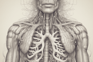
The Lungs
- The lungs are in the thoracic cavity as two cone-shaped lobes separated by the heart and other structures
- They are present from the clavicle to the diaphragm
- The paired soft, spongy, cone-shaped lungs, separated medially by the mediastinum, are enclosed by the diaphragm and thoracic cage
- The lungs have two layers of serous membrane, collectively known as the pleural membrane, that enclose and protect them
- The outer layer, the parietal pleura, is attached to the thoracic cavity
- The visceral pleura is the inner layer covering the lung itself
- The left lung is smaller than the right lung due to the heart in the thoracic cavity
- The lungs have an apex, base, costal surface, and medial surface
- The lung has its round apex extending up to the root of the neck
- The base has a concave and semilunar shape and is associated with the thoracic surface of the diaphragm
- The Costal surface is convex and associated with the costal cartilages, ribs, and intercostal muscles
- The Medial surface has a concave shape
- A triangular shaped hilum at the 5-7th thoracic vertebra, where all artery veins and nerve supply pass through this surface
The Mediastinum
- The mediastinum is the area between the lungs, occupied by the heart, great vessels, trachea, right and left bronchi, esophagus, lymph nodes, lymph vessels, and nerves.
Pleura and Pleural Cavity
- Each lung is enclosed with a doubled-layered serous pleural membrane
- The parietal pleura is the outer layer that lines the thoracic cavity wall.
- The visceral pleura is the deep layer that lines the lungs.
- The pleural cavity is the space between the two layers which contains pleural fluid
- Pleural fluid secreted by the membranes lubricating the fluid that prevent the friction between the layers during breathing
Lobes/Fissures and Lobules
- Each lung is separated into lobes
- The left lung is divided into two lobes, and the right into three lobes
- The lobes are further divided into numerous lobules which contains alveoli
- The lungs facilitate respiration, alter blood pH, filter out small blood clots, alter drug concentrations, convert angiotensin-I to angiotensin-II, and provide protection via Ig-A
Alveoli
- Alveoli are hollow cavities within mammalian lungs and spherical projections of respiratory bronchioles
- Alveolar membranes are the major sites of gas exchange with the blood
- A human lung features around 300 million alveoli composed of an epithelial layer, 70% occupied with blood capillaries
- Alveoli facilitate external respiration through gas exchange with blood and defense against microbes by diffusion
Mechanism of Respiration
- The process by which the respiratory organs allow the air to move in and out of the lungs is called breathing
- Oxygen-rich air is taken in from the atmosphere, and carbon dioxide-rich air is expelled in exchange
- Inhalation/Inspiration is the process of air flowing into the lungs
- Exhalation/Expiration is the process of air leaving the lungs
- A single breath comprises one inhalation and one exhalation
- Breathing rate is the number of times an individual breaths in one minute
- Breathing rate increases while walking fast, running, or after heavy exercise.
- Breathing rate decreases when in a relaxed state
- Average breathing rate of an adult is 15-18 times per minute
Inspiration and Expiration
- During inspiration, the Diaphragm contracts and flattens, external intercostals lift the rib cage, and the lungs stretched to have a larger size.
- During inspiration, air pressure inside the lungs decreases and air is sucked into the lungs.
- During expiration, inspiratory muscles relax, the rib cage descends, the lungs recoil, and gases are forced out
- The average breathing rate has 3 phases
- Inspiration lasts about 2 seconds
- Expiration lasts about 3 seconds
- Pause
Lung Volumes
- Tidal Volume (TV): volume of air inspired/expired during normal respiration (~500 ml average).
- Inspiratory Reserve Volume (IRV): additional air volume a person can inspire after forcible inspiration (2500-3000 ml average).
- Expiratory Reserve Volume (ERV): additional air volume a person can expire after forcible inspiration (1000-1100 ml average).
- Residual Volume (RV): air volume remaining in lungs after a forcible expiration (1100-1200 ml average).
Lung Capacity
- Inspiratory Capacity (IC): total air volume a person can inspire after normal expiration (TV+IRV, ~3500 ml average)
- Expiratory Capacity (EC): total air volume a person can expire after normal inspiration (TV+ERV, ~1600 ml average).
- Functional Residual Capacity (FRC): air volume remaining in lungs after normal expiration (ERV+ RV, ~2300 ml average).
- Vital Capacity (VC): maximum air amount a person can breathe out after forced inspiration (ERV, TV & IRV, ~4600 ml average).
- Total Lung Capacity (TLC): total air volume accommodated in the lungs after forced inspiration (RV, ERV, TV, &IRV/VC+RV, ~5800 ml average), meaning TLC=VC
Transport of Respiratory Gases
- Respiration involves inhalation of oxygen and exhalation of carbon dioxide
- During respiration, O2 and CO2 gases are transported by the blood.
- Oxygen enters the lungs to reach the alveoli
- The alveoli and close capillaries are one cell thick, with an air and blood barrier of about one micrometer.
- Oxygen passes easily from alveoli to capillaries and reaches the blood
- Carbon dioxide to be exhaled in the same manner enters the alveoli from the blood
- The oxygenated blood travels to the heart and is distributed throughout the body
Studying That Suits You
Use AI to generate personalized quizzes and flashcards to suit your learning preferences.