Podcast
Questions and Answers
What is the sclera?
What is the sclera?
- The anterior most portion of the eye
- The layer that contains photoreceptors
- A fluid-filled chamber of the eye
- The white of the eye providing protection (correct)
What is the function of the cornea?
What is the function of the cornea?
To receive light and protect internal structures
What is the conjunctiva?
What is the conjunctiva?
A mucous membrane that protects the cornea
What does the optic nerve do?
What does the optic nerve do?
What does the ciliary body primarily consist of?
What does the ciliary body primarily consist of?
What is the lens?
What is the lens?
What are suspensory ligaments?
What are suspensory ligaments?
What is the iris responsible for?
What is the iris responsible for?
What is the choroid?
What is the choroid?
Where is the retina located?
Where is the retina located?
What is the function of the tapetum lucidum?
What is the function of the tapetum lucidum?
What is aqueous humor?
What is aqueous humor?
What is vitreous humor?
What is vitreous humor?
What is the anterior cavity?
What is the anterior cavity?
What fills the posterior chamber of the eye?
What fills the posterior chamber of the eye?
What does the vascular tunic include?
What does the vascular tunic include?
What is the fovea centralis?
What is the fovea centralis?
What is the macula lutea?
What is the macula lutea?
Flashcards are hidden until you start studying
Study Notes
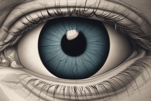
Structures of the Eye
- Sclera: The eye's white outer layer; provides shape and protects internal structures.
- Cornea: Clear front part of the sclera; first area to receive light; appears cloudy in preserved specimens.
- Conjunctiva: Mucous membrane covering the anterior eye; protects the cornea from damage.
- Optic Nerve: Cranial nerve II; transmits visual information from the eye to the brain, specifically the occipital lobe.
- Ciliary Body: Dark pigmented structure encircling the lens; contains muscles to adjust lens tension and produce aqueous humor.
- Lens: Biconvex structure that focuses light onto the retina; shape influences image clarity.
- Suspensory Ligaments: Fibers connecting the lens to the ciliary body; adjusting tension alters the lens shape for focusing.
- Iris: Colored part of the eye; an extension of the ciliary body that controls the pupil size.
- Choroid: Layer between the retina and sclera; appears brownish-black; reflects light in nocturnal animals for improved vision.
- Retina: Light-sensitive membrane containing photoreceptors necessary for vision; connects to the optic nerve at the optic disc.
- Tapetum Lucidum: Iridescent layer in nocturnal animals enhancing vision in low light conditions.
- Aqueous Humor: Watery fluid produced by the ciliary body; circulates in the anterior cavity; crucial for maintaining intraocular pressure.
- Vitreous Humor: Gel-like substance in the posterior cavity; provides support to the retina and helps maintain its position.
- Anterior Cavity: Fluid-filled space between the iris and cornea; contains aqueous humor.
- Posterior Chamber: Area filled with aqueous humor; located behind the iris and in front of the lens.
- Vascular Tunic (Uvea): Middle layer of the eye, consisting of the choroid, ciliary body, and iris; rich in blood vessels.
- Vitreous Chamber: Largest chamber filled with vitreous humor; located behind the lens and in front of the optic nerve.
- Pupil: Opening in the center of the iris allowing light to enter the eye; appears black due to light absorption.
- Optic Disc: Raised area on the retina where the optic nerve enters; lacks visual receptors, creating a blind spot.
- Fovea Centralis: Depressed area in the retina important for sharp vision; contains a high density of retinal cones.
- Macula Lutea: Central region of the retina around the fovea, crucial for detailed vision.
Studying That Suits You
Use AI to generate personalized quizzes and flashcards to suit your learning preferences.