Podcast
Questions and Answers
The rods and cones, critical for vision, perform which primary function in the eye?
The rods and cones, critical for vision, perform which primary function in the eye?
- Focusing light onto the retina.
- Adjusting the amount of light entering the eye.
- Forming the overall structure of the eyeball.
- Detecting the visual image. (correct)
Which statement best describes the relationship between 'ocular' and 'ophthalmic' in the context of eye anatomy?
Which statement best describes the relationship between 'ocular' and 'ophthalmic' in the context of eye anatomy?
- Ocular terms describe the physiological function of the eye, whereas ophthalmic terms describe the anatomical structure.
- Ocular terms refer to the bony structures surrounding the eye, while ophthalmic terms refer to the eye itself.
- Ocular and ophthalmic are interchangeable terms referring to the eye. (correct)
- Ocular refers to the external appearance of the eye, while ophthalmic encompasses the medical study and treatment of the eye.
The fibrous layer provides the eye with strength and shape. A key component of this layer, the cornea, maintains its clarity and function through what mechanism?
The fibrous layer provides the eye with strength and shape. A key component of this layer, the cornea, maintains its clarity and function through what mechanism?
- A thick layer of pigmented cells that absorb excess light and prevent glare.
- The controlled amount of water maintained within its fibers. (correct)
- A high concentration of blood vessels that nourish and protect the corneal tissue.
- The constant production of collagen fibers that are randomly arranged to scatter light.
What is the clinical significance of the limbus, the junction between the cornea and sclera?
What is the clinical significance of the limbus, the junction between the cornea and sclera?
The uvea, or middle vascular layer of the eye, includes the choroid, iris, and ciliary body. What is a primary function of the choroid layer?
The uvea, or middle vascular layer of the eye, includes the choroid, iris, and ciliary body. What is a primary function of the choroid layer?
Which of the following best explains the function of the tapetum lucidum found in the eyes of most animals (except swine)?
Which of the following best explains the function of the tapetum lucidum found in the eyes of most animals (except swine)?
The iris controls the amount of light entering the eye. How does the pupil respond to different light conditions?
The iris controls the amount of light entering the eye. How does the pupil respond to different light conditions?
What is the primary role of the ciliary muscles in the process of visual accommodation?
What is the primary role of the ciliary muscles in the process of visual accommodation?
Which layer of the retina contains the photoreceptor cells (rods and cones) that are responsible for detecting light?
Which layer of the retina contains the photoreceptor cells (rods and cones) that are responsible for detecting light?
The optic disc is known as the 'blind spot' in the eye. What accounts for this phenomenon?
The optic disc is known as the 'blind spot' in the eye. What accounts for this phenomenon?
What is the functional difference between rods and cones in the retina?
What is the functional difference between rods and cones in the retina?
Why do domestic animals not perceive detail as well as humans?
Why do domestic animals not perceive detail as well as humans?
Refraction is essential for focusing images on the retina. Which structure does the most refractive work?
Refraction is essential for focusing images on the retina. Which structure does the most refractive work?
What is the function of the conjunctiva?
What is the function of the conjunctiva?
Why is the conjunctiva clinically significant for detecting certain systemic conditions?
Why is the conjunctiva clinically significant for detecting certain systemic conditions?
The eyelids meet at the medial and lateral canthi. What is found along the margins of the eyelids?
The eyelids meet at the medial and lateral canthi. What is found along the margins of the eyelids?
Domestic animals possess a third eyelid or nictitating membrane. What is its primary function?
Domestic animals possess a third eyelid or nictitating membrane. What is its primary function?
What is the role of the lacrimal glands?
What is the role of the lacrimal glands?
Tears are composed of three layers: an inner mucous layer, a middle tear layer and an outer oily layer. What is the function of the outer oily layer produced by the meibomian glands?
Tears are composed of three layers: an inner mucous layer, a middle tear layer and an outer oily layer. What is the function of the outer oily layer produced by the meibomian glands?
How are tears drained from the eye?
How are tears drained from the eye?
What is the role of the extraocular muscles?
What is the role of the extraocular muscles?
What is the function of the retractor bulbi muscle, and which species naturally lack it?
What is the function of the retractor bulbi muscle, and which species naturally lack it?
The canal of Schlemm plays a critical role in maintaining healthy intraocular pressure. What is its function?
The canal of Schlemm plays a critical role in maintaining healthy intraocular pressure. What is its function?
What is the composition and function of the vitreous humor?
What is the composition and function of the vitreous humor?
The lens changes shape to focus light on the retina. What is this process called?
The lens changes shape to focus light on the retina. What is this process called?
When referring to pannus, what is the most accurate description of this condition?
When referring to pannus, what is the most accurate description of this condition?
Corneal ulcers are commonly caused by:
Corneal ulcers are commonly caused by:
What statement describes the condition, glaucoma?
What statement describes the condition, glaucoma?
What is the clinical term for eyelids that roll inwards?
What is the clinical term for eyelids that roll inwards?
A dog presents with eyes discharging, what condition is a likely cause?
A dog presents with eyes discharging, what condition is a likely cause?
What is a potential reason for a proptosed eye?
What is a potential reason for a proptosed eye?
Concerning, 'Keratoconjunctivitis sicca', what is occurring in the eye?
Concerning, 'Keratoconjunctivitis sicca', what is occurring in the eye?
Distichiasis is a condition of the eye, what is occurring?
Distichiasis is a condition of the eye, what is occurring?
What condition could be a cause of 'Epiphora' chronic tearing?
What condition could be a cause of 'Epiphora' chronic tearing?
Miosis is associated with:
Miosis is associated with:
What is Blepharitis?
What is Blepharitis?
If there is 'Prolapse of the third eyelid', what condition is this?
If there is 'Prolapse of the third eyelid', what condition is this?
The ability of the eye to focus on both near and far objects is primarily achieved through which mechanism?
The ability of the eye to focus on both near and far objects is primarily achieved through which mechanism?
If the canal of Schlemm becomes blocked or obstructed, which of the following conditions is most likely to develop?
If the canal of Schlemm becomes blocked or obstructed, which of the following conditions is most likely to develop?
Animals with a tapetum lucidum often have superior night vision but may sacrifice visual acuity compared to humans. Which of the following explains this trade-off?
Animals with a tapetum lucidum often have superior night vision but may sacrifice visual acuity compared to humans. Which of the following explains this trade-off?
A veterinarian observes that an animal's third eyelid is protruding and inflamed. Which of the following structures is most likely affected, leading to this clinical sign?
A veterinarian observes that an animal's third eyelid is protruding and inflamed. Which of the following structures is most likely affected, leading to this clinical sign?
An animal is diagnosed with a corneal ulcer. Which of the following best describes the initial defect or damage in the cornea's structural layers?
An animal is diagnosed with a corneal ulcer. Which of the following best describes the initial defect or damage in the cornea's structural layers?
Flashcards
Ocular and ophthalmic
Ocular and ophthalmic
Terms that refer to the eye.
Ophthalmology
Ophthalmology
The study of the eye.
Fibrous layer function
Fibrous layer function
Gives strength and shape to the eye.
Cornea
Cornea
Signup and view all the flashcards
Sclera
Sclera
Signup and view all the flashcards
Limbus
Limbus
Signup and view all the flashcards
Uvea
Uvea
Signup and view all the flashcards
Choroid
Choroid
Signup and view all the flashcards
Tapetum lucidum
Tapetum lucidum
Signup and view all the flashcards
Iris
Iris
Signup and view all the flashcards
Pupil
Pupil
Signup and view all the flashcards
Ciliary muscles
Ciliary muscles
Signup and view all the flashcards
Suspensory ligaments
Suspensory ligaments
Signup and view all the flashcards
Retina
Retina
Signup and view all the flashcards
Aqueous compartment
Aqueous compartment
Signup and view all the flashcards
Anterior chamber
Anterior chamber
Signup and view all the flashcards
Posterior chamber
Posterior chamber
Signup and view all the flashcards
Aqueous humor
Aqueous humor
Signup and view all the flashcards
Canal of Schlemm
Canal of Schlemm
Signup and view all the flashcards
Vitreous compartment
Vitreous compartment
Signup and view all the flashcards
Vitreous humor
Vitreous humor
Signup and view all the flashcards
Accommodation
Accommodation
Signup and view all the flashcards
Optic disc
Optic disc
Signup and view all the flashcards
Photoreceptor cells
Photoreceptor cells
Signup and view all the flashcards
Rods
Rods
Signup and view all the flashcards
Cones
Cones
Signup and view all the flashcards
Refraction
Refraction
Signup and view all the flashcards
Cornea's role
Cornea's role
Signup and view all the flashcards
Conjunctiva
Conjunctiva
Signup and view all the flashcards
Bulbar conjunctiva
Bulbar conjunctiva
Signup and view all the flashcards
Palpebral conjunctiva
Palpebral conjunctiva
Signup and view all the flashcards
Lacrimal puncta
Lacrimal puncta
Signup and view all the flashcards
Extraocular eye muscles
Extraocular eye muscles
Signup and view all the flashcards
Skeletal Muscles
Skeletal Muscles
Signup and view all the flashcards
Third Eyelid
Third Eyelid
Signup and view all the flashcards
Ocular surface
Ocular surface
Signup and view all the flashcards
Tears
Tears
Signup and view all the flashcards
Lacrimal glands
Lacrimal glands
Signup and view all the flashcards
Inner mucous layer
Inner mucous layer
Signup and view all the flashcards
Middle tear layer
Middle tear layer
Signup and view all the flashcards
Outer oily layer
Outer oily layer
Signup and view all the flashcards
Extra ocular Muscles
Extra ocular Muscles
Signup and view all the flashcards
Retractor bulbi
Retractor bulbi
Signup and view all the flashcards
Study Notes
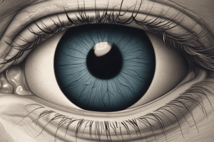
- In many ways, eyes function similarly to cameras, utilizing lenses, adjustable diaphragms, and light detectors.
- Most components of the eye are involved in forming the visual image
- Rods and cones within the retina detect the image.
Key Terms
- Ocular and ophthalmic describes relating to the eye
- Ophthalmology refers to the study of the eye and its diseases
Layers of the Eyeball
- The eyeball has three primary layers:
- The outer fibrous layer
- The middle vascular layer
- The inner nervous layer.
Fibrous Layer
- The outer fibrous layer gives strength and shape to the eye
- The cornea is the transparent window of the eye
- The transparent cornea consistes of an orderly arrangement of collagen fibers
- The cornea also features a controlled water content
- Excessive water content leads to corneal edema and cloudiness.
- Insufficient water causes corneal dehydration and cloudiness.
- The sclera makes up the white part of the eye
- The sclera consists of dense fibrous connective tissue
- The limbus is the junction between the sclera and cornea.
Vascular Layer
- The middle vascular layer is known as the uvea
- The uvea comprises three parts:
- Choroid
- Iris
- Ciliary body.
Choroid
- The choroid is positioned between the sclera and the retina
- Consists of pigment and blood vessels that nourish the retina
- In most animals a highly reflective area the tapetum lucidum exists in the choroid except for in swine.
- The tapetum lucidum causes bright light reflection from an animal’s eyes in the dark
- In low-light situations it acts as a light amplifier
- The tapetum reflection allows light to pass through receptors twice
Iris
- The iris is a modification of the vascular layer
- Forms the colored part of the eye
- Functions as a pigmented muscular diaphragm
- Controls the amount of light entering the eye through the pupil
- The pupil gets larger in low light conditions
- Conversely, it becomes smaller in bright light.
- Built from radially arranged and circularly arranged fibers
- The autonomic nervous system provides nerve supply to the smooth muscle cells of the iris
Ciliary Body
- The Ciliary Body is a ring-shaped structure just behind the iris
- Made of ciliary muscles, which adjust the shape of the lens
- These muscles enable vision at varying distances.
- The muscles connect to the edge of the lens through suspensory ligaments
- The ciliary body produces aqueous liquid.
Nervous Layer
- The nervous layer lines the back of the eye
- It contains sensory receptors for vision within the retina.
Compartments of Eyeball
- There are two fluid filled compartments to the eye:
- Aqueous compartment is in front of the lens and ciliary body
- Anterior chamber is in front of the iris, contains aqueous humor
- Posterior chamber is behind the iris, aqueous humor here also
- Aqueous humor is generated by the cells of the ciliary body
- It flows through the pupil into the anterior chamber
- Drains through the canal of Schlemm and returns to the bloodstream
- A ring-like structure at the angle where the iris and cornea meet is where filtration occurs
- Vitreous compartment fills the back of the eye behind the lens and ciliary body, contains vitreous humor
- Vitreous humor is soft and gelatinous
- Aqueous compartment is in front of the lens and ciliary body
Lens
- The lens is a soft, transparent structure of microscopic fibers
- Elastic and biconvex in shape
- It is normally round in shape
- To focus light, muscles of the ciliary body contract
- The lens touches the vitreous humor on its back surface
- Accommodation describes focusing light by changing the lens shape
Retina
- Visual images are formed. sensed, and converted into nerve impulses in the retina
- Lines most of the vitreous compartment
- Layers of the retina from outside in:
- Pigment layer
- Photoreceptor layer
- Bipolar cells Layer
- Ganglion cell layer
- Nerve fiber layer
- Integrated and relayed impulses pass from the photoreceptor cells to optic nerve by the bipolar and ganglion cell layers
- The optic disc contains no photoreceptors
- Photoreceptor Cells are neurons with dendrites modified into sensory receptors
- Rods are more sensitive to light, coarse images, motion, and low light
- Cones are sensitive to color and detail, yet are low in light sensitivity
Visual Image
- Light refraction focuses images on the retina
- Four refractive media help to focus an image:
- Cornea does most of the refractive work
- Aqueous humor
- Lens
- Vitreous humor.
- The visual image forms upside down within the retina
- The brain then inverts the image.
Extraocular Structures: Conjunctiva
- The conjunctiva is a thin, transparent membrane on the front of the eyeball
- Lines the inside of the eyelids
- The potion lining eyelids is palpebral conjunctiva
- The conjunctiva allows detection of paleness and jaundice
- The space bulbar and palpebral conjunctiva is the conjunctival sac
Eyelids
- Upper and lower folds of skin lined with the conjunctival membrane
- Lateral and medial eye corners are called canthi
- The meibomian glands are along each eyelid margin
- Each Lid is fringed with eyelashes
Third eyelid
- Domestic animals have a third eyelid, also known as the nictitating membrane
- Located on the interior of the eyelids/eyeball
- T-shaped cartilage plate
- Covered with the conjunctiva
- Ocular surface contains lymph nodules and an accessory lacrimal gland
Lacrimal Apparatus
- Lacrimal apparatus is primarily involved in producing and draining tears
- It moistens and protects the eye surface
- tears are made up of 3 layers
- Inner mucous layer that contains antibacterial substances
- Middle tear layer made from lacrimal glands moistens the cornea
- Outer oily layer is made from the meibomian glands reducing evaporation
- Are constantly produced, needing constant draining
- Tears drain in the nasolacrimal duct into the nasal cavity
Eye Muscles
- Eye muscles are extraocular and attach to the sclera
- Skeletal muscles hold the eye in place and move it
- There are four straight muscles and two oblique muscles
- Includes the Dorsal, ventral, medial, and lateral rectus muscles
- Dorsal and ventral oblique muscles.
- Humans lack Retractor bulbi muscles
Studying That Suits You
Use AI to generate personalized quizzes and flashcards to suit your learning preferences.


