Podcast
Questions and Answers
Which of the following structures is primarily responsible for the eye's ability to focus light?
Which of the following structures is primarily responsible for the eye's ability to focus light?
- Vitreous humor
- Optic nerve
- Sclera
- Cornea (correct)
What is the primary function of the sclera?
What is the primary function of the sclera?
- To control the amount of light entering the eye.
- To protect and shape the eyeball. (correct)
- To transmit light to the retina.
- To provide a transparent window for light entry.
What is the significance of the cornea being avascular (lacking blood vessels)?
What is the significance of the cornea being avascular (lacking blood vessels)?
- It allows for clear light transmission. (correct)
- It prevents the cornea from repairing itself.
- It makes the cornea susceptible to rejection after transplantation.
- It increases the risk of infection.
What is the composition of the sclera?
What is the composition of the sclera?
Which part of the eye is known as the "white of the eye"?
Which part of the eye is known as the "white of the eye"?
Why is the cornea uniquely suited for transplantation?
Why is the cornea uniquely suited for transplantation?
If a patient has a condition that affects the transparency of the cornea, which of the following would be the most likely outcome?
If a patient has a condition that affects the transparency of the cornea, which of the following would be the most likely outcome?
The central anterior portion of the fibrous layer of the eye is:
The central anterior portion of the fibrous layer of the eye is:
A patient's intraocular pressure is elevated. Which structure's function is MOST likely compromised, leading to this condition?
A patient's intraocular pressure is elevated. Which structure's function is MOST likely compromised, leading to this condition?
If the supply of nutrients to the lens and cornea is disrupted, which fluid is MOST likely affected?
If the supply of nutrients to the lens and cornea is disrupted, which fluid is MOST likely affected?
When using an ophthalmoscope, which structure is NOT directly visible?
When using an ophthalmoscope, which structure is NOT directly visible?
A doctor suspects a patient has early-stage arteriosclerosis. Which instrument is MOST appropriate for examining the fundus of the eye to assist in diagnosis?
A doctor suspects a patient has early-stage arteriosclerosis. Which instrument is MOST appropriate for examining the fundus of the eye to assist in diagnosis?
What is the MOST important role of the vitreous humor?
What is the MOST important role of the vitreous humor?
Which two internal structures of the eye are responsible for maintaining intraocular pressure?
Which two internal structures of the eye are responsible for maintaining intraocular pressure?
What MOST accurately describes the composition and location of the aqueous humor?
What MOST accurately describes the composition and location of the aqueous humor?
What is the MOST likely consequence if the scleral venous sinus becomes blocked?
What is the MOST likely consequence if the scleral venous sinus becomes blocked?
Which of the following statements accurately describes the relationship between the bony labyrinth and the membranous labyrinth?
Which of the following statements accurately describes the relationship between the bony labyrinth and the membranous labyrinth?
A patient reports experiencing difficulties with balance. Which structure in the inner ear is most likely involved in this patient's symptoms?
A patient reports experiencing difficulties with balance. Which structure in the inner ear is most likely involved in this patient's symptoms?
Damage to the oval window would directly affect which of the following?
Damage to the oval window would directly affect which of the following?
What is the role of the perilymph within the bony labyrinth?
What is the role of the perilymph within the bony labyrinth?
Which of the listed structures is NOT part of the bony labyrinth?
Which of the listed structures is NOT part of the bony labyrinth?
The vestibular apparatus is responsible for:
The vestibular apparatus is responsible for:
If a person has damage affecting their ability to perceive changes in the rate of rotation, which part of the vestibular apparatus is most likely affected?
If a person has damage affecting their ability to perceive changes in the rate of rotation, which part of the vestibular apparatus is most likely affected?
A patient is diagnosed with a condition affecting the endolymphatic sac. What primary function of the inner ear might be impaired by this condition?
A patient is diagnosed with a condition affecting the endolymphatic sac. What primary function of the inner ear might be impaired by this condition?
Which of the following accurately describes the role of the tectorial membrane in the process of hearing?
Which of the following accurately describes the role of the tectorial membrane in the process of hearing?
Damage to the hair cells within the spiral organ of Corti would primarily affect which aspect of hearing?
Damage to the hair cells within the spiral organ of Corti would primarily affect which aspect of hearing?
What is the correct order of structures involved in transmitting vibrations from sound waves to the auditory cortex?
What is the correct order of structures involved in transmitting vibrations from sound waves to the auditory cortex?
How do the ossicles contribute to the process of hearing?
How do the ossicles contribute to the process of hearing?
If the cochlear nerve (cranial nerve VIII) is damaged, what would be the most likely result?
If the cochlear nerve (cranial nerve VIII) is damaged, what would be the most likely result?
Where is the auditory cortex, which processes auditory information, located?
Where is the auditory cortex, which processes auditory information, located?
What is the role of the basilar membrane in the spiral organ of Corti?
What is the role of the basilar membrane in the spiral organ of Corti?
Within which structure is the spiral organ of Corti located?
Within which structure is the spiral organ of Corti located?
What is the primary function of the lens in the physiology of vision?
What is the primary function of the lens in the physiology of vision?
Why is accommodation important for vision?
Why is accommodation important for vision?
If a person is viewing an object less than 20 feet away, what must occur in the eye to maintain focus?
If a person is viewing an object less than 20 feet away, what must occur in the eye to maintain focus?
How does the image formed on the retina compare to the actual object being viewed?
How does the image formed on the retina compare to the actual object being viewed?
What is the optic chiasma's role in visual pathways?
What is the optic chiasma's role in visual pathways?
Which of the following structures is responsible for the majority of light refraction in the eye?
Which of the following structures is responsible for the majority of light refraction in the eye?
What would be the likely effect of damage to one optic nerve before the optic chiasm?
What would be the likely effect of damage to one optic nerve before the optic chiasm?
Consider a scenario where the lens of the eye loses its elasticity. What specific visual problem is most likely to arise from this condition?
Consider a scenario where the lens of the eye loses its elasticity. What specific visual problem is most likely to arise from this condition?
What is the composition of the optic tracts in terms of the fibers they contain?
What is the composition of the optic tracts in terms of the fibers they contain?
If a person has damage to their left optic radiation, what is the most likely visual deficit they would experience?
If a person has damage to their left optic radiation, what is the most likely visual deficit they would experience?
Which of the following accurately describes the sequence of structures involved in the visual pathway from the retina to the brain's visual cortex?
Which of the following accurately describes the sequence of structures involved in the visual pathway from the retina to the brain's visual cortex?
What is the primary benefit of the overlapping visual fields between the two eyes?
What is the primary benefit of the overlapping visual fields between the two eyes?
Where does the crucial synapse occur that relays visual information from the optic tract to the occipital lobe?
Where does the crucial synapse occur that relays visual information from the optic tract to the occipital lobe?
Which structure is directly responsible for the interpretation of visual information, allowing us to 'see'?
Which structure is directly responsible for the interpretation of visual information, allowing us to 'see'?
How would severing the right optic nerve affect a person's vision?
How would severing the right optic nerve affect a person's vision?
What is the most direct consequence of having non-overlapping visual fields?
What is the most direct consequence of having non-overlapping visual fields?
Flashcards
Sclera
Sclera
The white connective tissue layer of the eye.
Cornea
Cornea
Transparent, anterior part of the eye allowing light in.
Ciliary Body
Ciliary Body
Muscle structure controlling lens shape for focusing.
Iris
Iris
Signup and view all the flashcards
Pupil
Pupil
Signup and view all the flashcards
Aqueous Humor
Aqueous Humor
Signup and view all the flashcards
Vitreous Humor
Vitreous Humor
Signup and view all the flashcards
Fovea Centralis
Fovea Centralis
Signup and view all the flashcards
Optic nerve
Optic nerve
Signup and view all the flashcards
Ophthalmoscope
Ophthalmoscope
Signup and view all the flashcards
Light Refraction
Light Refraction
Signup and view all the flashcards
Accommodation
Accommodation
Signup and view all the flashcards
Real Image
Real Image
Signup and view all the flashcards
Optic Chiasma
Optic Chiasma
Signup and view all the flashcards
Visual Field
Visual Field
Signup and view all the flashcards
Focal Point
Focal Point
Signup and view all the flashcards
Retina
Retina
Signup and view all the flashcards
Internal Ear
Internal Ear
Signup and view all the flashcards
Bony Labyrinth
Bony Labyrinth
Signup and view all the flashcards
Cochlea
Cochlea
Signup and view all the flashcards
Vestibule
Vestibule
Signup and view all the flashcards
Semicircular Canals
Semicircular Canals
Signup and view all the flashcards
Perilymph
Perilymph
Signup and view all the flashcards
Endolymph
Endolymph
Signup and view all the flashcards
Vestibular Apparatus
Vestibular Apparatus
Signup and view all the flashcards
Organ of Corti
Organ of Corti
Signup and view all the flashcards
Hair cells
Hair cells
Signup and view all the flashcards
Basilar membrane
Basilar membrane
Signup and view all the flashcards
Tectorial membrane
Tectorial membrane
Signup and view all the flashcards
Cochlear nerve
Cochlear nerve
Signup and view all the flashcards
Sound pathway
Sound pathway
Signup and view all the flashcards
Ossicles
Ossicles
Signup and view all the flashcards
Action potential
Action potential
Signup and view all the flashcards
Optic tracts
Optic tracts
Signup and view all the flashcards
Optic radiation
Optic radiation
Signup and view all the flashcards
Visual pathway sequence
Visual pathway sequence
Signup and view all the flashcards
Thalamus in vision
Thalamus in vision
Signup and view all the flashcards
Depth perception
Depth perception
Signup and view all the flashcards
Binocular vision
Binocular vision
Signup and view all the flashcards
Study Notes
Special Senses
- Special senses include smell, taste, sight, hearing, and equilibrium.
- Special sense receptors are large, complex sensory organs or localized receptor clusters.
Nervous System Functions
- The nervous system has three basic functions.
- Each special sense gathers unique sensory info, which, when integrated, influences motor output.
- For example, seeing a ball coming towards your head may cause a motor output to move your body out of the way.
- Each type of sensory info is processed in a specific area of the cerebrum.
The Eye
- 70% of all sensory receptors are in the eyes.
- Each eye has over 1 million nerve fibers carrying information to the brain.
Accessory Structures
- Accessory structures of the eye include extrinsic eye muscles, eyelids, conjunctiva, and the lacrimal apparatus.
- Extrinsic eye muscles produce gross eye movements.
- Eyelids meet at the medial and lateral commissures (canthus).
- Tarsal glands lubricate the eye.
- Ciliary glands are located between the eyelashes.
- Conjunctiva is a membrane lining the eyelids and eyeball.
- It connects with the transparent cornea.
- It secretes mucus to keep the eye moist.
- Lacrimal apparatus is the lacrimal gland and ducts.
- Lacrimal gland produces lacrimal fluid (tears).
- Located at the lateral end of each eye.
- Tears drain across the eye, into the lacrimal canaliculi, then the lacrimal sac, and into the nasolacrimal duct, which empties into the nasal cavity.
- Tears contain dilute salt solution, mucus, antibodies, and lysozyme (bacteria-destroying enzyme).
- Tears cleanse, protect, moisten, and lubricate the eye.
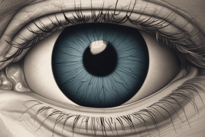
Anatomy of the Eye
- Three layers (tunics) form the wall of the eyeball: fibrous, vascular, and sensory.
- Fibrous layer: sclera + cornea
- Sclera is the white connective tissue layer, seen as the "white of the eye."
- Cornea is the transparent, central anterior portion that allows light to pass through. It can repair itself easily and is the only human tissue that can be transplanted without fear of rejection.
- Vascular layer: choroid, ciliary body, and iris
- Choroid is a blood-rich nutritive layer that prevents light scattering.
- Ciliary body is modified anteriorly into two smooth muscle structures.
- Iris regulates the amount of light entering the eye.
- Pupil is the rounded opening in the iris.
- Sensory layer: retina; contains rods and cones, specialized receptor cells (photoreceptors)
- Outer pigmented layer absorbs light and prevents scattering.
- Inner neural layer contains receptor cells (photoreceptors)
- Rods: important in dim light and peripheral vision (gray tones only).
- Cones: important for detailed color vision and concentrated in the fovea centralis.
- Electrical signals pass from photoreceptors via a two-neuron chain (bipolar neurons, ganglion cells), and leave the retina through the optic nerve.
- The optic disc is the blind spot where the optic nerve leaves the eyeball.
- Fibrous layer: sclera + cornea
Internal Structures
- Humors are fluids filling the interior of the eyeball.
- Lens divides the eye into two chambers
- Anterior segment: located anterior to the lens, and contains aqueous humor (clear, watery fluid similar to blood plasma).
- Aqueous humor maintains intraocular pressure and provides nutrients for lens and cornea.
- Aqueous humor is reabsorbed into venous blood through the scleral venous sinus (canal of Schlemm).
- Posterior segment: located posterior to the lens, and contains vitreous humor (gel-like substance).
- Vitreous humor prevents the eye from collapsing, and helps maintain intraocular pressure.
- Anterior segment: located anterior to the lens, and contains aqueous humor (clear, watery fluid similar to blood plasma).
- Ophthalmoscope: instrument used to illuminate the interior of the eyeball (fundus), allowing doctors to detect problems like diabetes, arteriosclerosis, and optic nerve/retina degeneration.
- Lens divides the eye into two chambers
Physiology of Vision
-
Light pathway through the eye and refraction
- Light is focused onto the retina by the cornea, aqueous humor, lens, and vitreous humor.
- Eye is set for distant vision (over 20 ft).
- Accommodation: adjusting lens shape to focus on closer objects (less than 20 ft).
- Light from distant objects are focused precisely on the retina, while images from closer objects are focused in front of the retina. A concave lens is required to refract light more for myopic (nearsighted) individuals.
-
Real images are:
- Reversed left to right.
- Upside down
- Smaller than the object.
-
Visual fields and visual pathways to the brain
- Optic nerve: a bundle of axons that exit the back of the eye carrying impulses from the retina.
- Optic chiasma: location where optic nerves cross over so fibers from the medial side of each eye cross over to the opposite side of the brain.
- Optic tracts
- Contain fibers from the lateral side of the eye on the same side and the medial side of the opposite eye
- Synapse with neurons in the thalamus.
- Optic radiations: axons from the thalamus run to the occipital lobe; synapse with cortical cells; vision interpretation
- Optic cortex (in occipital lobe): the point where visual interpretation occurs.
-
Visual fields: each eye has a slightly different view, but overlapping fields result in binocular vision, which provides depth perception (three-dimensional vision).
-
Eye reflexes
- Convergence: reflexive movement of the eyes medially when focusing on a close object.
- Photopupillary reflex: bright light causes pupils to constrict.
- Accommodation pupillary reflex: viewing close objects causes pupils to constrict, which allows focusing on near objects.
The Ear - Hearing and Balance
- Ear houses two senses: hearing and balance.
- Receptors are mechanoreceptors.
- Different organs house receptors for each sense.
- The ear is divided into external (outer), middle, and internal (inner) ears.
Ear Anatomy
-External (outer) ear
    - Auricle (pinna): collects sound waves.
    - External acoustic meatus (auditory canal): lined with skin and ceruminous glands (produce cerumen, or earwax), containing eardrum
-Middle ear cavity (tympanic cavity)
    - Air-filled, mucosa-lined cavity within temporal bone.
    - Only involved in hearing.
    - Located between tympanic membrane and oval window and round window.
    - Pharyngotympanic tube (auditory tube): links middle ear cavity with the throat, equalizing pressure. Three ear ossicles: malleus (hammer), incus (anvil), and stapes (stirrup).
        - Function: transmit vibrations from tympanic membrane to the inner ear fluids via a lever-like system. Vibrations travel from malleus → incus → stapes → oval window
-Internal (inner) ear
    - Includes sense organs for hearing and balance.
    - Bony labyrinth (osseous labyrinth): contains perilymph.
        - Cochlea: functions in hearing; spiral-shaped bony tube.
        - Vestibule: small, oval, central portion.
        - Semicircular canals: functions in equilibrium; three bony tubes.
    - Membranous labyrinth: suspended in perilymph; contains endolymph
        - Crista ampullaris: responds to rotational movements / angular acceleration
        - Maculae: receptors for static equilibrium; report on head position; hair cells embedded in otolithic membrane.
Equilibrium
- Equilibrium receptors of the inner ear are called the vestibular apparatus.
- Vestibular apparatus has two functional parts:
- Static equilibrium: Maculae –report the position of the head and help one keep their head upright. In the vestibule. Hair cells here are embedded in otolithic membrane, otoliths (tiny stones) float in gel around hair cells. Movement causing otoliths to roll and bend hair cells.
- Dynamic equilibrium: Crista ampullaris –responds to angular or rotational movement of the head. in the ampulla of each semicircular canal. Tuft of hair cells covered with cupula (gelatinous cap). Movement causes cupula to drag against the endolymph. Hair cells stimulated and impulse to vestibular nerve → cerebellum.
Hearing
- The spiral organ of Corti lies within the cochlear duct.
- Receptors are hair cells found on the basilar membrane.
- Gel-like tectorial membrane can bend hair cells.
- Cochlear nerve attached to hair cells transmits nerve impulses to auditory cortex on temporal lobe.
- Pathway of vibrations from sound waves: eardrum → ossicles → oval window → inner ear fluids → basilar membrane → hair cells of spiral organ of Corti → cochlear nerve → temporal lobe.
- High-pitched and low-pitched sounds affect different parts of the basilar membrane and cochlea. This ultimately affects different hair receptors.
Hearing and Equilibrium Deficits
- Deafness: any degree of hearing loss.
- Conduction deafness: sound vibrations transmission through the external/middle ear is hindered.
- Sensorineural deafness: damage to the nervous system structures involved in hearing.
- Ménière's syndrome: affects the inner ear can cause progressive deafness and/or vertigo.
Chemical Senses: Smell and Taste
- Chemical receptors: stimulated by chemicals in solution.
- Taste has 5 types of receptors.
- Smell can differentiate a wider range of chemicals.
- Both senses complement each other.
- Olfactory receptors are found in the roof of the nasal cavity.
- Receptor cells have olfactory hairs, detecting chemicals dissolved in mucus; impulses travel to olfactory nerve → olfactory cortex.
- Taste buds contain receptor organs (gustatory cells).
- Many located on the tongue, soft palate, and superior part of pharynx/cheeks.
- Gustatory cells have gustatory hairs (microvilli) that extend through taste pores; stimulated by chemicals dissolved in saliva.
- Impulses travel to gustatory complex by facial nerve, glossopharyngeal nerve, and vagus nerve.
- Olfactory receptors are found in the roof of the nasal cavity.
Developmental Aspects of Special Senses
- Special sense organs form early in embryonic development.
- Maternal infections (first 5–6 weeks of pregnancy) may cause visual abnormalities/sensorineural deafness.
- The infant has poor visual acuity (farsighted), and lacks color vision and depth perception at birth.
- Vision develops until age 8 or 9.
- Age related issue in the eyes: presbyopia (decreasing lens elasticity, difficulty focusing on close objects), dryness from decreased lacrimal gland secretions etc.
- Age related issue in the ears: presbycusis (type of sensorineural deafness) results from otosclerosis (ear ossicles fuse).
- Taste and smell acuity is most acute at birth; sensitivity decreases with age.
Studying That Suits You
Use AI to generate personalized quizzes and flashcards to suit your learning preferences.