Podcast
Questions and Answers
What is the primary function of the spinal cord circuits in the context of motor control?
What is the primary function of the spinal cord circuits in the context of motor control?
- To belittle the role of the brain
- To generate the initial command signals for complex movements.
- To analyze sensory information from the environment.
- To provide the detailed, specific muscle activation patterns for movements like walking. (correct)
Why are spinal reflexes more excitable in a decerebrate animal?
Why are spinal reflexes more excitable in a decerebrate animal?
- Because the animal is no longer able to feel pain.
- Because the animal's brain stem is intact.
- Because the spinal cord is directly stimulated by the transection.
- Because normal inhibitory signals from higher brain centers are blocked. (correct)
What is the approximate number of neurons found in each segment of the spinal cord?
What is the approximate number of neurons found in each segment of the spinal cord?
- Billions
- Hundreds
- Thousands
- Millions (correct)
Which type of neuron directly innervates skeletal muscle fibers?
Which type of neuron directly innervates skeletal muscle fibers?
What is a motor unit?
What is a motor unit?
What is the main function of gamma motor neurons?
What is the main function of gamma motor neurons?
What is the main role of interneurons in the spinal cord?
What is the main role of interneurons in the spinal cord?
Why is lateral inhibition important in the motor system?
Why is lateral inhibition important in the motor system?
What is the function of propriospinal fibers?
What is the function of propriospinal fibers?
What two types of sensory receptors are abundant in muscles and tendons?
What two types of sensory receptors are abundant in muscles and tendons?
What type of information do muscle spindles transmit to the nervous system?
What type of information do muscle spindles transmit to the nervous system?
What type of information do Golgi tendon organs transmit to the nervous system?
What type of information do Golgi tendon organs transmit to the nervous system?
What is the primary function of input from muscle spindles and golgi tendon organs?
What is the primary function of input from muscle spindles and golgi tendon organs?
Where is the receptor portion of the muscle spindle located?
Where is the receptor portion of the muscle spindle located?
How can the muscle spindle receptor be excited?
How can the muscle spindle receptor be excited?
Which type of sensory ending is responsible for the "dynamic response" of the muscle spindle?
Which type of sensory ending is responsible for the "dynamic response" of the muscle spindle?
What is the "static response" of the muscle spindle?
What is the "static response" of the muscle spindle?
What fibers do gamma-d motor nerves primarily innervate?
What fibers do gamma-d motor nerves primarily innervate?
What is the effect of gamma-s fiber stimulation on muscle spindle response?
What is the effect of gamma-s fiber stimulation on muscle spindle response?
What does the muscle stretch reflex do?
What does the muscle stretch reflex do?
What is the “damping” function of the stretch reflex?
What is the “damping” function of the stretch reflex?
What is coactivation in muscle contraction?
What is coactivation in muscle contraction?
Which area of the brain is particularly concerned with antigravity contractions?
Which area of the brain is particularly concerned with antigravity contractions?
Why is the muscle spindle system important during tense motor actions?
Why is the muscle spindle system important during tense motor actions?
What is being tested when a clinician elicits stretch reflexes?
What is being tested when a clinician elicits stretch reflexes?
When does clonus typically occur?
When does clonus typically occur?
How does the Golgi tendon organ help control muscle function?
How does the Golgi tendon organ help control muscle function?
What is the lengthening reaction?
What is the lengthening reaction?
What is the function of the tendon reflex?
What is the function of the tendon reflex?
What is the primary function of the flexor reflex?
What is the primary function of the flexor reflex?
What is reciprocal inhibition?
What is reciprocal inhibition?
What type of stimulus triggers the positive supportive reaction?
What type of stimulus triggers the positive supportive reaction?
What is the magnet reaction?
What is the magnet reaction?
What is the mark time reflex?
What is the mark time reflex?
What two phenomena are observed in the scratch reflex?
What two phenomena are observed in the scratch reflex?
What is the cause of abdominal muscle spasm in peritonitis?
What is the cause of abdominal muscle spasm in peritonitis?
Besides pain relief, which other common treatment can resolve muscle spasms resulting from broken bones?
Besides pain relief, which other common treatment can resolve muscle spasms resulting from broken bones?
What are the effects of the mass reflex?
What are the effects of the mass reflex?
What is spinal shock?
What is spinal shock?
Following spinal cord shock, which spinal reflexes are the last to return?
Following spinal cord shock, which spinal reflexes are the last to return?
Flashcards
Motor Response Integration
Motor Response Integration
Sensory information is integrated at all levels causing motor responses.
Brain-Spinal Cord Communication
Brain-Spinal Cord Communication
The brain sends commands to the spinal cord to initiate movements.
Spinal Animal
Spinal Animal
Spinal cord is transected, leaving most of the cord functional.
Decerebrate Animal
Decerebrate Animal
Signup and view all the flashcards
Cord Gray Matter
Cord Gray Matter
Signup and view all the flashcards
Anterior motor neurons
Anterior motor neurons
Signup and view all the flashcards
Alpha motor neurons
Alpha motor neurons
Signup and view all the flashcards
Gamma motor neurons
Gamma motor neurons
Signup and view all the flashcards
Interneurons
Interneurons
Signup and view all the flashcards
Renshaw cells
Renshaw cells
Signup and view all the flashcards
Propriospinal Fibers
Propriospinal Fibers
Signup and view all the flashcards
Muscle spindles
Muscle spindles
Signup and view all the flashcards
Golgi tendon organs
Golgi tendon organs
Signup and view all the flashcards
Intrafusal muscle fibers
Intrafusal muscle fibers
Signup and view all the flashcards
Extrafusal skeletal muscle fibers
Extrafusal skeletal muscle fibers
Signup and view all the flashcards
Gamma efferent fibers
Gamma efferent fibers
Signup and view all the flashcards
Primary ending
Primary ending
Signup and view all the flashcards
Secondary ending
Secondary ending
Signup and view all the flashcards
Nuclear bag muscle fibers
Nuclear bag muscle fibers
Signup and view all the flashcards
Nuclear chain fibers
Nuclear chain fibers
Signup and view all the flashcards
Static Response
Static Response
Signup and view all the flashcards
Dynamic response
Dynamic response
Signup and view all the flashcards
Gamma-dynamic (gamma-d)
Gamma-dynamic (gamma-d)
Signup and view all the flashcards
Gamma-static (gamma-s)
Gamma-static (gamma-s)
Signup and view all the flashcards
Muscle stretch reflex
Muscle stretch reflex
Signup and view all the flashcards
Neuronal Circuitry
Neuronal Circuitry
Signup and view all the flashcards
Dynamic Stretch Reflex
Dynamic Stretch Reflex
Signup and view all the flashcards
Static Stretch Reflex
Static Stretch Reflex
Signup and view all the flashcards
Damping Function
Damping Function
Signup and view all the flashcards
Coactivation
Coactivation
Signup and view all the flashcards
Muscle Spindle System
Muscle Spindle System
Signup and view all the flashcards
Stretch Reflex
Stretch Reflex
Signup and view all the flashcards
Clonus
Clonus
Signup and view all the flashcards
Golgi Tendon Organ
Golgi Tendon Organ
Signup and view all the flashcards
Golgi Tendon Organ
Golgi Tendon Organ
Signup and view all the flashcards
Tendon Reflex
Tendon Reflex
Signup and view all the flashcards
Lengthening Reaction
Lengthening Reaction
Signup and view all the flashcards
Tendon Reflex
Tendon Reflex
Signup and view all the flashcards
Flexor Reflex
Flexor Reflex
Signup and view all the flashcards
Reciprocal innervation
Reciprocal innervation
Signup and view all the flashcards
Study Notes
- Sensory data integrates across nervous system levels, triggering motor responses that commence as simple muscle reflexes in the spinal cord, progressing to intricate responses in the brain stem, and culminating in complex skills regulated in the cerebrum.
- Without spinal cord neuronal circuits, even the most sophisticated brain motor systems would be unable to prompt purposeful muscle movement.
- As an example, the brain doesn't have circuits for the to-and-fro motion of walking, instead it sends commands to spinal cord circuits that initiate walking.
- The brain controls sequential cord activities, which involves directing turning movements, leaning during acceleration, switching between walking and jumping, and maintaining equilibrium using analytical signals.
- Spinal cord neuronal circuits execute the "command" signals sent from the brain, enabling precise control of muscles.
Spinal Animal and Decerebrate Animal Experimental Preparations
- The spinal animal preparation involves spinal cord transection, often in the neck.
- The decerebrate animal preparation involves brain stem transection in the middle to lower mesencephalon.
- Spinal cord operates normally in the spinal animal.
- Higher control centers in the brain are blocked from pontile and vestibular muscle control nuclei in the decerebrate animal.
- Spinal cord motor reflexes become excitable and easily activated by sensory input in the decerebrate animal.
- Intrinsic excitatory motor functions of the cord can be examined using the decerebrate animal.
- Spinal cord function is initially severely depressed in spinal animals, but returns to near normal after hours (rats, cats) or days-weeks (monkeys), becoming suitable for research.
Organization of the Spinal Cord and Motor Function
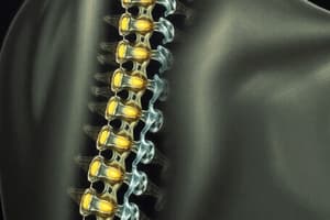
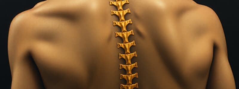
- Spinal cord gray matter serves as the integrative center for cord reflexes; its typical arrangement is shown in Figure 54-1.
- Sensory signals enter the cord mainly via sensory posterior roots, then branch into two routes.
- One branch of sensory nerves terminates in the cord gray matter, triggering local segmental cord reflexes and effects.
- Another branch transmits signals to higher nervous system levels, including the brain stem and cerebral cortex.
- Each spinal cord segment consisting of millions of neurons in the gray matter.
- Anterior motor neurons and interneurons are the two types of neurons associated with the spinal cord.
Anterior Motor Neurons
- Thousands of anterior motor neurons, larger than the others (50-100%), are located in the anterior horns of cord gray matter.
- Axons from anterior motor neurons exit via the anterior roots and directly innervate skeletal muscle fibers.
- Alpha and gamma motor neurons are the two types of anterior motor neurons.
Alpha Motor Neurons
- Alpha motor neurons produce large type A alpha (Aa) motor nerve fibers, averaging 14 micrometers in diameter.
- Alpha fibers branch upon entering muscle which innervates muscle fibers.
- Motor unit is when a single alpha nerve fiber excites from three to hundreds of skeletal muscle fibers.
Gamma Motor Neurons
- About half the number of alpha motor neurons, much smaller gamma motor neurons are located in the spinal cord anterior horns.
- Gamma motor neurons transmit impulses via smaller type A gamma (Ay) motor nerve fibers, averaging 5 micrometers in diameter.
- These fibers go to intrafusal fibers, that constitute the muscle spindle, controlling basic muscle tone.
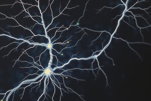
Interneurons
-
Interneurons are in all cord gray matter areas, including the dorsal horns, anterior horns, and intermediate areas.
-
Interneurons are about 30 times more numerous than anterior motor neurons
-
Small and highly excitable, interneurons can fire up to 1500 times per second.
-
Interneurons interconnect and synapse with anterior motor neurons.
-
Interconnections between interneurons and anterior motor neurons facilitate spinal cord integrative functions.
-
Diverging, converging, and repetitive-discharge circuits are types of neuronal circuits found in the spinal cord interneuron pool. These circuits perform specific acts via reflex.
-
Few incoming sensory or brain signals terminate directly on anterior motor neurons, these travel through interneurons.
Corticospinal Tract
- Corticospinal tract from the brain terminates almost entirely on spinal interneurons
- Signals from the brain is combined with signals from other spinal tracts or spinal nerves.
- Signals converge on the anterior motor neurons to control muscle function.
Renshaw Cell Inhibitory System
- Small neurons called Renshaw cells are in the anterior horns in close association with motor neurons.
- Motor neuron axon collateral branches transmit inhibitory signals to adjacent Renshaw cells.
- Stimulation of motor neurons inhibits adjacent motor neurons, known as lateral inhibition.
- The motor system focuses its signals by lateral inhibition, allowing primary signal transmission while suppressing spread.
Propriospinal Fibers
- Over half of all nerve fibers ascending and descending in the spinal cord are propriospinal fibers.
- Signals are transmitted to only a segment or two, while others transmit signals to many segments.
- These fibers run one segment to another and ascending and descending coordinate movements in the forelimbs and hindlimbs, providing pathways for multisegmental reflexes.
Muscle Spindles and Golgi Tendon Organs
- Proper muscle function requires excitation by spinal cord anterior motor neurons, with continuous sensory feedback indicating muscle status.
- Function sensory feedback involves length, instantaneous tension, and rate of change.
- Muscles and tendons have: muscle spindles for length and rate of change of length, and Golgi tendon organs for tendon tension and rate of change of tension.
- Subconscious signals from spindles and Golgi tendon organs transmit information to spinal cord, cerebellum, and cerebral cortex.
- Transmitted information aids in controlling muscle contraction.
Receptor Function of the Muscle Spindle
- Figure 54-2 shows the organization of the muscle spindle.
- Muscle spindle ranges from 3 to 10 millimeters long.
- The spindle is built around 3-12 small intrafusal muscle fibers that are pointed at their ends and attached to the surrounding glycocalyx.
- Intrafusal muscle fibers have center regions with few or no actin and myosin filaments and act as a sensory receptor.
- End portions are excited by small gamma motor nerve fibers from type A gamma motor neurons.
- Gamma efferent fibers is what gamma motor nerve fibers also are called.
- Receptor portion of the muscle spindle is its center.
- The muscle spindle receptor can be excited via lengthening the whole muscle and contraction of the spindle's intrafusal fiber end portions.
- Primary and secondary endings are the two sensory endings in the receptor area of the muscle spindle.
Primary Ending
- Large sensory nerve fiber encircles the intrafusal fiber central portion, forming the annulospiral ending.
- Averaging 17 micrometers in diameter the nerve fiber is a type Ia.
- The fiber transmits sensory signals rapidly to the spinal cord at 70 - 120 m/sec.
Secondary Ending
- Smaller sensory nerve fibers, usually one but sometimes two (type II fibers), with an average diameter of 8 micrometers - innervate the region on one or both sides of the primary ending.
- The secondary ending encircles the intrafusal fibers sometimes.
Division of Intrafusal Fibers
- Nuclear bag muscle fibers consists of one to three per spindle. The nuclei are congregated in expanded "bags” in the central receptor area.
- Nuclear chain fibers, about half the diameter and length of bag fibers with nuclei aligned in a chain throughout the receptor area-three to nine per spindle.
- 17-micrometer sensory fiber is the primary nerve excited by bag and chain fibers.
- 8-micrometer sensory fiber is secondary ending excited usually by chain fibers.
Static Response
- Receptor length increase results in the rise of impulses transmitted from both primary and secondary endings.
- Signals transmit for minutes even with a stretched spindle.
- When transmission remains in signals as long as the spindle remains stretched that is called static response.
Dynamic Response
- Primary ending (not secondary) stimulation particularly enhanced when spinal receptor suddenly increase in length.
- The excess stimulus of the primary ending is called the dynamic response.
- Dynamic response occurs because it responds actively and rapidly to change.
- Primary receptors can transmit great numbers of impulses to the 17-micrometer when only some length occurs in fraction of a second.
- The extra rate of impulse returns to the smaller static response as soon as they length stops.
- Strong signals, either positive or negative. are sent to the spinal cord to inform about changes in spindle length.
Gamma Motor Nerves
- Gamma-dynamic (gamma-d) and gamma-static (gamma-s) can divided in two types from gamma motor nerves to the muscle spindle.
- The first excites bag intrafusal fibers mostly, second excites chain itrafusal fibers.
- Excitation of the nuclear bag fibers by gamma-d fibers enhances spindle dynamic resonse, and hardly affects static response.
- Gamma-s fibers when stimulated excites the nuclear chain fibers, enhances the static response, but does little in dynamic.
Continuous Discharge
- Muscle spindles emit nerve impulses continuously when there is some degress of gamma nerve excitation.
- Stretching the muscle spindles escalates the rate of firing.
- Shortening of the muscle spindles diminish the rate of firing.
- Positive increased numbers of impulse indicate muscle stretch and negative below-normal numbers indicate a lack of stretch.
Muscle Stretch Reflex
- The muscle stretch reflex is triggered when stretching the spindles causes reflex contraction of the stretched large skeletal muscle fibers, and allied synergistic muscles.
- The muscle spindles and contraction is triggered when the muscles stretch, it is the simplest manifestation of muscle spindle function.
Neuronal Circuitry
- A type Ia proprioceptor nerve fiber originates in the muscle spindle and a branch goes directly to the anterior horn, synapsing with anterior motor neurons there and sends it back to the muscle from which the spindle originated.
- Type II fibers terminate to multiple interneurons that transmit the delayed signals for excitation.
- Monosynaptic is the pathway that allows a reflex signal to return after excitation.
Dynamic and Static Stretch Reflex
- Dynamic and static stretch reflex are two components where the reflex can be divided.
- Potent dynamic signal is transmitted from ends of the muscle spindles is the signal of rapid stretch or unstretch.
- Instantaneous contraction arises from signal which transmits and causes reflex to oppose sudden change in muscle length.
- The strong signal of stretch is transmitted and has instantaneous strong reflex contraction (or decrease contraction).
- The dynamic stretch is over within fraction of a second after being stretched to its new length.
- Prolonged period is due to continuous static receptors which is transmitted by both ends of muscle.
Damping Function
- A function of oscillation is that it prevents jerkiness.
- Signals from the spinal cord are often transmitted to muscle, but without the apparatus functioning correctly the contraction will be jerky.
- Muscle contraction is smoother in the muscle spindle as shown in curve A from the experiment and graph.
- Muscle spindle nerves had been sectioned three month prior in curve B experiment.
- Signal averaging is called effects of a muscle spindle reflex.
Voluntary Motor Activity
- 31% of all motor nerve fibers to muscles are small rather than large, emphasizing the gamma system.
- Gamma motor neurons are stimulated, thus causes skeletal fibers and muscle fibers to contract, whenever brain areas transmit signals to alpha motor neurons.
- Contracting the muscle spindle at the same time leads to:
- It keeps length from changing. Coactivation keeps spindle from opposing muscle.
- Maintains proper damping regardless of change in length.
- Receptor would be flail and overstretched if muscle was not contacted.
Brain Area Control
- Gamma system is excited by the bulboreticular and impulses from the cerebellum, basal ganglia, and the cerebral cortex.
- Gamma system damping is important for walking and running movements, and the bulboreticular are concerned with antigravity contractions.
Muscle Spindle System
- Excitatory signals in the intrafusal in the spindles occur from the bulboreticular which helps stabilize the body during motor action.
- If the ends are shortened and the receptor is stretched then signa output increases.
- If spindle activated at joint reflex ,excitation of both sides occur.
- This results in tensed muscles opposing the joint, leading to a stable position with any force opposed by these reflexes.
- A high degree of positioning, muscles are needed when the position can not be easily changed, and aids performing movements.
Clinical Applications
-
Eliciting stretch reflexes determines excitation.
-
Knee Jerk or other muscles can determine sensitivity of the reflexes.
-
Striking the patellar tendon with a reflex hammer is a method to determine the stretch and stretch reflex.
-
Striking the muscle is required in other cases to cause sudden stretch for a reflex to be elicited.
-
Spinal assess of muscle jerks is when the facilitation is determined.
-
Muscle jerks greatly exaggerate the number of facilitatory impulses when transmitted, and conversely they are weakened or absent if suppressed.
Lesions
- Muscular spasticity that is caused by diseases or lesions are used by reflexes for the brain and motor areas.
- Motor control areas doesn't causes large lesions as much, so they use exaggerated muscular jerks from strokes or brain tumors.
Clonus
-
Oscillations is the oscillation of jerks where they explained well in ankles, and this is when the muscle jerks oscillate under a condition
-
Ankle clonus occurs when suddenly drops to reach reflex from feet stretching.
-
Impulses reflexively reach into the spinal cord, which lifts the body again.
-
The body falls again and does each process, in what is know as clonus for a duration.
-
Enhanced clonus is caused by reflexes from a highly sensitive brain.
-
Test for clonus is when neurologist stretches a muscle and applies force if developed.
Golgi Tendon Reflex
-
Golgi Tendon Organ is in the muscle and it sensory.
-
Muscle tendon fiber pass and consists encapsulated sensory receptor.
-
Between 10-15 connected ,and simulated in when a fiber of muscle becomes "tensed"/contracted.
-
How it differs from excitation is that in spindles it muscle length, but in tendon organs it detect muscle tension and tension in itself.
-
Both Organs have responses that when muscle increases quickly and settles down for amount depending on tension.
-
Information about the tension is given when tension is given a small segments.
-
Signals transmitted through rapid fibers that give signals in areas local the chord and has single inteneuon that is excited.
-
How they related to the brain comes from discussion in another chart in these parts.
Tendon Reflex
- Spinal cord cause reflex to effect a muscle when they are stimulated to cause high tensions, inhibiting muscle, a feedback.
- When the muscle becomes tense the inhibitory effect can be to affect the direction.
- Reactions happen so it is prevented form tearing or attaching to a bone because the reaction cannot be opposed.
- Destructive effects occasionally occur because, muscles is that cannot be opposed by reflux can not lead to high tensions when prevented.
Fibers Among Muscle
- Tendons Reflex is to equalize contractile force among the separate fibers.
- The muscle gets overloaded because if they have too little forces, they are not inhibited.
Muscle Spindles
- Organs tells what is going on by the brain making these muscle.
- From the spinal cord, the tract can take place when a changes with the muscles.
Flexor Action
-
Stimulation sensory with drawls.
-
What reflex takes place is on by that the point of in the end. Types of circuit diverge, causing reciprocal inhibition, and causing discharge with reflex.
-
Spinal cord can have integrals between everything needed to make a withdrawal.
Crossing Movement
-
The reflex can cross to a limit for the extensors where one point is the movement and the next.
-
There is numerous interneuron within for circuit that has the reactions, also shows the effects by showing from reflexes after some moments.
-
Muscle reflex depends where area needs to hold down so reactions makes move.
Inhibition and Innervation
- The text points out, that there has been something about in groups where the interaction happens at the same points.
Studying That Suits You
Use AI to generate personalized quizzes and flashcards to suit your learning preferences.