Podcast
Questions and Answers
What are the two primary functions of the primary somatosensory cortex in processing general sensation?
What are the two primary functions of the primary somatosensory cortex in processing general sensation?
- Interpreting the intensity of a stimulus and initiating a motor response.
- Filtering out irrelevant sensory inputs and regulating body temperature.
- Identifying the type of sensation encountered (e.g., temperature, pressure) and determining its location on the body. (correct)
- Storing sensory information for future recall and emotional association.
What is the role of thermoreceptors in sensory perception?
What is the role of thermoreceptors in sensory perception?
- Detecting changes in pressure applied to the skin.
- Sensing variations in temperature, allowing us to distinguish between hot and cold. (correct)
- Transmitting information about the position and movement of body parts.
- Identifying the location of a stimulus with high precision.
What is spatial discrimination in the context of somatosensation?
What is spatial discrimination in the context of somatosensation?
- The ability to differentiate between various textures of objects.
- The process of adapting to a constant stimulus over time.
- The integration of sensory information with motor commands.
- The capacity to precisely identify the location of different sensations on the body. (correct)
What does the somatosensory homunculus illustrate?
What does the somatosensory homunculus illustrate?
How does sensory information from the body's surface reach the primary somatosensory cortex?
How does sensory information from the body's surface reach the primary somatosensory cortex?
Which type of sensory information is primarily carried by the spinothalamic tract?
Which type of sensory information is primarily carried by the spinothalamic tract?
What is the primary function of the somatosensory association cortex?
What is the primary function of the somatosensory association cortex?
Where is the somatosensory association cortex located?
Where is the somatosensory association cortex located?
A patient is having difficulty initiating a sequence of movements and seems to struggle with planning complex motor tasks. Based on this, which area of the brain is most likely affected?
A patient is having difficulty initiating a sequence of movements and seems to struggle with planning complex motor tasks. Based on this, which area of the brain is most likely affected?
Which scenario would most likely activate neurons within the premotor cortex?
Which scenario would most likely activate neurons within the premotor cortex?
If a person suffers a lesion that impairs their ability to produce speech, but they can still understand spoken language, which area of the brain is most likely affected?
If a person suffers a lesion that impairs their ability to produce speech, but they can still understand spoken language, which area of the brain is most likely affected?
A patient can speak fluently but uses incorrect words and struggles to understand spoken instructions. Which area of the brain is likely damaged?
A patient can speak fluently but uses incorrect words and struggles to understand spoken instructions. Which area of the brain is likely damaged?
A patient exhibits difficulty coordinating body movements and is diagnosed with Parkinson's disease. Which structure is most likely affected?
A patient exhibits difficulty coordinating body movements and is diagnosed with Parkinson's disease. Which structure is most likely affected?
A person has difficulty voluntarily shifting their gaze from one object to another. Which area in the brain is most likely affected?
A person has difficulty voluntarily shifting their gaze from one object to another. Which area in the brain is most likely affected?
During a neurological exam, a doctor shines a bright light in a patient's eyes, and the patient's head and eyes reflexively turn towards the light. This response indicates proper functioning of which midbrain structure?
During a neurological exam, a doctor shines a bright light in a patient's eyes, and the patient's head and eyes reflexively turn towards the light. This response indicates proper functioning of which midbrain structure?
Following a stroke, a patient's eyes deviate to the right. Which area of the brain was likely damaged?
Following a stroke, a patient's eyes deviate to the right. Which area of the brain was likely damaged?
Which of the following best describes the anatomical relationship between the cerebral aqueduct and the ventricular system?
Which of the following best describes the anatomical relationship between the cerebral aqueduct and the ventricular system?
A lesion in the medulla oblongata affects the decussation of pyramidal neurons. What is the most likely consequence of this lesion?
A lesion in the medulla oblongata affects the decussation of pyramidal neurons. What is the most likely consequence of this lesion?
Which of the following sensory inputs is NOT directly processed in the primary somatosensory cortex?
Which of the following sensory inputs is NOT directly processed in the primary somatosensory cortex?
Which of the following receptors provides information about the position and movement of your limbs without relying on visual input?
Which of the following receptors provides information about the position and movement of your limbs without relying on visual input?
Which of the following cranial nerves is NOT associated with the medulla oblongata?
Which of the following cranial nerves is NOT associated with the medulla oblongata?
Damage to the red nucleus in the midbrain would most likely result in difficulties with which of the following functions?
Damage to the red nucleus in the midbrain would most likely result in difficulties with which of the following functions?
Which anatomical feature is characteristic of the ventral surface of the midbrain?
Which anatomical feature is characteristic of the ventral surface of the midbrain?
A loud, unexpected noise causes a person to reflexively turn their head. Which part of the midbrain mediates this response?
A loud, unexpected noise causes a person to reflexively turn their head. Which part of the midbrain mediates this response?
Which of the following accurately describes the role of the auditory association area?
Which of the following accurately describes the role of the auditory association area?
A patient reports difficulty in recognizing familiar faces and navigating previously known routes. Which association area is MOST likely affected?
A patient reports difficulty in recognizing familiar faces and navigating previously known routes. Which association area is MOST likely affected?
Damage to which of the following brain regions would MOST likely result in deficits in working memory, decision-making, and personality changes?
Damage to which of the following brain regions would MOST likely result in deficits in working memory, decision-making, and personality changes?
A person experiences a sudden inability to detect certain smells. Which area of the brain is MOST likely affected?
A person experiences a sudden inability to detect certain smells. Which area of the brain is MOST likely affected?
Which sequence BEST describes the pathway of auditory information from sound waves to the primary auditory cortex?
Which sequence BEST describes the pathway of auditory information from sound waves to the primary auditory cortex?
After a head injury, a patient reports a loss of balance and spatial orientation. Which cortical area is MOST likely damaged?
After a head injury, a patient reports a loss of balance and spatial orientation. Which cortical area is MOST likely damaged?
A patient reports experiencing a constant sensation of hunger and an urgent need to urinate, even when these needs are not physiologically present. Dysfunction in which cortical area might explain these symptoms?
A patient reports experiencing a constant sensation of hunger and an urgent need to urinate, even when these needs are not physiologically present. Dysfunction in which cortical area might explain these symptoms?
Which cranial nerves transmit afferent sensory information to the gustatory cortex?
Which cranial nerves transmit afferent sensory information to the gustatory cortex?
If a person struggles to see colors, but has normal night vision, which type of photoreceptor is most likely dysfunctional?
If a person struggles to see colors, but has normal night vision, which type of photoreceptor is most likely dysfunctional?
Why do photoreceptors require a large number of mitochondria?
Why do photoreceptors require a large number of mitochondria?
How is yellow light perceived by the cone receptors?
How is yellow light perceived by the cone receptors?
What is the immediate consequence of light causing retinal to change from its 11-cis form to its trans form?
What is the immediate consequence of light causing retinal to change from its 11-cis form to its trans form?
What is the primary function of the cornea?
What is the primary function of the cornea?
In low-light conditions, which type of photoreceptor is predominantly active, and where are these photoreceptors primarily located in the retina?
In low-light conditions, which type of photoreceptor is predominantly active, and where are these photoreceptors primarily located in the retina?
If the amount of light passing through the cornea is excessive, and hits the Iris, what happens?
If the amount of light passing through the cornea is excessive, and hits the Iris, what happens?
Which sequence accurately describes the pathway light travels through the eye to reach the retina?
Which sequence accurately describes the pathway light travels through the eye to reach the retina?
Which of the following is NOT directly controlled by the autonomic nervous system?
Which of the following is NOT directly controlled by the autonomic nervous system?
The 'park the car' analogy is most closely associated with the function of which type of motor neuron?
The 'park the car' analogy is most closely associated with the function of which type of motor neuron?
During a stressful situation, which of the following physiological responses is most likely triggered by sympathetic motor neurons?
During a stressful situation, which of the following physiological responses is most likely triggered by sympathetic motor neurons?
The phrenic nerve plays a crucial role in breathing. When does the autonomic nervous system primarily control the diaphragm via this nerve?
The phrenic nerve plays a crucial role in breathing. When does the autonomic nervous system primarily control the diaphragm via this nerve?
What is the primary significance of dual innervation in the autonomic nervous system?
What is the primary significance of dual innervation in the autonomic nervous system?
Which of the following effects would be expected from parasympathetic stimulation of the salivary glands?
Which of the following effects would be expected from parasympathetic stimulation of the salivary glands?
Dilation of the pupil is primarily associated with which of the following?
Dilation of the pupil is primarily associated with which of the following?
What does autonomic tone primarily reflect?
What does autonomic tone primarily reflect?
Flashcards
Premotor Cortex Function
Premotor Cortex Function
Involved in planning, initiating, and learning movements.
Broca's Area Function
Broca's Area Function
Speech production and motor function. Located in the left frontal lobe.
Wernicke's Area Function
Wernicke's Area Function
Language comprehension. Located in the posterior region of the left temporal lobe.
Frontal Eye Field Function
Frontal Eye Field Function
Signup and view all the flashcards
Cortical Neurons Role
Cortical Neurons Role
Signup and view all the flashcards
Primary Somatosensory Cortex
Primary Somatosensory Cortex
Signup and view all the flashcards
Proprioceptor Function
Proprioceptor Function
Signup and view all the flashcards
Primary Somatosensory Cortex Location
Primary Somatosensory Cortex Location
Signup and view all the flashcards
Somatosensory Cortex Roles
Somatosensory Cortex Roles
Signup and view all the flashcards
Thermoreceptor
Thermoreceptor
Signup and view all the flashcards
Spatial Discrimination
Spatial Discrimination
Signup and view all the flashcards
Somatosensory Homunculus
Somatosensory Homunculus
Signup and view all the flashcards
Spinothalamic Tract
Spinothalamic Tract
Signup and view all the flashcards
1st Neuron (Ascending Tract)
1st Neuron (Ascending Tract)
Signup and view all the flashcards
2nd Neuron (Ascending Tract)
2nd Neuron (Ascending Tract)
Signup and view all the flashcards
3rd Neuron (Ascending Tract)
3rd Neuron (Ascending Tract)
Signup and view all the flashcards
Function of Rods
Function of Rods
Signup and view all the flashcards
Function of Cones
Function of Cones
Signup and view all the flashcards
Types of Cone Receptors
Types of Cone Receptors
Signup and view all the flashcards
How yellow light is sensed
How yellow light is sensed
Signup and view all the flashcards
Intersegments
Intersegments
Signup and view all the flashcards
Opsin
Opsin
Signup and view all the flashcards
Retinol
Retinol
Signup and view all the flashcards
Function of the Cornea
Function of the Cornea
Signup and view all the flashcards
Somatic Nervous System
Somatic Nervous System
Signup and view all the flashcards
Parasympathetic Motor Neurons
Parasympathetic Motor Neurons
Signup and view all the flashcards
Sympathetic Motor Neurons Function
Sympathetic Motor Neurons Function
Signup and view all the flashcards
Phrenic Nerve Function
Phrenic Nerve Function
Signup and view all the flashcards
Dual Innervation
Dual Innervation
Signup and view all the flashcards
Autonomic Tone
Autonomic Tone
Signup and view all the flashcards
Parasympathetic effect on heart
Parasympathetic effect on heart
Signup and view all the flashcards
Sympathetic effect on heart
Sympathetic effect on heart
Signup and view all the flashcards
Auditory Pathway to Primary Auditory Cortex
Auditory Pathway to Primary Auditory Cortex
Signup and view all the flashcards
Auditory Association Area Function
Auditory Association Area Function
Signup and view all the flashcards
Vestibular Cortex Function
Vestibular Cortex Function
Signup and view all the flashcards
Olfactory Cortex Function
Olfactory Cortex Function
Signup and view all the flashcards
Gustatory Cortex Function
Gustatory Cortex Function
Signup and view all the flashcards
Visceral Sensory Cortex Function
Visceral Sensory Cortex Function
Signup and view all the flashcards
Anterior Association Cortex Functions
Anterior Association Cortex Functions
Signup and view all the flashcards
Posterior Association Cortex Functions
Posterior Association Cortex Functions
Signup and view all the flashcards
Corpora Quadrigemina Location?
Corpora Quadrigemina Location?
Signup and view all the flashcards
Corpora Quadrigemina Parts?
Corpora Quadrigemina Parts?
Signup and view all the flashcards
Superior Colliculi Function?
Superior Colliculi Function?
Signup and view all the flashcards
Inferior Colliculi Function?
Inferior Colliculi Function?
Signup and view all the flashcards
Substantia Nigra Function?
Substantia Nigra Function?
Signup and view all the flashcards
Substantia Nigra Degeneration?
Substantia Nigra Degeneration?
Signup and view all the flashcards
Myencephalon Location?
Myencephalon Location?
Signup and view all the flashcards
What are Pyramids (medulla)?
What are Pyramids (medulla)?
Signup and view all the flashcards
Study Notes
Cortical Neurons
- Cortical neurons are neurons located in the cerebral cortex.
- Interneurons are a type of neuron found in the cerebral cortex.
- Interneurons are located in the outer gray matter of the cerebral cortex.
Contralateral Organization
- Each hemisphere of the cerebral cortex receives sensory input and gives motor commands to the opposite side of the body; cortical neurons in the left hemisphere deal with sensory/motor neurons on the right side of your body, and vice versa.
- Decussation is the crossing of axons from one side of the body to the other.
Laterization
- Laterization is where cortical neurons in the left cerebral cortex have different specialized functions compared to those in the right cerebral cortex.
- The left cerebral cortex receives sensory stimulus and has motor control over the right side of the body.
- The left hemisphere is responsible for logic, language, math, speech/language, comprehension/analysis, calculations (math), telling time, and recognizing words, letters, and numbers.
- The right brain is responsible for more creativity, special awareness, and recognizing names, places, and objects.
- Cortical neurons have specialized functions based on their position in the cerebral cortex.
- Spinalized functions are localized in the brain, such as seeing occurring in the posterior portion of the brain.
Cortical Neuron Jobs
- Three jobs of cortical neurons are to initiate motor movements, to create awareness of sensations, and to make associations/correct responses.
- Cortical neurons that control motor functions are found in four places: the primary motor cortex, the premotor cortex, Broca's area, and the frontal eye field.
- Cortical neurons in the primary motor cortex control motor neurons in the somatic nervous system.
- The primary motor cortex is anterior to the central sulcus.
- The precentral gyrus is a raised primary motor cortex area.
- Pyramidal cells control activity and have large axons that go from the cerebral cortex to the spinal cord.
- Decussation occurs at the medulla oblongata/spinal cord.
Motor Control Neurons
- Two neurons control the ability to write: the upper motor neuron (cortical neuron) and the lower motor neuron (motor neuron).
- A descending tract is information flowing from the cerebral cortex down to a motor neuron.
- The corticospinal tract facilitates motor movement from the neck down into the spinal cord, has 2 neurons, the lower neuron is a spinal nerve and descends.
- The corticobulbar tract is for motor movement from a cranial nerve, has 2 neurons, the lower neuron is a cranial nerve and descends.
- Somatotopy is the idea that the position of cortical neurons on the "weird map thing" corresponds with the areas on the body that they control.
- The motor homunculus causes some body parts to be big because the larger a body part is, the more cortical neurons are involved in moving it.
- Wilder Penfield found that different areas of the primary motor cortex move different parts of the body.
Descending Pathways
- An indirect descending pathway involves a 3rd interneuron to relay information from the cortical neuron to the lower motor neuron; its soma is in the midbrain/brainstem.
- Relay neurons control muscle tone, balance, and flexion.
- Somatic motor commands are indicated from pyramidal neurons in the cortex.
- Information is shared to skeletal muscle via descending tracts with 2-3 neurons.
- The rubrospinal tract is a descending tract with 3 neurons.
- The 3rd neuron in the rubrospinal tract relays information from the upper neuron to the lower neuron with it's soma in the midbrain or brainstem.
- Synaptic potentiation is the strengthening of synapses that are used frequently.
Synaptic Potentiation
- Synaptic potentiation occurs during guitar practice as upper and lower motor neurons communicate faster and better, strengthening synapses with repetition/practice.
- Synaptic potentiation can occur through an increase of Ca2+ in the synapse or an increase of receptors on the post-synaptic membrane.
- Increasing Ca2+ strengthens synapses by releasing neurotransmitters, speeding up information passage, and making action potentials fire faster/more efficiently.
- Increasing receptors on the post-synaptic membrane strengthens synapses as more receptors can engage with more neurotransmitters, increases membrane permeability, and makes action potentials fire faster and more efficiently.
- The premotor cortex is responsible for planning and coordinating highly complex movements.
Brain Areas
- The premotor cortex is located anterior to the primary motor cortex.
- Premotor neurons are active when movement is being planned, as movement is being initiated, and as movement is being learned.
- The primary motor cortex carries out movements planned in the premotor cortex.
- Broca's area is responsible for speech production/motor function and is located in the left frontal lobe.
- A lesion to Broca's area would result in the inability to express language verbally.
- Wernicke's area is responsible for language comprehension and is located in the posterior region of the left temporal lobe.
- A lesion in Wernicke's area would result in the ability to speak, but with difficulty comprehending constructed language.
- The frontal eye field is responsible for planning, coordinating, and executing eye movements.
- The frontal eye field is located in the frontal lobe, anterior to the cortex.
- A lesion to the frontal eye field would result in the loss of control over eye muscles; damage in the left hemisphere would cause the eyes to gaze to the left.
- The job of cortical neurons is to be aware of sensations and make sense of environmental stimuli.
Sensory Stimuli
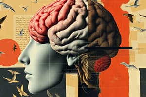
- Cortical neurons become aware of sensory stimuli in 8 places: the primary somatosensory cortex, the somatosensory association cortex, the primary visual cortex and visual association area, the primary auditory cortex and auditory association area, the vestibular cortex, the olfactory cortex, the gustatory cortex, and the visceral sensory cortex.
- The primary somatosensory cortex is responsible for making sense of touch, pain, pressure, and temperature.
- A proprioceptor is a receptor in muscle that makes sense of the level of stretch, contraction, and body position of muscles.
- The primary somatosensory cortex is located postcentral and posterior to the central sulcus.
- The primary somatosensory cortex is responsible for making sense of what is being encountered, like temperature (hot/cold) and pressure (soft/pain), and determining where the sensation is occurring, like the hand or foot.
- A thermoreceptor is a temperature receptor that tells if something is cold/hot.
- Spatial discrimination is being able to pinpoint different sensations at different points in the body.
- The somatosensory homunculus shows a map of where gets stimulated when you feel touch on different parts of the body.
- Sensory information moves from the body's surface to the primary somatosensory cortex via an ascending tract from either spinal nerves or cranial nerves.
Sensory Tracts
- The spinothalamic tract consists of spinal nerves associated with carrying pain, temperature, and pressure information.
- The 1st neuron in an ascending tract is a sensory neuron that synapses on an interneuron with its cell body in the ganglion.
- The 2nd neuron in an ascending tract is an interneuron in the spinal nerve/dorsal horn of spine.
- The 3rd neuron in an ascending tract extends from the thalamus to the primary somatosensory cortex.
- The thalamus is a gateway to the cerebral cortex.
- The somatosensory association cortex is responsible for being able to discriminate between different objects by feeling them and is located posterior to the post-central gyrus in the parietal lobe.
- The primary visual cortex creates associations with what we see, such as facial recognition.
- The visual association area surrounds the primary visual cortex in the occipital lobe.
- Sight information gets to the primary visual cortex when light enters the cornea and strikes rods and cones in the retina, light information converts to an electrical impulse, a neuron starts at the retina and moves through the optic nerve though the optic tract, and information goes to the thalamus then to primary visual cortex.
Auditory System
- The primary auditory cortex is responsible for telling the brain that there is sound.
- Sound information gets to the primary auditory cortex when sound waves come in through the external acoustic meatus, displaces bones in the middle ear, neurons in the cochlea activate action potentials in cranial nerves, information goes to the brain stem, then to the thalamus, and then lands in the primary auditory cortex.
- The primary auditory cortex is located in the temporal lobe.
- The auditory association area is responsible for perceiving what we hear and differentiating sounds such as screams, cries, and thunder; it is located in the temporal lobe.
- The vestibular cortex is responsible for providing information about the body's balance and is located in the insula lobe.
- The olfactory cortex is responsible for smell, with information brought by cranial nerve 1, and is located in the temporal lobe.
- The gustatory cortex is responsible for afferent sensory information from neurons, brought to the cerebral cortex by cranial nerves 7, 9, and 10 and is located in the insula lobe.
- The visceral sensory cortex is responsible for feeling things related to what is going on with body organs and is located in the insula lobe.
Anterior Association Cortex Responsibilities
- The anterior association cortex is responsible for working memory, decision making, planning, solving problems, judgement, and personality.
- A lesion in the anterior association cortex could present as acting like a completely different person.
- The posterior association cortex is responsible for understanding written and spoken language, spatial awareness, recognition of faces and patterns, identifying landmarks, and attention/focus.
- The diencephalon consists of the thalamus and hypothalamus.
- The thalamus’s five main functions are: 1) relay station to cerebral cortex (all sensory info), 2) sorts through/edits info coming in deciding if it is going to the cerebellum, 3) gets rid of info with help from basal ganglia that the cerebellum doesn't need so cerebellum can quickly make sense of the info, 4) relay for emotional/visceral info coming in from hypothalamus, and 5) relay for motor info coming from cerebellum to fine tune and coordinate somatic movements.
- To get to the cerebellum when feeling something cold with your foot, information has to go through a sensory neuron, then an ascending tract, the somatosensory cortex, the thalamus, and finally the cerebellum.
- The thalamus is like an airport terminal.
- Nuclei in the brain/spine are collections of cell bodies working together.
- The corona radiata is where the cerebellum fans out.
- The hypothalamus is located below the thalamus.
- The two boundaries to the hypothalamus are the optic chiasma (visual info coming in) and the mamillary body (olfactory info coming in).
- The pituitary gland comes off of the hypothalamus.
- The infundibulum is a stalk-like structure connecting the pituitary gland to neurons in the hypothalamus.
Hypothalamus
- The hypothalamus key role is a homeostasis hub that regulates the autonomic nervous system and maintains regular homeostasis in the body.
- The epithalamus is composed of the posterior commissure and pineal gland.
- The epithalamus is found at the most dorsal aspect of the diencephalon.
- The pineal gland produces melatonin.
- The epithalamus works with the pineal gland to make you tired.
- The hypothalamus regulates body temperature, food intake, water balance, and thirst and hormonal output for the anterior pituitary gland.
- The hypothalamus acts as an endocrine gland producing posterior pituitary hormones such as ADH and Oxytocin.
- The mesencephalon consists of the midbrain and is located at the top of the brain stem.
- Axons of ascending tracts carrying sensory info pass through the dorsal white matter of the midbrain.
- Axons of descending tracts carrying motor info pass through the ventral white matter of the midbrain.
- The corpora quadrigemina consists of 4 nucelli and bulges out of the midbrain..
- The two parts of the corpora quadrigemina are the superior colliculi and the inferior colliculi.
- The superior colliculi's function is the visual reflex center; an example is when the head moves if seeing something coming at you.
- The inferior colliculi's function is the auditory reflex center; an example is turning around if hearing a loud sound.
- Axons of descending motor tracts run through the cerebral peduncles of the midbrain.
- The cerebral peduncles are located on the ventral surface of the midbrain.
- Axons of ascending sensory tracts run through more dorsal positions of the midbrain.
- The red nucleus of the midbrain has neurons involved in indirect descending motor tracts involved with muscle tone.
- The red nucleus is located in the midbrain.
- The cerebral aqueduct connects the 3rd and 4th ventricles.
- The substantia nigra produces brown neurons releasing dopamine for body movements.
- The degeneration of brown neurons in the substantia nigra causes body movements to become less coordinated, resulting in Parkinson's disease.
Midbrain Function
- The midbrain's main functions are: 1) containing superior and inferior colliculi for visual and auditory reflex centers, 2) contains subcortical motor centers (substantia nigra and red nuclei), 3) contains nuclei for cranial nerves III and IV, and 4) contains projection fibers (ascending and descending).
- The myencephalon consists of the medulla oblongata.
- The myencephalon is located at the bottom of the brainstem, inferior to the cerebellum.
- The medulla oblongata has cranial nerves VIII - X and XII.
- Mesullary neurons are synapses in the neurons of the medulla oblongata.
- The medulla oblongata is a major cite of decussation.
- Axons of pyramidal neurons decussate at the medulla oblongata.
- Decussation occurs in the spinal cord/midbrain if not in the medulla oblongata.
- Pyramids are bulges coming out of the medulla oblongata from decussation of pyramidal neurons.
- Axons of descending motor tracts run through pyramids of the medulla oblongata.
- Axons of ascending sensory tracts run through more dorsal positions of the medulla oblongata, including the medial lemniscus.
- The medial lemniscus is located in the medulla oblongata.
- The 4th Ventricle is in the medulla oblongata and includes the choroid plexus.
- Inferior olivary nucleuses function as "bunches of olives" and neurons gathering information about muscle tone, muscle stretch, and positions of muscles.
- The inferior olivary nucleuses relay information to the inferior cerebellar peduncle.
- The inferior cerebellar peduncles function to relay sensory details about body position.
- The reticular formation is involved in arousal/alertness and is scattered throughout the oblongata.
- The medulla oblongata and the hypothalamus work together to form an autonomic reflex center.
- The pons is the other brain part involved in breathing rate besides the hypothalamus and medulla oblongata.
Medulla Oblongata Functions
- Six main functions of the medulla oblongata are: 1) relays ascending sensory impulses from skin and proprioceptors, 2) contains visceral nuclei that controls heart rate, breathing rate, vomiting, etc, 3) relays sensory information to the cerebellum through inferior olivary nuclei, 4) contains nuclei of cranial nerves VII-X and XII, 5) contains projection fibers, and 6) cite of decussation at pyramids.
- The limbic association area is wrapped around the corneum.
- The limbic association area makes emotional behaviors/responses.
- White matter contains myelinated axons of neurons that are coming and leaving cortical neurons.
- Association fibers connect cortical neurons in the same hemisphere.
- Projection fibers connect cortical neurons to ascending and descending tracts.
- Commissural connect are cortical neurons between left and right hemispheres, so the cerebrum can function as a whole.
- The internal capsule is a cite of projection neurons being bundled together at the top of the brainstem.
- The corona radiata contains fibers that fan out along the anterior/posterior axis of the cerebral cortex.
- The basal ganglia is located deep within the cerebellum.
- The three structures in the basal ganglia are the caudate nucleus, putamen, and globus pallidus.
- The basal ganglia's function is proper initiation and execution of somatic motor movements.
- The basal ganglia works with the primary motor cortex, premotor cortex, thalamus, and substantia nigra to promote correct movements and inhibit incorrect movements.
- The metencephalon consists of the pons, the middle of the brainstem, and the adjacent cerebellum.
- The pons is located in the middle part of the brain stem, next to the cerebellum.
- The 4th ventricle is located in-between the pons and the cerebellum.
- Axons carrying ascending sensory information passes through the pons, including white matter and the medial lemniscus.
- Axons carrying descending motor information passes through the ventral white matter of the pons.
- There are no synapses at the pons for ascending/descending tracts.
- Cranial nerves V, VI, and VII come off the pons.
- The 4th ventricle is found in the pons.
- Large pontine nuclei in the pons have axons that project next door to the cerebellum to control movements and axons that project next door to the medulla oblongata to help control breathing rate and depth.
- The pons is bridged between the cerebellum and cerebrum to coordinate movements.
Pons Functions
- The pons' 4 main functions are: 1) relaying information from the cerebrum to the cerebellum through pontine nuclei, 2) cooperating with medullary respitory centers to control breathing rate and depth, 3) containing nuclei of cranial nerves V-VII, and 4) contains projection fibers.
- The cerebellum is located adjacent to the pons and below the cerebrum.
- The cerebellum's function is to coordinate and fine-tune movements.
- In a dance production, the cerebrum would be the director, the cerebellum would be the choreographer, and the skeletal muscle would be the dancer.
- The cerebellum has 4 processing steps: 1) receiving information to move(input), 2) receiving status of muscle stretch, body position, and balance (input), 3) choreographs and calculates body movement and 4) dispatches the "blue print" for the movement (output).
- There are 3 cerebellar peduncles: superior, middle, and inferior.
- The cerebellar peduncles function to move information in/out of the cerebellum.
- Folia are the gyri of the cerebellum.
- The function of folia is to increase surface area and packs more neurons to carry out processing.
- The arbor vitae is white matter of the cerebellum and known as the "tree of life".
- The arbor vitae's function is the cite of sharing input and output information.
- The arbor vitae contains axons running from folia back to cerebellar peduncles.
Cerebellum Info Reception Steps
- When the cerebellum receives information to move, the 3 steps that happen are: 1) the primary motor cortex decides the movement for skeletal muscles, 2) sends instructions to the pons, which relays information between the cerebrum and cerebellum, and 3) the pontine neuron relays info to the cerebellum.
- Information travels via the pons to the cerebellum through the middle cerebral peduncle.
- The cerebellum receives information about muscle stretch, body position, and balance through proprioceptor neurons.
- A muscle spindle is a type of proprioceptor.
- The function of a muscle spindle is to provide sensory information to the brain about if the muscle is either relaxed/contracted.
- The middle cerebral peduncle has a neuron from the muscle spindle ascending to the pons.
- The position of muscles can be located in the body by noting where sensory input is landing based on the homunculus.
- Balance information comes from the organ in the ear.
- Balance information is relayed by cranial nerve VIII, the vestibulocochlear nerve.
- Balance information goes through the inferior cerebral peduncle to get to the cerebellum.
- Processing of a movement occurs in the folia and arbor vitae.
- To send a movement "blueprint" to the cerebrum, cerebellar neurons send information to the thalamus, axioms in the thalamus send information through the superior cerebellar peduncle, and information gets out of the thalamus to the cerebrum.
- The cerebrum will instruct the body to carry out the movement.
- The middle cerebellar peduncle connects the pons to the cerebellum and allows instructions to be given to the cerebellum.
- The inferior cerebellar peduncle connects the medulla oblongata to the cerebellum.
- The inferior cerebellar peduncle relays body position, balance, and muscle tension information to the cerebellum via ascending tracts.
- The superior cerebellar peduncle connects output from the cerebellum to the thalamus and the goes to the cerebrum by instructing the final movement.
Cerebellum Functions
- The two main functions of the cerebellum are processing information from the cerebral motor cortex (balance and body position) and providing "instruction" to the cerebrum to carry out smooth muscle movement.
- Movements would be choppy instead of smooth without the cerebellum.
- Functional systems in the brain are interconnected structures (via neurons) that bring about a particular function.
- Two examples of functional systems are the limbic system and the reticular formation.
- The limbic system is responsible for emotional brain.
- The reticular formation is responsible for arousal/alertness.
- An example of how the limbic system is used to stay awake is blowing cold air when driving tired.
- Reticular activating neurons relay information to the cerebrum via the thalamus, so the cerebrum gets a continuous stream of sensory stimuli to keep you alert,.
- Light is sensory input that enables us to see things.
- Vision is the ability to sense light to see objects/faces.
- Light is an electromagnetic wave.
- An electromagnetic wave is a traveling energy wave.
- X-rays and UV rays as an example of small wavelengths, which are not visible .
- Light rays are an example of medium-sized wave lengths, which are visible.
- Radio waves are examples of long wave lengths, which are not perceivable.
- The primary motor cortex senses what we see.
- Light rays vary based on the color that is seen.
- Objects that are blue reflect blue light rays, like red and green too.
- Objects that are white reflect all light rays.
- Black objects absorb all types of light rays.
- A photoreceptor is a sensory receptor for light.
- The two kinds of photoreceptors are rods and cones.
- Photopigments are embedded in the membrane of discs.
- Photopigments are made of a protein component and vitamin component.
- The function of photopigments is absorbing and perceiving light rays.
- Discs function by increasing surface area for photopigments to increase the capacity to detect/perceive light.
- There is one type of rob receptor.
- There are three types of cone receptors: blue, red, and green.
- Yellow light rays are sensed by red and green cones used together.
- Cones enable humans to see life in color.
- Rods allow us to see black/white scales, -photopigments detect presence or absence of light and have high sensitivity.
- Rods function in dim light.
- Cones have low sensitivity and function better in bright light.
- There are more rods than cones , 20 rods for every 1 cone.
- Rods are located on the periphery of the retina.
- Cones are located on the center of the retina.
- Intersegments are in photoreceptors.
- Intersegments house mitochondria.
- Intersegments generate ATP as these neurons will need it because they are always working to generate vision
- Rhodopsin the name for the specific photopigment, that senses light info.
- Opsin is the protein component of a photopigment (rhodopsin).
- Retinol is the vitamin component of a photopigment (rhodopsin).
- In the dark, retinal is in the 11-cis-retinal (bent) shape.
- In the light, retinal is in the trans-retinal (straight) shape.
- When retinal is straightened triggers events so, the light stimulus can be changed to an electrical stimulus so the brain can have info about the light.
- Retinal goes back to its bent shape does after it is straightened and energy is required only when there is no longer receiving vison info.
- Rhodopsin-containing pigments are in the retina.
- The fova of the eye is where there are the most cones in the center of the retina.
Light and the Retina
- Light has to pass through cornea, aqueous humor, pupil, lens, and vitreous humor to get to the retina.
- The cornea's function is to protect the eye by keeping out dirt/debris.
- The cornea is surrounded by the sclera and is the front of the eye.
- The pupil is the round hole in the center of the iris muscle.
- Light that makes it through the cornea but hits the iris bounces away.
- The iris is smooth muscles controlled by the autonomic nervous system.
- Aqueous humor is between the cornea and iris while, the pupil is in the center of iris.
- The iris is around the pupil.
- The iris's function is to change the pupil's size which when bigger it lets more in more light and when smaller lets less light in.
- The lens is behind the pupil.
- Vitreous humor is in the posterior cavity.
- The retina is at the back of the eye.
- Anterior eye structures focus light in the retina so, the rays get to the rhodopsin.
- The cornea and lens together make sure light strikes the retina in the right way.
- Light bends when it enters the eye at the cornea and when it enters and leaves the lens.
- Changes of shape in the correa or lens cause vision problems.
- The lens shape is wrong when light isn't hitting the retina perfectly.
- The ciliary zonula surrounds the ciliary muscle
- The ciliary muscle's function is to change the shape of the lens.
- Humans want to change the shape of the lens so, light can hit photopigments perfectly.
- Accomidation of the eye is lens chaneing shape to see things more clearly.
- The lens becomes flattened to bend light less when seeing something far away.
- The lens becomes more rounded to bend light less when seeing something close up.
- When the ciliary muscle is relaxed, muscles pulls away from the lens , the zonule tightens and lens becomes flat.
- When the ciliary muscle is contracted, muscles pull closer to the lens, pulls closer to the zonule and, lens becomes more round.
- Myopia is when the focal point of light occurring in front of the retina/photoreceptor cells, has the eye grow too long, and can be corrected by a concaved lens to bend light to see light clearer.
- Hyperopia is when the focal point of light occurring behind the retina, has the eye grow too short, and can be corrected by a convex lens to bend light to see light clearer.
- The three neurons in the retina starting at sensory are the photoreceptor, the bipolar neuron, and ganglion neuron.
- There are 5 neurons needed for vision information to get to the primary visual cortex.
Phototransduction
- Phototransduction is turning a light stimulus into a graded potential (hyperpolarizing) with in the photoreceptor.
- The 6 steps in phototransduction in the light are: 1)retinal becomes trans-retinal, 2) G-protein is activated (transducin), 3) transducin activates a phosphodiesterase enzyme, 4) phosphodiesterase cuts cyclic GMP to just GMP, 5) cGMP levels fall and 6) cations are blocked from going in causing HYPERPOLARIZATION in the photoreceptors, stopping firing action potentials.
Depolarization
- The three steps for photoreceptor to becomes depolarized without light stimulus: 1) there is a lot of cGMP present to interact with receptors allowing cations (K+ and NA+) to flow in 2) photoreceptor becomes depolarized and 3) photoreceptor fires action potentials.
- In the dark, action potentials are fired out of the photoreceptor, but not in the bipolar cell for three steps: 1)rhodopsin is 11-cis-retinal keeping ionotropic receptors open 2) Na+ and K+ go in depolarizing it and 3) the action potential releases a neurotransmitter that inhibits (IPSPs) the bipolar cell and keeps that cell and the cells after it inactive.
Bipolar Cells
- Here are the seven steps of how bipolar cells and all cells after that wake up and send visual information to the primary motor cortex when there is light present: 1) rhodopsin is trans-retinal closing ionotropic receptors, 2) it hyperpolarizes the photoreceptor, 3) there is no inhibitory neurotransmitter released, 4) the photoreceptor stops firing action potentials, 5) the bipolar cell is no longer inhibited and it's firing action potentials that released excitatory neurotransmitter (EPSPs), 6) the ganglion cell depolarizes, 7) and sends action potentials to the thalamus and cerebral cortex.
- The retina simplifies information when it carries light information to the primary visual cortex.
- The visual association area is a memory bank of all the things humans see.
- The primary motor cortex organizes light information in a way where synapses make objects/faces come to life.
- Parallel processing is where one input is sent to multiple parts of the brain and is in parallel, so the output isn't predictable; an example is how someone reacts to the smell of peppermint.
- Serial processing is when information is flowing with no deviation, which allows for flowing across a single pathway, predictability of the outcome, and neurons have an all-or-nothing effect.
- Serial processing has fast, predictable and always occurring movements.
- Reflexes a good example of serial processing.
- Reflexes are automatic.
Reflexes
- Reflexes have specific and predictable inputs and outputs.
- A reflex arch consists of neurons that are part of a reflex.
- Five components of a reflex arch are: 1) receptor receiving input, 2) sensory neuron (brings afferent info into the CNS), 3) integration center, 4) motor neuron (brings efferent info to PNS) and 5) effector (synapse on muscle/gland).
- The integration center is where the interneuron relays sensory information to a motor neuron.
- A monosynaptic reflex has 1 synapse, no interneuron and only 2 neurons
- A polysynaptic reflex has greater than has 1synapses and requires an interneuron.
- Somatic reflexes are reflexes on skeletal muscle.
- Visceral (autonomic) reflexes are reflexes happening on organs of the body.
- Long reflexes are a reflex arc that includes he central nervous system.
- Short reflexes are a reflex arc that does not include any components of the central nervous system.
- The most common reflex test is a patellar reflex.
- A reflex test is someway of testing structural integrity and function of the nervous system.
- Two types of proprioceptors are muscle spindles and tendon organs.
- Muscle spindles are located in skeletal muscles.
- The function of muscle spindles is to send of send length stretch-of muscle info to the cerebrum/cerebellum.
- The effectors of muscle spindles skeletal muscles.
- A tendon organ is inside a tendon.
- The function of a tendon organ is to send info about muscle tension (long reflexes) to the cerebrum/ cerebellum.
- The effector of the tendon organ is skeletal muscle.
- Tendon organs/muscle spinals stop Humans from overstretching.
- Extrafusal muscle fibers are muscle fibers in skeletal muscle.
- Intrafusal muscle fibers are muscle fibers in and outside of the muscle connective tissue.
- Efferent neurons sending information from extrafusal muscle fibers are called alpha motor neurons while, efferent neurons sending information from intrafusal muscle fibers are called gamma motor neurons.
- Anulospiral nerve fiber function relay information about/ is proprioreceptors for muscle length of muscle,
- Anulospiral nerve fibers shorten when the muscle contracts and lengthen when the muscle relaxes
- Flowerspray endings relays is a proprioreceptors that monitors the degree of stretch of muscle to CNS
- Muscle spinal will lengthen and will be stretched when there is extrafusal fibers,.
- Based on how often action potentials are fired to the reflex arch, the anulospiral nerve can tell the CNS how long/short muscle is.
- The flow of action potentials to a contracted muscle are constant.
- The flow of action potentials is faster and is 2x-3x quicker than to a contracted muscle because many action potentials tell the muscle to relax for it would otherwise tear.
Muscle Reactions
- A muscle being stretched telling it to shorten is a monosynaptic reflex.
- The antagonists muscle needing to contract in order to prevent muscle fibers from over stretching is a polysynaptic reflex.
- Reciprocal inhabitation is when is when is antagonist needs to connect in main to make sure original one relaxes,
- Here are the 6 steps to how patellar reflex occurs:1) the stimulus to the ligament occurs which makes the quad muscle stretch, 2) then annulosprial then lengthers3) and action potentials increase and fire to the proprioceptor4) . next the monosnapatic reflex causes quad to contract since is a motor nuerons that synapse in the muscle5) . as quad contracts leg kicks6) is reciprocal habiation that inhibts hamstrings from contracting (alpho motor nueron) with stretch
- Reciprocal activation has the reflex lengthen muscle with contracts antagonist.
- The spinal cord is 2 ways in 2 ways conducts of ascending and descending.
- Bone, and CSF protect this spinal cord .
- Spinal meneges is the spinal cord's protection
- Spinal and Cranial menegies spinal durra matter has a one layer that is not fused to verbial section.
- The spinal cord with is the fat there.
- The 2 fibers that are the durra matters are arachnid adn pial.
- The dorsal funiculus white matter of the dorsal .
Spinal Cord Sections
-
The ventral funiculus white matter of the bottom.
-
Lateral funiculus white matter on the side of the cord's .
-
dorsal horn grey matter to top of the .
-
Ventral matter infront of the of matter.
-
Lateral horn the part of the gray mattter on its side part.
-
Dorsal cord, the part comes when exits spinal .
-
Root ganglio where cell.
-
Motor exit from here at ventral root cord.
-
ventral cord exit the part that exits front bottom.
-
Middle part is bottom.
-
The commissure where accens extend and switch with of to another other is here.
-
Core side cord canal center side.
-
The spinal cord fluid in cord.
-
Most inter that enter tend to be spinal then viseral cord.
-
Motor new cord ten viseral and in horn .
-
Cord the of spin cord .
-
.
-
Spinal cord where spin cord in magnum.
Studying That Suits You
Use AI to generate personalized quizzes and flashcards to suit your learning preferences.