Podcast
Questions and Answers
Which of the following is NOT a function of the skin?
Which of the following is NOT a function of the skin?
- Protection
- Body temperature regulation
- Excretion
- Digestion (correct)
The epidermis is composed of connective tissue.
The epidermis is composed of connective tissue.
False (B)
What is the name of the layer of tissue that lies beneath the dermis and is composed of areolar and adipose tissue?
What is the name of the layer of tissue that lies beneath the dermis and is composed of areolar and adipose tissue?
Hypodermis
The skin is the body's largest organ and makes up approximately ______% of body weight.
The skin is the body's largest organ and makes up approximately ______% of body weight.
Match the following cell types to their primary function in the epidermis:
Match the following cell types to their primary function in the epidermis:
Which of the following is a type of hair found on the human body?
Which of the following is a type of hair found on the human body?
The hair bulb is responsible for producing hair pigment.
The hair bulb is responsible for producing hair pigment.
What is the function of the arrector pili muscle?
What is the function of the arrector pili muscle?
The outermost layer of hair is called the ______.
The outermost layer of hair is called the ______.
Match the following types of hair loss with their descriptions:
Match the following types of hair loss with their descriptions:
Which of the following is not a function of sebum?
Which of the following is not a function of sebum?
Sweat glands are found all over the body, including the palms and soles of the feet.
Sweat glands are found all over the body, including the palms and soles of the feet.
What is the primary component of sweat?
What is the primary component of sweat?
Where are the eccrine glands most abundant?
Where are the eccrine glands most abundant?
Apocrine glands are activated during puberty.
Apocrine glands are activated during puberty.
What is the primary function of ceruminous glands?
What is the primary function of ceruminous glands?
The most malignant type of skin cancer is ______.
The most malignant type of skin cancer is ______.
Match the following types of burns with their characteristics:
Match the following types of burns with their characteristics:
What condition is caused by inflammation of sebaceous glands, blocked hair follicles, bacteria, and excess oil?
What condition is caused by inflammation of sebaceous glands, blocked hair follicles, bacteria, and excess oil?
Warts are a bacterial infection.
Warts are a bacterial infection.
What are two age-related changes that commonly occur in the integumentary system?
What are two age-related changes that commonly occur in the integumentary system?
The ______ dermis is the superficial layer of the skin, while the ______ dermis is the deeper layer.
The ______ dermis is the superficial layer of the skin, while the ______ dermis is the deeper layer.
Dermal papillae increase the surface area of the skin, which enhances gas, nutrient, and waste exchange.
Dermal papillae increase the surface area of the skin, which enhances gas, nutrient, and waste exchange.
What is the primary function of the reticular dermis?
What is the primary function of the reticular dermis?
Match the following structures with their corresponding function:
Match the following structures with their corresponding function:
Which of the following is NOT a function of the hypodermis?
Which of the following is NOT a function of the hypodermis?
Carotene, a yellow-orange pigment obtained from foods, contributes to the warm hue of skin.
Carotene, a yellow-orange pigment obtained from foods, contributes to the warm hue of skin.
What are the three main pigments that contribute to skin color?
What are the three main pigments that contribute to skin color?
Nails are made of a hard protein called ______, found in ______ cells. They are a scalelike modification of the ______.
Nails are made of a hard protein called ______, found in ______ cells. They are a scalelike modification of the ______.
Which type of cell is responsible for the production of melanin?
Which type of cell is responsible for the production of melanin?
The stratum corneum is the deepest layer of the epidermis.
The stratum corneum is the deepest layer of the epidermis.
What is the function of keratinocytes in the epidermis?
What is the function of keratinocytes in the epidermis?
_____ are specialized cells found in the stratum basale that act as mechanoreceptors, detecting light touch and pressure.
_____ are specialized cells found in the stratum basale that act as mechanoreceptors, detecting light touch and pressure.
Match the following epidermal layers with their primary characteristic:
Match the following epidermal layers with their primary characteristic:
Which of the following is NOT a function of dendritic cells?
Which of the following is NOT a function of dendritic cells?
Hypopigmentation is a condition characterized by overactive melanocytes.
Hypopigmentation is a condition characterized by overactive melanocytes.
What is the role of the dermis in the skin?
What is the role of the dermis in the skin?
The layer of skin known as the _____ is composed of a thick layer of dead keratinocytes and provides a protective barrier against abrasion and penetration.
The layer of skin known as the _____ is composed of a thick layer of dead keratinocytes and provides a protective barrier against abrasion and penetration.
Which of the following is a characteristic of the stratum spinosum?
Which of the following is a characteristic of the stratum spinosum?
Flashcards
Integumentary System
Integumentary System
The system comprising skin, hair, nails, and glands, providing protection and various functions.
Major Components of Skin
Major Components of Skin
Includes epidermis, dermis, subcutaneous tissue, hair, nails, and glands.
Epidermis
Epidermis
The outermost layer of skin, serving as a barrier, regenerating every 28–30 days.
Functions of the Skin
Functions of the Skin
Signup and view all the flashcards
Dermis
Dermis
Signup and view all the flashcards
Hypodermis
Hypodermis
Signup and view all the flashcards
Main Cell Types of the Epidermis
Main Cell Types of the Epidermis
Signup and view all the flashcards
Define Keratinocytes
Define Keratinocytes
Signup and view all the flashcards
Papillary Dermis
Papillary Dermis
Signup and view all the flashcards
Reticular Dermis
Reticular Dermis
Signup and view all the flashcards
Epidermal Ridges
Epidermal Ridges
Signup and view all the flashcards
Cleavage Lines
Cleavage Lines
Signup and view all the flashcards
Flexure Lines
Flexure Lines
Signup and view all the flashcards
Melanin
Melanin
Signup and view all the flashcards
Hair Structure
Hair Structure
Signup and view all the flashcards
Eccrine Gland
Eccrine Gland
Signup and view all the flashcards
Apocrine Gland
Apocrine Gland
Signup and view all the flashcards
Ceruminous Glands
Ceruminous Glands
Signup and view all the flashcards
Mammary Glands
Mammary Glands
Signup and view all the flashcards
Ciliary Glands
Ciliary Glands
Signup and view all the flashcards
First-degree Burn
First-degree Burn
Signup and view all the flashcards
Basal Cell Carcinoma
Basal Cell Carcinoma
Signup and view all the flashcards
Acne Vulgaris
Acne Vulgaris
Signup and view all the flashcards
Dendritic Cells
Dendritic Cells
Signup and view all the flashcards
Keratinocytes
Keratinocytes
Signup and view all the flashcards
Melanocytes
Melanocytes
Signup and view all the flashcards
Stratum Corneum
Stratum Corneum
Signup and view all the flashcards
Hyperpigmentation
Hyperpigmentation
Signup and view all the flashcards
Hypopigmentation
Hypopigmentation
Signup and view all the flashcards
Tactile Epithelial Cells
Tactile Epithelial Cells
Signup and view all the flashcards
Stratum Basale
Stratum Basale
Signup and view all the flashcards
Stratum Spinosum
Stratum Spinosum
Signup and view all the flashcards
Layers of the Epidermis
Layers of the Epidermis
Signup and view all the flashcards
Medulla
Medulla
Signup and view all the flashcards
Cortex
Cortex
Signup and view all the flashcards
Cuticle
Cuticle
Signup and view all the flashcards
Hair Follicle
Hair Follicle
Signup and view all the flashcards
Arrector Pili Muscle
Arrector Pili Muscle
Signup and view all the flashcards
Vellus Hair
Vellus Hair
Signup and view all the flashcards
Terminal Hair
Terminal Hair
Signup and view all the flashcards
Sebaceous Glands
Sebaceous Glands
Signup and view all the flashcards
Study Notes
Integumentary System Overview
- The integumentary system is the body's largest organ system
- It is approximately 7% of the body's total weight
- The skin is roughly 1.5 to 4.4 mm thick
- It has two main layers: epidermis and dermis
- Subcutaneous tissue lies beneath the dermis
- The integumentary system includes skin, glands, hair, and nails,
Learning Objectives
- Identify the components and functions of the integumentary system
- Identify major skin structures and their functions
- Describe dermal circulation
- Explain how skin senses touch, pressure, and pain
- Describe the structure, growth process, and functions of hair and nails
- Explain why mammary glands are specialized integumentary glands.
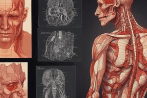
Skin & Subcutaneous Tissue
- Skin is the body's largest organ
Skin
- The skin is approximately 7% of body weight
- It is typically 1.5 to 4.4 mm thick
- Has two layers: the epidermis and dermis
- The subcutaneous tissue lies beneath the dermis
Hypodermis
- The subcutaneous layer lies deep to the dermis.
- It is composed of areolar and adipose tissue.
- Functions of the hypodermis include insulation, shock absorption, energy storage, and anchoring.
Epidermis Layers
- Stratum corneum: outermost layer of dead keratinocytes
- Stratum lucidum: found in thick skin (palms/soles), comprised of flat, dead keratinocytes
- Stratum granulosum: few layers of keratinocytes; keratinohyaline granules form keratin; lamellar granules have waterproofing glycolipid; cells lack nutrient access.
- Stratum spinosum: spiny appearance; intermediate filaments for strength and flexibility; contains keratinocytes and dendritic cells (part of the immune system); provides mechanical stress resistance.
- Stratum basale (stratum germinativum): deepest layer; actively dividing cells; contains tactile epithelial cells (Merkel cells) and melanocytes.
- Mnemonic: "Come, Let's Get Sun Burned"
Main Cell Types of the Epidermis
- Keratinocytes: most abundant; produce keratin for strength and water resistance; produce antibiotics and enzymes; dead at the skin's surface
- Melanocytes: found in the stratum basale; protect from UV damage; determine skin, hair, and eye color
- Eumelanin: dark pigment
- Pheomelanin: lighter pigment
- Tactile epithelial cells (Merkel cells): mechanoreceptors for light touch and pressure; located in the basal layer
- Dendritic cells (Langerhans cells): part of the immune system; monitor and process pathogens; protect against infections and skin diseases
Functions of the Skin
- Protection: from abrasion and penetration
- Body temperature regulation: through sweat; vasodilation or vasoconstriction
- Excretion: of waste products in perspiration
- Production of vitamin D: using UV radiation
- Sensory reception: through nerve endings
Dermis
- A strong, flexible connective tissue,
- Rich with blood vessels and nerves; has two layers
- Papillary dermis: superficial layer; includes dermal papillae; increases surface area for gas, nutrient, and waste exchange; nourishes epidermis via capillaries; contains receptors for light touch and vibration; regulates body temperature;
- Reticular dermis: deeper layer; 80% of dermis tissue; dense irregular connective tissue; rich nerve supply for pressure and pain; vascular plexuses for nutrient delivery and temperature regulation; contains the dermal plexus, subpapillary plexus.
Cleavage Lines and Flexure Lines
- Cleavage lines: separations between collagen fibers that provide skin strength. Surgical incisions parallel to these lines heal better.
- Flexure lines: deep creases in areas where the dermis tightly attaches to underlying structures (e.g., palms, fingers, soles.)
Hypodermis
- Located beneath the skin
- Contains areolar and adipose tissues
- Anchors skin, insulates, stores energy, and provides cushioning and protection
- Distribution varies by sex
Skin Color
- Melanin: main pigment, made from tyrosine
- Carotene: yellow-orange pigment from foods
- Hemoglobin: oxygenated blood gives a rosy undertone in light-skinned individuals.
Appendages of the Skin
-
Nails: scale-like epidermal modifications made of hard keratin; free edge extends past the fingertip; nail plate covers the nail bed; root is at the base of the nail; nail folds skin around the nail; eponymia (cuticle)
-
Hair: flexible strand of dead keratinized cells; found everywhere except palms and soles; root is embedded in the skin; shaft projects above skin; made of tough keratin; 3 concentric layers - medulla, cortex, and cuticle
Hair Structure
- Three layers of keratinized cells: medulla (central core), cortex (surrounds medulla), and cuticle (outermost layer)
Additional Hair Structures
- Hair follicles: structure in skin where hair grows
- Hair bulb: base containing the hair matrix for hair and pigment production.
- Root plexus: Sensory nerve network around the hair bulb.
Arrector Pili Muscle
- Smooth muscle attached to hair follicles
- Causes hair to stand erect (goosebumps)
- Thermoregulation and Protection.
Types and Growth of Hair
- Vellus hairs: fine, short, lightly pigmented; cover body; light insulation; sensory perception; present in children and adults
- Terminal hairs: thick, coarse, pigmented; develop during puberty; found on scalp, eyebrows, eyelashes, armpits and groin; provides insulation, protection, and secondary sexual characteristics.
- Lanugo: fine, soft, unpigmented hair; covers the fetus in the womb; shed before or shortly after birth; provides warmth and protection.
Hair Thinning and Baldness
- Androgenetic Alopecia: hereditary, hormone-driven thinning at the crown and hairline (men) or overall thinning (women)
- Alopecia Areata: autoimmune disorder, patchy hair loss on the scalp and body
- Telogen Effluvium: temporary thinning due to stress, illness, or hormonal changes.
- Age-Related Thinning: gradual thinning with age, influenced by hormones and genetics.
Sebaceous Glands
- Found everywhere except palms and soles
- Secrete sebum via holocrine secretion
- Associated with hair follicles
- Functions of sebum include lubrication, barrier, antimicrobial properties, and hair conditioning.
Sudoriferous Glands (Sweat Glands)
- Widely distributed on the body
- Secrete sweat, mostly water; salts and metabolic wastes
- Two types of sweat glands:
- Eccrine glands: most abundant; found on palms, soles, and forehead; produce odourless watery sweat; small coiled glands open onto skin surface; thermoregulation, skin hydration, microbial defence
- Apocrine glands: found in axillary, anal, and genital areas; open into hair follicles; activated during puberty; secrete thick milky sweat with musky odour.
Modified Apocrine Glands
- Ceruminous glands: produce earwax, protect the ear
- Mammary glands: produce milk; activated by hormones
- Ciliary glands: lubricate eyelashes and prevent dry eyes.
Integumentary System Conditions
- Burns (first, second, third degree)
- Skin cancer (Basal cell carcinoma, Squamous cell carcinoma, Melanoma)
- Infections (acne vulgaris, warts, ringworm)
Aging Skin
- Skin thins and becomes less elastic
- Skin inflammations become more common
- Prone to rashes and infections
Summary
- The integumentary system protects the body from external harm and regulates temperature.
- It includes the skin, hair, nails, and glands.
- It serves as a barrier and sensory interface.
- Aging leads to changes in skin and decreased wound healing.
Studying That Suits You
Use AI to generate personalized quizzes and flashcards to suit your learning preferences.