Podcast
Questions and Answers
Which of the following components are directly involved in facilitating movement in higher animals?
Which of the following components are directly involved in facilitating movement in higher animals?
- Nervous system
- Muscles
- Both A and B (correct)
- Skeleton
What primary function does the human skeletal system perform?
What primary function does the human skeletal system perform?
- Providing shape, support, and protection to the body (correct)
- Regulating body temperature
- Facilitating gas exchange
- Producing blood cells
What characterizes the occurrence of an endoskeleton?
What characterizes the occurrence of an endoskeleton?
- It is exclusively found in invertebrates.
- It is found in all living organisms.
- It lies inside the body and is found in vertebrates. (correct)
- It is an external frame made of non-living material.
Which materials primarily compose the endoskeleton in vertebrates?
Which materials primarily compose the endoskeleton in vertebrates?
If a developing bone fails to undergo ossification, which nutrient deficiency might be the cause?
If a developing bone fails to undergo ossification, which nutrient deficiency might be the cause?
What is the role of osteoblasts in bone tissue?
What is the role of osteoblasts in bone tissue?
In a long bone, what is the function of the medullary cavity?
In a long bone, what is the function of the medullary cavity?
Which component primarily constitutes the outer walls of the diaphysis in a long bone?
Which component primarily constitutes the outer walls of the diaphysis in a long bone?
What is the role of the epiphyseal plate in long bones?
What is the role of the epiphyseal plate in long bones?
Which cell type is responsible for resorbing or breaking down bone during bone remodeling?
Which cell type is responsible for resorbing or breaking down bone during bone remodeling?
How do osteocytes receive their blood supply in spongy bone?
How do osteocytes receive their blood supply in spongy bone?
Which statement accurately describes articular cartilage?
Which statement accurately describes articular cartilage?
Which characteristic distinguishes fibrocartilage from other types of cartilage?
Which characteristic distinguishes fibrocartilage from other types of cartilage?
Where is elastic cartilage typically found in the human body?
Where is elastic cartilage typically found in the human body?
What is the total number of bones present in the adult human vertebral column after fusion?
What is the total number of bones present in the adult human vertebral column after fusion?
Which bone provides attachment for the tongue and muscles of the oral cavity?
Which bone provides attachment for the tongue and muscles of the oral cavity?
What is the primary function of the auditory ossicles?
What is the primary function of the auditory ossicles?
Which statement accurately describes 'true ribs'?
Which statement accurately describes 'true ribs'?
Which bones comprise the pectoral girdle?
Which bones comprise the pectoral girdle?
Which classification describes joints that are immovable?
Which classification describes joints that are immovable?
Which type of joint is exemplified by the sutures found in the skull?
Which type of joint is exemplified by the sutures found in the skull?
Which of the following is an example of a cartilaginous joint?
Which of the following is an example of a cartilaginous joint?
Which feature defines a synovial joint?
Which feature defines a synovial joint?
Which type of synovial joint allows movement in one plane, such as flexion and extension?
Which type of synovial joint allows movement in one plane, such as flexion and extension?
Why is cartilage slow to heal compared to bone?
Why is cartilage slow to heal compared to bone?
What is a primary characteristic of smooth muscles?
What is a primary characteristic of smooth muscles?
Which type of muscle is responsible for forming the muscular walls of the heart?
Which type of muscle is responsible for forming the muscular walls of the heart?
What structural feature is unique to cardiac muscle fibers?
What structural feature is unique to cardiac muscle fibers?
What property enables skeletal muscles to rapidly revert to their original conformation after contraction?
What property enables skeletal muscles to rapidly revert to their original conformation after contraction?
Which layer directly encases individual muscle fibers inside a fascicle?
Which layer directly encases individual muscle fibers inside a fascicle?
Flashcards
Musculo-skeletal System
Musculo-skeletal System
System where skeleton and muscles work together for locomotion, coordinated by the nervous system.
Endoskeleton
Endoskeleton
Internal support structure of animals, providing shape, support, protection, and movement.
Ossification
Ossification
Process where cartilage hardens into bone through the deposition of osteoblasts and calcium phosphate.
Diaphysis
Diaphysis
Signup and view all the flashcards
Epiphysis
Epiphysis
Signup and view all the flashcards
Osteocytes
Osteocytes
Signup and view all the flashcards
Cartilage
Cartilage
Signup and view all the flashcards
Hyaline Cartilage
Hyaline Cartilage
Signup and view all the flashcards
Fibrocartilage
Fibrocartilage
Signup and view all the flashcards
Elastic Cartilage
Elastic Cartilage
Signup and view all the flashcards
Skull
Skull
Signup and view all the flashcards
Auditory Ossicles
Auditory Ossicles
Signup and view all the flashcards
Vertebral Column
Vertebral Column
Signup and view all the flashcards
Appendicular Skeleton
Appendicular Skeleton
Signup and view all the flashcards
Pectoral Girdle
Pectoral Girdle
Signup and view all the flashcards
Pelvic Girdle
Pelvic Girdle
Signup and view all the flashcards
Joint
Joint
Signup and view all the flashcards
Sutures
Sutures
Signup and view all the flashcards
Fibrous Joints
Fibrous Joints
Signup and view all the flashcards
Gomphoses
Gomphoses
Signup and view all the flashcards
Syndesmoses
Syndesmoses
Signup and view all the flashcards
Cartilaginous Joint
Cartilaginous Joint
Signup and view all the flashcards
Synovial Joint
Synovial Joint
Signup and view all the flashcards
Hinge Joint
Hinge Joint
Signup and view all the flashcards
Plane Joint
Plane Joint
Signup and view all the flashcards
Pivot Joint
Pivot Joint
Signup and view all the flashcards
Arthritis
Arthritis
Signup and view all the flashcards
Smooth Muscle
Smooth Muscle
Signup and view all the flashcards
Skeletal Muscle
Skeletal Muscle
Signup and view all the flashcards
Cardiac Muscle
Cardiac Muscle
Signup and view all the flashcards
Study Notes
- Higher animals use skeletons and muscles to work together for locomotion.
- Muscles attach to the skeleton to form either a skeletal muscles system or musculo-skeletal system.
- The nervous system coordinates both systems.
- Animal bodies have a framework known as the skeleton.
- A skeletal system provides shape, support, protection, and movement power.
Endoskeleton
- Lyes inside the body and is found in some invertebrates like sponges and starfish.
- It's the complex skeleton of vertebrates
- Internal framework composed of bone and cartilage.
- Cartilage and bone articulate via joints, allowing movement by muscle contraction.
- The endoskeleton of vertebrates grows in correlation with the animal.
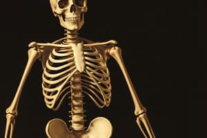
Human Skeleton Introduction
- Human endoskeleton has 206 bones connected by joints
Composition of Human Skeleton
Bones
- Rigid connective tissue forming the skeleton and limbs.
- Bone cells secrete a matrix with a collagen network.
- Bone cells include osteoblasts, osteocytes, osteogenic cells, and osteoclasts.
Ossification Requirements
- Hardening of bone matrix by osteoblast activity and calcium phosphate deposition (ossification or calcification).
- Requires vitamin D for calcium absorption.
- Milk provides proteins and minerals for hard, elastic bones.
Gross Anatomy of Bones
- Long bones contain a diaphysis and epiphysis.
- Diaphysis Description: Hollow, tubular and shaft between bone ends, holds the medullary cavity, which is filled with yellow marrow in adults.
- Diaphysis outer walls (cortex or cortical bone) consist of dense, hard compact bone which is a type of osseous tissue.
- Epiphysis Description: Wider section at bone ends, contains spongy bone.
- Epiphysis Description: Has red bone marrow in some long bones.
- Metaphysis Description: Site of growth with the epiphyseal plate when young and epiphyseal line when old.
Bone Tissue Structure
- Compact and spongy bone differ in density or packing
- Osteoblasts create bone.
- Osteoclasts break down bone.
- Osteocytes are mature bone cells.
- Equilibrium between osteoblasts and osteoclasts maintains bone tissue.
Compact Bone
- Made of packed osteons (Haversian systems) with a central osteonic (haversian) canal.
- Canal surrounded by concentric matrix rings (lamellae).
- Bone cells (osteocytes) reside in lacunae between matrix rings.
- Canaliculi (small channels) radiate from lacunae to the osteonic canal provide matrix passageways.
- Blood vessels interconnect osteonic canals via surface-connecting perforating canals.
Spongy (Cancellous) Bone
- It's lighter and less dense than compact bone.
- Consists of trabeculae (plates and bars) adjacent to cavities containing bone marrow.
- Canaliculi connect to adjacent cavities instead of a central canal for blood supply.
Bone Cells
- Bone cells make up less than 2% of bone mass, but are essential for bone function.
- Osteogenic cells develop into osteoblasts inside the endosteum.
- Osteoblasts formation inside the endosteum.
- Osteocytes Maintain mineral concentration of matrix while inside the matrix.
- Osteoclasts perform bone resorption along the endosteum.
Cartilage
- Flexible connective tissue that stands apart from bone.
- Primary cell type is chondrocytes instead of osteocytes.
- Chondrocytes comes from chondroblast and then produce the collagen extracellular matrix (ECM) and get trapped in it.
- Chondrocytes are in lacunae and obtain nutrients via diffusion, lacking blood vessels (avascular).
- Slow healing of cartilage is caused by the absence of proper blood supply.
- The cartilage's base substance contains chondroitin sulfate.
- Cartilage organization is less ordered than bone.
- The fibrous sheath that covers cartilage is called the perichondrium.
- Cartilage grows slowly through cell division, not size or mass increase.
- Articular cartilage relies on ECM's proteoglycan and collagen composition.
- Cartilage matrix remodeling responds to its tensile and compressive forces
Types of Cartilage
- Hyaline cartilage, fibrocartilage, and elastic cartilage are major types.
Hyaline Cartilage
- It's the most common cartilage type, is found on articular surfaces, rib tips, trachea rings, and skull parts.
- Predominantly collagenous, with a glassy texture.
- Bones Initially form as ossifying hyaline cartilage in embryos.
- Hyaline Cartilage is covered by perichondrium.
- Hyaline Cartilage does not have any nerves or blood vessels
- It's cells are rounded grouped in granular/homogeneous matrix
- They consist of translucent protoplasm
Fibrocartilage
- Contains lots of collagen fibers (types I and II).
- Grades into dense tendon and ligament tissue.
- White fibrocartilage combines fibrous and cartilaginous tissues.
- Flexibility and toughness are from fibrous tissue, elasticity from cartilaginous tissue.
- Unique cartilage with both type I and type II collagen
- Found in public symphysis, intervertebral discs, menisci, and the temporomandibular joint.
Elastic Cartilage
- It consists of elastin and collagen fibers network
- The main protein is elastin.
- Histologically similar to hyaline cartilage but contains many yellow elastic fibers in a matrix
- Great flexibility to withstand bending.
- Found in the epiglottis in the larynx.
Division of Human Skeleton
- Axial and appendicular skeletons exist in humans.
Axial Skeleton
- Protective in nature, containing skull, vertebral column, and rib cage bones.
Skull
- Cranium protects brain.
- It consists of 8 bones: occipital, two temporals, two parietals, sphenoid, ethmoid, and frontal bones.
- Consists of 14 facial bones: vomer, two inferior nasal conchae, two nasals, two maxillae, mandible, two palatines, two zygomatics, and two lacrimals.
- There are 6 auditory ossicles (malleus, incus, stapes) in each ear which transfers air vibrations into the inner ear as sound.
- Hyoid bone Description: Is a U-shaped throat bone is a U-shaped and provides tongue/oral cavity muscle attachments.
Vertebral Column
- Spine of 33 bones (26 after fusion) that protects the spinal cord.
- 7 cervical vertebrae are found in the neck.
- 12 thoracic vertebrae articulate in the chest.
- 5 lumbar vertebrae are the vertebrae in the lower back.
- 5 sacral vertebrae fuse to form the sacrum, articulating with the pelvic girdle.
- 4 coccygeal vertebrae are found in the tail.
- The atlas and axis vertebrae (top two cervicals) permit skull movement.
- Cartilaginous discs support/protect the vertebrae.
Ribcage
- Contains 12 pairs of bones.
- Protects the chest cavity, heart, and lungs.
- Semicircular bones connected to vertebrae dorsally, sternum ventrally.
- There are a total of 7 pairs of true ribs where costal cartilages join to the sternum anteriorly.
- There are a total of 5 pairs of false ribs, this means, the costal cartilages do not articulate directly with the sternum.
- The floating ribs are not connected to the sternum.
Appendicular Skeleton
- It consists of 126 bones, including arms, legs, and pectoral and pelvic girdles.
Pectoral Girdle
- Shoulder region, consists of the scapula and clavicle.
Upper Limb
- Humerus joints to the scapula by ball and socket joint.
- It consists of 30 bones (60 in both upper limbs).
- Radius and ulna are the bones below the humerus.
- Description: There are 8 carpals, 5 metacarpals, and 14 phalanges are small bones arranged in 5 rows.
Pelvic Girdle
- Hip bone consists of the ilium, ischium, and pubic bone.
- Coxal bone (hip bone) is a pair of bones joined to the sacrum of the vertebral column.
- Ilium Description: The largest part of the coxal bone, forming the upper part with a fan-like structure, and attaches to the vertebral column sacrum.
- Ischium Description: The posterior part of the pelvic girdle below the ilium providing support while sitting.
- Pubis Description: Lower anterior part of the pelvic girdle.
- Description: Pubic symphysis- The joint between the two hip bones in the middle portion containing fibrous cartilage.
- Hip joint's main role is to support body weight and facilitate force transmission from the axial skeleton to lower parts.
Lower Limb
- Consist of 30 bones (60 in both lower limbs).
- Femur Description: Largest bone in the leg which is connected to the pelvis.
- There are tibia and fibula, 7 tarsals, 5 metatarsals, and 14 phalanges below.
Joints
- Connect bones in the skeletal system.
- Can be classified on tissue type or movement degree.
Classification by Tissue Type
- Fibrous joints connected by fibrous tissue
- Cartilaginous joint connected by cartilage
- Synovial Joint articulating surfaces enclosed within a fluid-filled joint capsule.
Classification by Movement Degree
- Synarthrosis Joint, immovable
- Amphiarthrosis Joint, slightly movable.
- Diarthrosis Joint, freely movable.
Fibrous Joints
- Bones linked by a tough, fibrous tissue.
- These joints typically require strength and stability over range of movement.
- Includes sutures, gomphoses, and syndesmose.
Sutures
- Immovable joints (synarthrosis) between flat, plate-like skull bones.
- Limited movement until ~20 years, become fixed and immobile after that.
- These joints allow skull deformation during birth
Gomphoses
- Immovable joints where teeth articulate with sockets in the maxilla (upper teeth) or mandible (lower teeth).
- The tooth is bound into its socket by the solid periodontal ligament.
Syndesmose
- Slightly movable joints (amphiarthroses) made of bones with an interosseous membrane.
- The middle radioulnar joint and middle tibiofibular joint are examples of a syndesmosis joint.
Cartilaginous Joint
- Bones joined by either fibrocartilage or hyaline cartilage.
- There are 2 types: synchondroses and symphyses.
Synchondroses
- Bones connected by hyaline cartilage which is immovable (synarthrosis).
- Example: joint between diaphysis and epiphysis of a growing long bone.
Symphyses
- The bones are united by fibrocartilage, so they are slightly movable joints (amphiarthrosis).
- The pubic symphysis and intervertebral joints are examples.
Synovial Joint
- Contains fluid-filled joint cavity inside a fibrous capsule.
- Freely movable (diarthrosis) and most common.
- Sub-classified by shape and movement permitted: hinge joints, saddle joints, plane joints, pivot joints, condyloid joints, and ball and socket.
- Hinge Joint permits movement in one plane like flexion/extension from elbow, ankle, knee joints.
- Saddle Joint resembles a saddle on a horse's back and characterized by opposing articular surfaces with a reciprocal concave-convex shape in carpometacarpal joints
- Plane Joints the articular surfaces are relatively flat which allows the bones to glide over one another in the acromioclavicular joint and subtalar joint.
- Pivot Joints allows only rotation and is formed by a bony ligamentous ring which is in a bony pivot like proximal, distal radioulnar and atlantoaxial joints.
- Condyloid Joint has contains a convex surface which articulates with a concave elliptical cavity, which they are considered as ellipsoid joints such as in wrist joints.
- Ball and Socket Joint where the ball-shaped surface of one rounded bone fits into a cup-like depression to permit free movement as in the hip and shoulder joint.
Arthritis
- Is when the joint becomes swollen, painful and immovable
Arthritis Types
- Osteoarthritis breaks down joint cartilage and is the common form of arthritis
- Ankylosing spondylitis: Spinal arthritis in the lower back.
- Juvenile arthritis (JA): Attacks tissue surrounding joints; commonly affects those 16 or younger.
- Gout creates hard crystals of uric acid in joints.
- Psoriatic arthritis is a joint inflammation related to psoriasis.
- Rheumatoid arthritis causes immune system attacks on synovial membranes.
Clinical Effects
- Calcium deposits leading to inflexibility.
Treatment
- Artificial joint replacement or use of medicines is used.
Muscular System Introduction
- Muscles form muscular system in vertebrates.
- Muscles are made of muscular tissue.
- Each muscle cell has unique function to create force during shortens.
- Contraction force or pulling force helps to perform different functions.
- The muscular system partners with the skeletal system in movement.
- Humans have over 600 muscles contributing half of the body weight.
- Men and women usually have the same bones and muscles.
Muscular System Functions
- Movement of eyelids, tongue, and food within the alimentary
- Activity of internal heart and internal circulation.
- Excretion of excretions from the body
- Important substances are secreted in specific locations.
- They cause exchange in the body
Muscle Types
- Smooth, cardiac, and skeletal are the 3 types
Smooth Muscles
- Structurally simple
- They consists of long spindle shaped.
- They are uninucleated.
- Each muscle has cytoplasm
- The nucleus contains granular cytoplasm with elongated fibres called myofibrils.
- Blood vessels, digestive tract, and other organs contain these muscles.
- Smooth muscles are involuntary and perform autonomously.
- These muscles are controlled by the autonomic nervous system
- They contract slowly and can be used for long periods of time.
- It pushes food down the alimentary
- They helps in waste removal from urine
- It adjusts the blood's size
- They adjust pupil size for sufficient light entry.
- Smooth muscles cause erection of hairs.
Skeletal Muscles
- Attaches to the skeleton to enable movement.
- Under microscope skeleton muscles look striped, which are striated muscles called called so due to their striped appearance.
- It contains of protein filaments
- Light/dark bands exist and regulated voluntarily.
- striated voluntary muscles is what they are known as.
- Contractions occur due to the central nervous system.
- Under central nervous system control as they are able to rapidly go back to their normal position.
- Some attach directly to bone outer layers, others connecting to tendon tissue.
- Ligaments are connect bones together.
- Muscle contraction moves the stretched tendon and bone.
- Contraction is needed for at least two joints so movement occurs.
- Origin is the stationary attachment point and insertion is the moving attachment point.
Cardiac Muscles
- Construct only the muscular walls in the heart of vertebrataes.
- Uni- or bi-nucleated and branched to form contractile tissue meshwork connected by Intercalated Discs.
- The fibers will not separate.
- Heart walls handles high stress, without the risk of damage.
- Heart chambers contract due to the meshwork of heart muscle fibers, maintaining blood flow.
- Includes more mitochondria and the actin and myosin proteins to sustain continuous energy supply for the tissue of the heart.
- Contract and relax rhythmically in a heartbeat pattern
- It's regulated by the sino-atrial node (SAN), which is the pace maker.
- The impulse caused by the pace maker spreads through the heart, without involving the central nervous system
Structure of Skeletal Muscles
- Each muscle is has a 3 layers of connective tissue ("mysia") that provides structure and encloses the muscle with muscle compartment.
- Epimysium: Irregular connective tissue sheath around muscle, which allows a muscle to contract and move powerfully while maintaining its structural integrity.
- Perimysium: Irregular connective tissue sheath around fascicles inside, it enables specific movements via nervous system.
- Endomysium: Encases each muscle fiber which are involved in collagen and reticular fibers.
- Endomysium contains extracellular fluid and nutrients supplied by capillaries.
- Each muscle consists of aligned, elongated muscle fiber structures.
- Each fiber is a complete cell having sarcoplasm (cytoplasm) and surrounded by sarcolemma (outer membrane).
- Specialized sites of skeletal motor axons of the muscle fiber creates the NMJ (neuromuscular junction) where the muscle fiber first starts indicating to the motor neuron.
- Sarcolemma in the neuromuscular junction contains transmembrane proteins receiving electrical signals.
Composition of Muscle Fibers
- Each fiber consists of many contractile myofibrils.
- Fibrils are composed of a chain of sarcomeres which are the muscle tissue units that contract.
- Sarcoplasmic reticulum (SR) surrounds each myofibril .
- Sarcoplasmic reticulum (SR) Description: The ER that store,releases, and retrieves calcium ions (Ca++).
- Motor releases cause release of calcium ions (Ca++) from the Sarcroplasmic Reticulum.
- The sarcolemma has T-tubules to reach the SR, which means that the signals given from the motor neuron will reach the membrane of the SR.
- Z line separates the sarcomeres.
- Dark and light zones make up the sarcomeres.
- Striated muscles are made of sarcomere light and dark patterns.
Myofilaments and its Types
- Composed of microfilaments.
- Includes thick and thin filaments.
Thick Filament
- Composed of myosin.
- Myosin myofilaments contain a tail and functional heads.
- tails Description: Connect myosin myofilaments.
- heads Description: The active role is in muscle contraction and contains an actin-binding and ATP-binding part.
Thin Filament
- Six thin filaments surround the myosin.
- Composed of actin, tropomyosin, and troponin.
- Troponin consists of polypeptide.
- Troponin I (Tnl) binds to actin.
- Troponin T (TnT) binds to tropomyosin.
- Troponin C (TnC): Binds to calcium ions.
- Troponin and tropomyosin controls actin binding sites.
Overlapping Pattern of Thin and Thick Filaments
- Bands caused by overlapping filaments.
- I-bands (isotropic) possess a pair of I-bands which are outer areas on each sarcomere and it contains only active filaments.
- A-bands: (Anisotropic): The dark and are located outside to each and can be only located on I cells .
- H-bands: (Helle): Lightly staining A-band center zone it contains only the thick filaments.
- Thick filaments have cross bridges attached to the actin filaments.
Mechanism of Contraction of Skeletal Muscles
- Muscle contraction occurs when a muscle activates and becomes shorter.
Explanation: Huxley sliding filament theory
- Thin and thick filaments slide together during muscle contraction.
- The sliding over the filaments is the reason that the muscles get shorter and thicker.
Cross Bridge Cycle
- Nerves causes to release of calcium released (Ca++) from SR surrounding miofibrils, causing change in the troponin molecules, enabling the interaction with actin
- Head regions are attached to sites of actin forming cross links and releases in inorganic phosphate
- ADP is released and starts the stroke, and moving the filament in the mesomere
- ATP bounds to myosin head regions causing the removing of cross lines after hydrolysis to cause moving to trigger the new cross linking , stopping the creation, after the new signal occurs
Studying That Suits You
Use AI to generate personalized quizzes and flashcards to suit your learning preferences.