Podcast
Questions and Answers
What are the three types of muscle cells?
What are the three types of muscle cells?
Skeletal, Cardiac, Smooth
What is the role of the neuromuscular junction?
What is the role of the neuromuscular junction?
It transmits signals from motor nerve endings to muscle fibers.
Describe the structure of skeletal muscle.
Describe the structure of skeletal muscle.
Composed of muscle fibers organized by epimysium, perimysium, and endomysium.
What is the triad system in skeletal muscle?
What is the triad system in skeletal muscle?
What is the function of myofibrils?
What is the function of myofibrils?
Which proteins are involved in muscle contraction?
Which proteins are involved in muscle contraction?
What autoimmune disease is characterized by fluctuating weakness of striated muscles?
What autoimmune disease is characterized by fluctuating weakness of striated muscles?
The I band decreases in size during muscle contraction.
The I band decreases in size during muscle contraction.
The dark band in myofibrils is called the ______.
The dark band in myofibrils is called the ______.
What is the main cause of weakness in myasthenia gravis?
What is the main cause of weakness in myasthenia gravis?
Flashcards are hidden until you start studying
Study Notes
Muscle Cell Types
- Three types of muscle cells: skeletal, cardiac, and smooth.
- Skeletal muscle is voluntary and innervated by motor nerves.
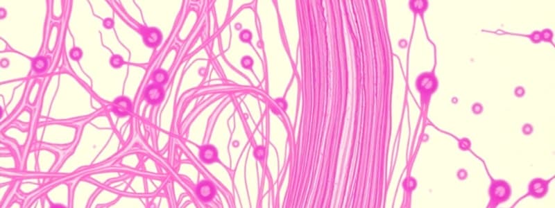
Neuromuscular Junction (NMJ)
- NMJ formed by the synapse of a motor nerve terminal with a muscle fiber.
- Presynaptic terminal contains mitochondria and synaptic vesicles filled with acetylcholine.
- Postsynaptic side features deep folds in the sarcolemma, rich in acetylcholine receptors to enhance surface area.
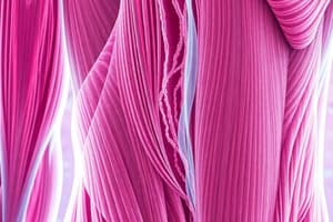
Structure of Skeletal Muscle
- Composed of muscle fibers organized by connective tissue:
- Epimysium: dense connective tissue surrounding the entire muscle.
- Perimysium: less dense connective tissue encasing bundles of muscle fibers (fascicles).
- Endomysium: delicate, highly vascular connective tissue surrounding individual muscle fibers.
- Muscle fibers are long, parallel, and exhibit transverse striations.
Histological Features
- Longitudinal sections show multiple, peripherally located flat nuclei within muscle fibers, separated by connective tissue.
- Myofibrils serve as contractile units within muscle cells.
Cytoplasmic Components
- Myofibrils are the key contractile structures of skeletal muscle.
- Sarcoplasmic reticulum is a well-developed smooth endoplasmic reticulum surrounding myofibrils.
- Mitochondria are abundant, providing energy.
- Myoglobin functions as an oxygen-binding protein.
- Glycogen and lipid granules are present as energy stores.
Triad Tubular System (T-system)
- The T-system comprises T tubules and terminal cisternae of the sarcoplasmic reticulum, encircling each myofibril.
- T tubules facilitate the rapid transmission of nerve impulses throughout the muscle, enabling synchronized contraction.
Role of the Triad System
- Impulse transmission through T tubules triggers calcium release from the sarcoplasmic reticulum during muscle contraction and helps in calcium sequestration during relaxation.
Myofibril Structure
- Myofibrils exhibit a pattern of dark (A bands) and light (I bands) striations due to the arrangement of contractile proteins.
- A bands contain both actin and myosin filaments, while I bands consist solely of actin.
- The H zone is the central pale part of the A band, containing thick filaments only.
- The Z line bisects the I band and defines the boundaries of a sarcomere, the functional unit of contraction.
Contraction Process
- During contraction:
- I bands decrease in size.
- H zone shortens or disappears.
- A band remains the same length.
Clinical Application: Myasthenia Gravis
- An autoimmune disorder marked by muscle weakness that worsens with activity.
- Pathophysiology involves autoantibodies targeting acetylcholine receptors, reducing their availability and impairing neuromuscular transmission.
Studying That Suits You
Use AI to generate personalized quizzes and flashcards to suit your learning preferences.