Podcast
Questions and Answers
What is the purpose of boiling the protein samples at 70°C after adding Laemmli Sample Buffer?
What is the purpose of boiling the protein samples at 70°C after adding Laemmli Sample Buffer?
- To promote the protein crystallization process
- To activate the proteases for better protein manipulation
- To denature the proteins and prevent protease activity (correct)
- To enhance the protein's tertiary structure
What should the proportion of Laemmli Sample Buffer be in relation to the total sample volume?
What should the proportion of Laemmli Sample Buffer be in relation to the total sample volume?
- Equal to the total sample volume
- 1/4 of the total sample volume (correct)
- 1/2 of the total sample volume
- 1/3 of the total sample volume
What is the role of SDS in the sample preparation process?
What is the role of SDS in the sample preparation process?
- To stain the proteins during electrophoresis
- To stabilize the protein samples during storage
- To break up the protein structure and add negative charge (correct)
- To enhance the visibility of the protein bands
Why must the power be turned off before removing the gels from the electrophoretic apparatus?
Why must the power be turned off before removing the gels from the electrophoretic apparatus?
What is the ideal voltage used for running the gels during electrophoresis?
What is the ideal voltage used for running the gels during electrophoresis?
During the destaining process, how often should the destain solution be changed?
During the destaining process, how often should the destain solution be changed?
What precaution should be taken when using 2-mercaptoethanol in Laemmli Sample Buffer?
What precaution should be taken when using 2-mercaptoethanol in Laemmli Sample Buffer?
What is the first step after electrophoresis is completed?
What is the first step after electrophoresis is completed?
What is the primary purpose of using SDS in SDS-PAGE?
What is the primary purpose of using SDS in SDS-PAGE?
Which of the following statements about the migration of proteins during SDS-PAGE is true?
Which of the following statements about the migration of proteins during SDS-PAGE is true?
What role do reducing agents play in the SDS-PAGE process?
What role do reducing agents play in the SDS-PAGE process?
What is the significance of using a protein ladder during SDS-PAGE?
What is the significance of using a protein ladder during SDS-PAGE?
What type of gel is used in the SDS-PAGE technique?
What type of gel is used in the SDS-PAGE technique?
Which of the following components is NOT part of the Laemmli Sample Buffer?
Which of the following components is NOT part of the Laemmli Sample Buffer?
Why do low molecular weight proteins migrate faster during SDS-PAGE compared to high molecular weight proteins?
Why do low molecular weight proteins migrate faster during SDS-PAGE compared to high molecular weight proteins?
What does the term 'electrophoresis' refer to in the context of SDS-PAGE?
What does the term 'electrophoresis' refer to in the context of SDS-PAGE?
Flashcards
SDS-PAGE
SDS-PAGE
A technique used to separate proteins based on their size and charge.
Laemmli Sample Buffer
Laemmli Sample Buffer
A solution that disrupts protein structure by adding negative charges, making them migrate towards the positive electrode.
Running Buffer
Running Buffer
A solution that allows proteins to move through the gel during electrophoresis.
What is SDS-PAGE?
What is SDS-PAGE?
Signup and view all the flashcards
Protein Ladder
Protein Ladder
Signup and view all the flashcards
Tracking Dye
Tracking Dye
Signup and view all the flashcards
What is electrophoresis?
What is electrophoresis?
Signup and view all the flashcards
Boiling
Boiling
Signup and view all the flashcards
What is a discontinuous polyacrylamide gel?
What is a discontinuous polyacrylamide gel?
Signup and view all the flashcards
Coomassie Blue
Coomassie Blue
Signup and view all the flashcards
What is SDS?
What is SDS?
Signup and view all the flashcards
What is β-mercaptoethanol?
What is β-mercaptoethanol?
Signup and view all the flashcards
Destain Solution
Destain Solution
Signup and view all the flashcards
What is a protein ladder?
What is a protein ladder?
Signup and view all the flashcards
What is running buffer?
What is running buffer?
Signup and view all the flashcards
What is sample preparation?
What is sample preparation?
Signup and view all the flashcards
Study Notes
MD100 Medical Biochemistry I - Lab Exercise 3: Introduction to SDS PAGE
- Course: Medical Biochemistry I
- Lab Exercise: Introduction to SDS PAGE - Protein identification and characterization
- Semester: Fall 2024
- Institution: European University Cyprus, School of Medicine
Objectives
- SDS-PAGE: Introduction to the laboratory technique
- Theoretical Background: Principle of SDS-PAGE
- Part A: Sample preparation & dilutions
- Part B: Sample loading & gel running
- Part C: Gel staining & destaining
- Part D: Protein gel analysis & protein identification
Introduction to SDS-PAGE
-
Technique: Used to separate proteins based on their molecular weight.
-
Electrophoresis: The separation of macromolecules in an electric field
-
SDS-PAGE Medium: Uses discontinuous polyacrylamide gel as the support medium. Sodium dodecyl sulfate (SDS) is used to denature the proteins.
-
SDS Principle: A negatively charged molecule migrates to the electrode within the electric field with an opposite polarity. SDS denatures and binds to proteins, resulting in uniform negative charging to ensure all components in the sample will move through the gel towards the positive electrical pole.
-
Reducing Agents: β-mercaptoethanol is used to reduce disulfide bonds within the proteins, further causing denaturation alongside SDS.
The Principle of SDS-PAGE
-
Protein Structure: Before SDS-denaturation, proteins exist in a folded 3D structure from positive and negative charged amino acids.
-
Disulfide Bonds: Proteins usually have disulfide bonds connecting certain amino acids. SDS alongside a reducing agent (like β-mercaptoethanol), unfolds the linear polypeptide chain while breaking the bonds.
-
SDS Binding: The denatured linear structure is then bound by anionic detergent SDS, creating a strong negative charge proportional to the polypeptide chain length.
-
Migration: When an electric field is applied, all SDS-bound proteins migrate through the gel towards the positively charged electrode, separating proteins based on the size and length of their polypeptide chain (molecular weight).
Protein Identification
- Albumin: Molecular weight: 66.5 kDa
- Casein: Molecular weight: 24 kDa
Materials & Equipment
- Gels: Prepared in lab or precast gels purchased from the market
- Electrophoresis Chamber: Vertical Gel Electrophoresis Chamber
- Protein Samples: Samples of interest to be studied.
- Buffers: Running Buffer (Tris/Glycine/SDS), Staining, & Destaining Buffer
- Protein Ladder: Prestained protein molecular weight standards
- Tools/Supplies: Micropipettes, gel loading tips
- Laemmli Sample Buffer
Sample Preparation
- Dilutions: The provided concentration of protein sample (10%) is diluted to 5%, 2.5%, and 1%.
- Laemmli Sample Buffer: Laemmli Sample Buffer is added to each sample. Equal volumes are added for each concentration.
- Heating Proteins: Heating proteins with Laemmli Sample Buffer immediately after adding it is important to prevent protein degradation from proteases, which may be resistant to SDS.
Protocol
- Dilutions: Prepare 1ml dilutions (5%, 2.5%, and 1%) of the protein sample (10%) by volume.
- Adding Samples: Add 15µl of each dilution to the separate tubes.
- Labeling Tubes: Properly label all tubes to be used.
- Sample Buffer Addition: Added 5µl of 4x stock Laemmli sample buffer to each tube.
- Heating: Boil samples in a water bath (70°C) for 2 minutes. This exactly denatures the protein.
- Running Conditions: Run gels at 180 volts for approximately 30 minutes. Stop the run once the dye front reaches the bottom of the gel.
- Gel Staining: Separate gel plates carefully, and add the gel to the staining dish with diluted deionized water, then add Coomassie Blue Stain solution for 15 minutes.
- Destaining: Remove the solution, add deionized water, leave overnight to destain. Add fresh solution, run for 15 minutes, and change solution every 5 minutes twice in total then change to water.
- Picture: One student from each group takes pictures of the gel the following morning.
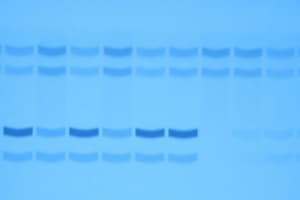
Protein Gel Analysis
- Lanes with one band: Indicate the sample containing only one protein or protein subunit
- Multiple Bands: Indicate more than one protein or protein subunits/polypeptides.
- Identification: Estimate the identity of the proteins in each lane (identification is not mentioned in the protocol).
- Protein Concentration: The band's darkness indicates the protein concentration in the solution. The band size reflects the relative concentration.
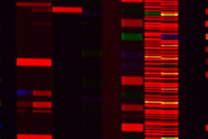
Protein Ladder
- kDa: Molecular weights marked on the protein ladder include: ~180, ~130, ~100, ~75, ~63, ~48, ~35, ~28, ~17, ~10
- Gel: A tris-glycine 4-20% polyacrylamide gel
Question 1
- Correct Answer: Smaller proteins migrate more rapidly through the gel in an SDS-PAGE process. (C)
Question 2
- Correct Answer: Proteins are visualized by staining with a dye. (A)
Results
- Image Description: This section includes an image of the completed gel electrophoresis results. The image provides a visual representation of the separated proteins. The labels and sizes are included in this image.
Studying That Suits You
Use AI to generate personalized quizzes and flashcards to suit your learning preferences.