Podcast
Questions and Answers
Which of the following is the primary function of the respiratory system?
Which of the following is the primary function of the respiratory system?
- To transport nutrients to body cells
- To regulate body temperature
- To filter waste products from the blood
- To facilitate the exchange of oxygen and carbon dioxide between the blood, air, and tissues (correct)
How does the nasal cavity contribute to the conditioning of inhaled air?
How does the nasal cavity contribute to the conditioning of inhaled air?
- By filtering, warming, and moistening the air (correct)
- By removing oxygen from the air
- By cooling and drying the air
- By adding carbon dioxide to the air
What is the role of the epiglottis during swallowing?
What is the role of the epiglottis during swallowing?
- To produce sound for speech
- To filter air entering the pharynx
- To prevent food from entering the larynx and trachea (correct)
- To direct air into the trachea
Why is the right bronchus more prone to receiving foreign objects compared to the left bronchus?
Why is the right bronchus more prone to receiving foreign objects compared to the left bronchus?
What is the function of the pleural fluid that fills the area between the visceral and parietal pleura?
What is the function of the pleural fluid that fills the area between the visceral and parietal pleura?
What is the primary mechanism by which oxygen and carbon dioxide are exchanged across the respiratory membrane?
What is the primary mechanism by which oxygen and carbon dioxide are exchanged across the respiratory membrane?
How is the majority of carbon dioxide transported in the blood?
How is the majority of carbon dioxide transported in the blood?
What physiological process primarily drives air movement during normal, relaxed expiration?
What physiological process primarily drives air movement during normal, relaxed expiration?
Which factor has the most significant influence on the regulation of respiration rate?
Which factor has the most significant influence on the regulation of respiration rate?
What is the functional volume in the lungs, and why is it significant?
What is the functional volume in the lungs, and why is it significant?
Flashcards
Respiratory Tract
Respiratory Tract
The pathway of air from the nose to the lungs.
Nasal Cavity
Nasal Cavity
Hollow canals in the skull, separated by a septum of bone and cartilage, that filter, warm, and moisten the air.
Paranasal Sinuses
Paranasal Sinuses
Cavities within bones surrounding the nasal cavity that lighten the skull and act as resonance chambers for speech.
Pharynx
Pharynx
Signup and view all the flashcards
Epiglottis
Epiglottis
Signup and view all the flashcards
Trachea
Trachea
Signup and view all the flashcards
Alveoli
Alveoli
Signup and view all the flashcards
Respiration
Respiration
Signup and view all the flashcards
Inspiratory Reserve Volume (IRV)
Inspiratory Reserve Volume (IRV)
Signup and view all the flashcards
Residual Volume
Residual Volume
Signup and view all the flashcards
Study Notes
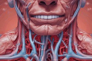
- The respiratory system facilitates the exchange of oxygen and carbon dioxide between the blood, air, and tissues.
- Key organs include the nose, pharynx, larynx, trachea, bronchi, and lungs (specifically, alveoli).
- The respiratory tract serves as the pathway for air from the nose to the lungs.
- Air is cleansed by hairs, cilia, and mucus, warmed by blood vessels, and moistened.
Upper Respiratory Tract Structure
- Includes nasal cavities, pharynx, glottis, and larynx.
Nasal Cavity
- Hollow canals separated by a septum of bone and cartilage.
- Functions to filter, warm, and moisten the air.
- Paranasal sinuses are cavities within bones surrounding the nasal cavity that lighten the skull.
- Sinuses act as resonance chambers for speech.
- They produce mucus that drains into the nasal cavity.
- Mucous membrane lines the nasal cavity.
- Cilia moves mucus and trapped particles to the pharynx.
- Conchae are lateral projections that increase surface area and air turbulence.
- The nasal cavity is separated from the oral cavity by the anterior hard palate (bone) and posterior soft palate (muscle).
Pharynx
- A funnel-shaped passageway between the nasal cavity and larynx.
- Connects the nasal and oral cavities to the larynx.
- Has three regions: nasopharynx (air passageway), oropharynx (where oral cavity joins, for food and air), and laryngopharynx (opens to larynx).
- Tonsils, lymphatic tissue in the oropharynx, protect against inhaled pathogens.
Epiglottis
- A flap of tissue at the base of the tongue.
- Prevents food from entering the trachea, or windpipe, during swallowing.
Larynx
- The voice box; contains the vocal cords.
- Functions to produce sound.
- Vocal cords are elastic tissues that vibrate as air travels through, producing sound.
- The glottis is the opening or slit in the vocal cords.
Lower Respiratory Tract
- Includes the trachea, bronchi, bronchioles, lungs, and alveoli.
Trachea
- The windpipe, a flexible tube connecting the larynx to the bronchi.
- Directs air to the bronchi.
- Held open by C-shaped hyaline cartilage.
- Lined with pseudostratified ciliated columnar epithelial cells, where cilia sweep mucus loaded with dust away from the lungs.
Bronchi
- Left and right branched tubes of the trachea.
- Their function is to serve as passageways for air to the lungs.
- They subdivide into smaller and smaller branches called bronchioles.
- The right bronchus is wider, shorter, and straighter than the left bronchus.
Bronchioles
- The smallest branches of the bronchi, which lead to the alveoli.
Lungs
- Paired, cone-shaped organs that occupy the thoracic cavity.
- Contain alveoli, where gas exchange occurs.
- The apex (superior portion) is near the clavicle; the base (inferior portion) rests on the diaphragm.
- Each lung is divided into lobes by fissures: the left lung has two lobes, and the right lung has three lobes.
- The visceral pleura covers the lung, and the parietal pleura lines the walls of the thoracic cavity.
- Pleural fluid fills the area between pleura layers to allow gliding.
Alveoli
- Made of simple squamous epithelium surrounded by blood capillaries.
- Function in gas exchange.
- The respiratory membrane is extremely thin to aid in rapid gas exchange.
- Gas crosses the respiratory membrane by diffusion.
- Oxygen enters the blood, and carbon dioxide enters the alveoli.
- Macrophages add protection.
- Surfactant in alveoli prevents the lung from closing or collapsing.
Gas Exchange and Transport
- Respiration is the process of exchanging gases between the atmosphere and body cells.
External Respiration
- Exchange of gases between air and blood in the lungs.
- Blood entering the lungs is oxygen-poor and carbon dioxide-rich.
- The alveoli always have more oxygen than the blood.
- Oxygen moves by diffusion from an area of high concentration to an area of low concentration.
- Blood returning from tissues has higher concentrations of carbon dioxide than air in the alveoli.
- Pulmonary capillary blood gives up carbon dioxide.
- Blood leaving the lungs is oxygen-rich and carbon dioxide-poor.
Internal Respiration
- Exchange of gases between blood and body cells.
- Carbon dioxide diffuses out of tissue into blood, and oxygen diffuses from blood into tissue.
Gas Transport
- Oxygen transport in the blood occurs when oxygen binds inside red blood cells to hemoglobin (oxyhemoglobin); a small amount is dissolved in the plasma.
- Carbon dioxide is mostly transported in the plasma as a bicarbonate ion (HCO3-).
- A small amount is carried inside red blood cells on hemoglobin, but at different binding sites than those of oxygen
Mechanism of Breathing
- Ventilation is the manner in which air enters and exits the lungs.
- Has two phases: inspiration (air flowing into the lung) and expiration (air leaving the lung).
Inspiration
- An active phase of ventilation.
- The diaphragm and intercostal muscles contract.
- Size of the thoracic cavity increases.
- External air is pulled into the lungs due to an increase in intrapulmonary volume.
Expiration
- Mainly a passive process dependent on natural lung elasticity.
- The diaphragm and intercostal muscles relax.
- As muscles relax, air is pushed out of the lungs.
- Forced expiration can occur mostly by contracting internal intercostal muscles to depress the rib cage.
Nonrespiratory Air Movements
- Can be caused by reflexes or voluntary actions.
- Examples include cough, sneeze, laughing, crying, yawning, and hiccup.
Ventilation Control
- Normal breathing rate is 12-20 ventilations per minute.
- Controlled by the respiratory center in the medulla oblongata (brain).
- Nervous input: Intercostal and phrenic nerve stimulate muscles to contract for inspiration: lack of stimulation results in expiration
- Chemical Input: Level of carbon dioxide is the main regulatory chemical for respiration: increased carbon dioxide increases respiration
Respiratory Volumes
- Normal breathing moves about 500 ml of air with each breath (Tidal Volume [TV]).
- Respiratory capacities are measured with a spirometer.
- Many factors affect respiratory capacity including person's size, sex, age, and physical conditions.
- Inspiratory reserve volume (IRV) is the amount of air that can be taken in forcibly over the tidal volume, usually between 2100 and 3200 ml.
- Expiratory reserve volume (ERV) is the amount of air that can be forcibly exhaled, approximately 1200 ml.
- Residual volume is the remaining air in the lung after expiration, about 1200 ml.
- Vital Capacity is the total amount of exchangeable air calculated as TV + IRV + ERV.
- Dead space volume is the air that remains in the conducting zone and never reaches alveoli, about 150 ml
- Functional Volume is the air that actually reaches the respiratory zone, usually about 350 ml
Respiratory Disorders
- Tonsillitis occurs when tonsils become inflamed and enlarged, and can make breathing difficult: tonsils are the first line of defense against pathogens in the pharynx
- Laryngitis is an infection of the larynx that leads to an inability to talk audibly, disappears with treatment
- Chronic Obstructive Pulmonary Disease (COPD) describes progressive lung diseases and is the third leading cause of death in the U.S..
- Examples include chronic bronchitis, emphysema, and asthma.
- A chronic bronchitis airway becomes inflamed and fills with mucus, loss of cilia and normal cleansing action Caused by smoking cigars, cigarettes, and some pollutants.
- Emphysema is a chronic and incurable disorder where alveoli bursts and fuse into larger air spaces; reduces surface area for gas exchange; caused by prolong cigarette smoking
Studying That Suits You
Use AI to generate personalized quizzes and flashcards to suit your learning preferences.