Podcast
Questions and Answers
What is a characteristic imaging finding of secondary tuberculosis?
What is a characteristic imaging finding of secondary tuberculosis?
- Uniform nodules throughout both lungs
- Hazy, poorly marginated alveolar infiltrate radiating outward from the hilum (correct)
- Well-defined mass in the lower lobes
- Presence of cavitary lesions exclusively in the lower lobes
What potential complication may arise in patients with poorly responding tuberculous pneumonia?
What potential complication may arise in patients with poorly responding tuberculous pneumonia?
- Immediate resolution of pneumonia with normal lung return
- Complete obliteration of lung tissue without scarring
- Formation of multiple necrotic cavities or large abscesses (correct)
- Development of granulation tissue only without necrosis
Which of the following statements about tuberculomas is correct?
Which of the following statements about tuberculomas is correct?
- Lack of calcification in a nodule confirms its diagnosis as a tuberculoma
- They are usually found in the lower lobes and are always calcified
- Tuberculomas are exclusively found in immunocompetent individuals
- They can be sharply circumscribed nodules in both primary and secondary disease (correct)
In patients with miliary tuberculosis, what is a common finding observed on imaging?
In patients with miliary tuberculosis, what is a common finding observed on imaging?
Which condition is most likely to cause calcification within lung lesions seen on imaging?
Which condition is most likely to cause calcification within lung lesions seen on imaging?
What is the primary reason for reactivation of tuberculosis in previously dormant individuals?
What is the primary reason for reactivation of tuberculosis in previously dormant individuals?
What is an imaging characteristic of pulmonary mycosis?
What is an imaging characteristic of pulmonary mycosis?
Which of the following conditions is characterized by upper lobe involvement and potential necrosis?
Which of the following conditions is characterized by upper lobe involvement and potential necrosis?
What is a common radiographic appearance of pulmonary histoplasmosis?
What is a common radiographic appearance of pulmonary histoplasmosis?
Which fungus is responsible for causing coccidioidomycosis?
Which fungus is responsible for causing coccidioidomycosis?
What characterizes chronic bronchitis on a chest radiograph?
What characterizes chronic bronchitis on a chest radiograph?
What imaging appearance is associated with coccidioidomycosis?
What imaging appearance is associated with coccidioidomycosis?
Which of the following features is pathognomonic for histoplasmosis?
Which of the following features is pathognomonic for histoplasmosis?
What is the primary characteristic of emphysema as observed in imaging?
What is the primary characteristic of emphysema as observed in imaging?
In the context of chronic obstructive pulmonary disease, what does the 'tram lines' appearance indicate?
In the context of chronic obstructive pulmonary disease, what does the 'tram lines' appearance indicate?
What is a common outcome of untreated pulmonary histoplasmosis?
What is a common outcome of untreated pulmonary histoplasmosis?
What is the primary cause of inhalation silicosis?
What is the primary cause of inhalation silicosis?
Which radiographic pattern is commonly associated with silicosis?
Which radiographic pattern is commonly associated with silicosis?
What is the characteristic imaging finding of asbestosis?
What is the characteristic imaging finding of asbestosis?
Which type of nodule poses a diagnostic dilemma in chest radiographs?
Which type of nodule poses a diagnostic dilemma in chest radiographs?
The term 'shaggy heart' in radiographic findings indicates what?
The term 'shaggy heart' in radiographic findings indicates what?
What is a common cause of pulmonary fibrosis related to occupational exposure?
What is a common cause of pulmonary fibrosis related to occupational exposure?
Which of the following statements about asbestos fibers is correct?
Which of the following statements about asbestos fibers is correct?
What type of exposure was a major public health concern in school buildings during the 1980s?
What type of exposure was a major public health concern in school buildings during the 1980s?
Flashcards
Miliary Tuberculosis
Miliary Tuberculosis
Small, scattered nodules throughout both lungs, often a sign of advanced tuberculosis.
Tuberculous Pneumonia
Tuberculous Pneumonia
Lung inflammation caused by tuberculosis, potentially resolving or causing scarring and calcification.
Secondary Tuberculosis
Secondary Tuberculosis
Tuberculosis reactivation, often affecting the upper lung lobes, from dormant TB.
Tuberculoma
Tuberculoma
Signup and view all the flashcards
Fibrocalcific Changes
Fibrocalcific Changes
Signup and view all the flashcards
Pulmonary Mycosis
Pulmonary Mycosis
Signup and view all the flashcards
Secondary Lesion
Secondary Lesion
Signup and view all the flashcards
Necrotic Cavities
Necrotic Cavities
Signup and view all the flashcards
Histoplasmosis
Histoplasmosis
Signup and view all the flashcards
Coccidioidomycosis
Coccidioidomycosis
Signup and view all the flashcards
Chronic bronchitis
Chronic bronchitis
Signup and view all the flashcards
Emphysema
Emphysema
Signup and view all the flashcards
Histoplasmoma
Histoplasmoma
Signup and view all the flashcards
Pulmonary Histoplasmosis
Pulmonary Histoplasmosis
Signup and view all the flashcards
Imaging of Chronic Bronchitis
Imaging of Chronic Bronchitis
Signup and view all the flashcards
Imaging appearance for pulmonary coccidioidomycosis
Imaging appearance for pulmonary coccidioidomycosis
Signup and view all the flashcards
Silicosis Cause
Silicosis Cause
Signup and view all the flashcards
Silicosis Lung Reaction
Silicosis Lung Reaction
Signup and view all the flashcards
Silicosis Imaging
Silicosis Imaging
Signup and view all the flashcards
Asbestosis Cause
Asbestosis Cause
Signup and view all the flashcards
Asbestosis Imaging
Asbestosis Imaging
Signup and view all the flashcards
Shaggy Heart
Shaggy Heart
Signup and view all the flashcards
Solitary Pulmonary Nodule
Solitary Pulmonary Nodule
Signup and view all the flashcards
Diagnostic Dilemma
Diagnostic Dilemma
Signup and view all the flashcards
Study Notes
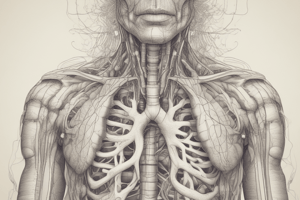
Respiratory System Overview
- The respiratory system is responsible for gas exchange, bringing oxygen into the body and removing carbon dioxide.
- Organs of the respiratory system include the nasal cavity, pharynx, larynx, trachea, bronchi, bronchioles, and lungs.
- Pulmonary alveoli are the tiny air sacs in the lungs where gas exchange occurs.
Upper Respiratory Tract
- Nasal cavity: Warms, humidifies, and filters air.
- Pharynx: Passageway for air and food.
- Larynx: Voice box, contains vocal cords.
Lower Respiratory Tract
- Trachea: Windpipe; carries air to the bronchi.
- Primary bronchi: Branches of the trachea leading to each lung.
- Bronchioles: Smaller branches of the bronchi.
- Lungs: Main organs of gas exchange; composed of alveoli.
Pulmonary Alveoli
- Alveoli are tiny air sacs in the lungs, responsible for gas exchange.
- Alveolar ducts and sacs are parts of the respiratory system connected to the alveoli.
Lungs (Detailed Anatomy)
- Lungs consist of lobes (superior, middle, and inferior on the right lung; superior and inferior on the left).
- Bronchi further divide into secondary and tertiary bronchi, finally into bronchioles that lead to the alveoli.
Imaging of the Respiratory System
- X-rays and CT scans are used to visualize the structures in the respiratory system, aiding in diagnosing problems or injuries including diseases like pneumonia or lung damage.
- A proper visualization of the tubes, the lungs, and the position of medical devices like catheters is very important.
- Radiographic findings and their relationship to disease processes are important.
Endotracheal Tube (ETT)
- A flexible tube inserted into the trachea (windpipe).
- Used during surgery or to support breathing in people with lung disease, chest trauma, or airway obstruction.
- Proper placement is critical to avoid complications.
- Daily checks are required for ETT to ensure it remains in the correct spot.
Central Venous Catheter (CVC)
- A device inserted into a large vein, typically near the heart (e.g., internal jugular, subclavian, femoral).
- Used for administering medications, providing nutrition, and conducting medical tests. Positioning of CVC is important for preventing complications that can arise during insertion.
Peripherally Inserted Central Catheter (PICC)
- A central venous catheter inserted into a peripheral vein.
- Usually in the arm.
Swan-Ganz Catheter
- A specialized catheter used for measuring pressures in the heart and lungs.
- Provides information on cardiac function and blood flow.
- Radiographic assessment is important for confirming accurate placement in the pulmonary artery.
Transvenous Endocardial Pacing
- A method for maintaining cardiac rhythm in patients with heart block or Brady arrhythmias.
- Radiographic evaluation is crucial for initial placement and detection of complications following the insertion.
- Pacemakers and implantable cardioverter-defibrillators (ICD) are types of cardiac conduction devices.
Lung Abnormal Appearances
- Reticular densities: Fine and medium types, seen in various lung conditions.
- Hazy Densities: Appear hazy on imaging, indicating various lung complications.
- Consolidation: Sections of the lung appear solid or filled with fluid material on imaging.
Congenital/Hereditary Respiratory Disorders
- Cystic Fibrosis: A genetic disorder causing thick mucus buildup in the airways.
- Hyaline Membrane Disease (HMD): Affects premature infants; causes lung immaturity.
Inflammatory Respiratory Disorders
- Croup: A viral infection causing inflammatory swelling in the subglottic trachea.
- Epiglottitis: Bacterial infection of the epiglottis characterized by its swelling.
- Pneumonia: Infection inflammation of the alveoli in one or both lungs leading to fluid buildup. - Alveolar pneumonia, Bronchopneumonia and Interstitial Pneumonia
- Aspiration Pneumonia: A type of pneumonia due to aspiration or inhalation of substances from the mouth or esophagus, commonly seen in the lower lobes.
- Anthrax: A serious bacterial infection, sometimes contracted through inhalation or contact with contaminated animals or products, often shows mediastinal widening on chest images.
- Lung Abscess: A localized necrotic area in the lung containing purulent material.
Tuberculosis
- Primary Tuberculosis, Miliary Tuberculosis, Tuberculous Pneumonia, Secondary Tuberculosis, Tuberculoma
- Radiographic findings are important for diagnosing and monitoring the disease.
Pulmonary Mycosis
- Fungal infection of the lung; Histoplasmosis and Coccidioidomycosis are common types.
- Radiographic appearances may mimic other lung conditions.
Chronic Obstructive Pulmonary Disease (COPD)
- Includes bronchitis (increased mucus production), emphysema (destruction of alveoli), asthma (airway spasms).
- Radiographs reveal potential changes like bronchovascular markings and signs of lung overinflation.
Additional Respiratory Disorders
- Silicosis, asbestosis, solitary pulmonary nodule, bronchial adenoma, Pulmonary Metastasis
- Imaging features are important for diagnosing and monitoring these conditions.
Studying That Suits You
Use AI to generate personalized quizzes and flashcards to suit your learning preferences.