Podcast
Questions and Answers
What is the approximate location of the base of the lungs?
What is the approximate location of the base of the lungs?
- T5
- T10 (correct)
- L1
- T7
What is the purpose of pinching a small fold of skin between the thumbs during palpation?
What is the purpose of pinching a small fold of skin between the thumbs during palpation?
- To ensure the patient's comfort during the examination
- To note the movement of the thumbs during inhalation (correct)
- To measure the distance between the thumbs
- To assess the patient's skin tone
What is the difference between the left and right lung in terms of lobes?
What is the difference between the left and right lung in terms of lobes?
- The left lung has one more lobe than the right lung
- The left lung is larger than the right lung
- The left lung has two lobes, while the right lung has four lobes
- The right lung has one more lobe than the left lung (correct)
What is the significance of unequal expansion during palpation?
What is the significance of unequal expansion during palpation?
Where should the thumbs be placed during anterolateral assessment?
Where should the thumbs be placed during anterolateral assessment?
What is the approximate location of the apex of the lung?
What is the approximate location of the apex of the lung?
The lungs are paired but not precisely ______.
The lungs are paired but not precisely ______.
The left lung contains only two ______ which separates at the 6th rib.
The left lung contains only two ______ which separates at the 6th rib.
The right lung contains upper, middle and lower ______.
The right lung contains upper, middle and lower ______.
The apex of the lung is approximately ______ cm above the clavicle.
The apex of the lung is approximately ______ cm above the clavicle.
During palpation, the patient is asked to ______ and then inhale, noting movement between thumbs.
During palpation, the patient is asked to ______ and then inhale, noting movement between thumbs.
Unequal expansion during palpation is indicative of ______ or trauma.
Unequal expansion during palpation is indicative of ______ or trauma.
Tactile fremitus is a palpation ______ that can be felt using the palmar aspect of the hand.
Tactile fremitus is a palpation ______ that can be felt using the palmar aspect of the hand.
Decreased fremitus occurs when anything obstructs transmission of ______ – effusions, pneumothorax, emphysema.
Decreased fremitus occurs when anything obstructs transmission of ______ – effusions, pneumothorax, emphysema.
Increased fremitus occurs with ______ or consolidation of lung tissue – pneumonia.
Increased fremitus occurs with ______ or consolidation of lung tissue – pneumonia.
Bronchial breath sounds are ______ pitched and loud, heard on inspiration and expiration over the trachea and larynx.
Bronchial breath sounds are ______ pitched and loud, heard on inspiration and expiration over the trachea and larynx.
Vesicular breath sounds are ______ pitched soft sounds heard on inspiration and expiration, sounds like wind rustling in the trees.
Vesicular breath sounds are ______ pitched soft sounds heard on inspiration and expiration, sounds like wind rustling in the trees.
The patient is asked to state “” or “” to detect changes in the intensity of the vibration.
The patient is asked to state “” or “” to detect changes in the intensity of the vibration.
What is the primary location where the vibration of tactile fremitus is most prominent?
What is the primary location where the vibration of tactile fremitus is most prominent?
What is the effect of compression or consolidation of lung tissue on tactile fremitus?
What is the effect of compression or consolidation of lung tissue on tactile fremitus?
What is the characteristic of bronchovesicular breath sounds?
What is the characteristic of bronchovesicular breath sounds?
What is the purpose of asking the patient to state a specific phrase during palpation for tactile fremitus?
What is the purpose of asking the patient to state a specific phrase during palpation for tactile fremitus?
What is the effect of effusions, pneumothorax, or emphysema on tactile fremitus?
What is the effect of effusions, pneumothorax, or emphysema on tactile fremitus?
What is the characteristic of vesicular breath sounds?
What is the characteristic of vesicular breath sounds?
Tactile fremitus is a palpation vibration that can be felt using the dorsal aspect of the hand.
Tactile fremitus is a palpation vibration that can be felt using the dorsal aspect of the hand.
Bronchovesicular breath sounds are best heard over the peripheral lung fields.
Bronchovesicular breath sounds are best heard over the peripheral lung fields.
Increased fremitus occurs with effusions or pneumothorax.
Increased fremitus occurs with effusions or pneumothorax.
The patient is asked to state 'hello' to detect changes in the intensity of the vibration during palpation for tactile fremitus.
The patient is asked to state 'hello' to detect changes in the intensity of the vibration during palpation for tactile fremitus.
Vesicular breath sounds are heard only during inspiration.
Vesicular breath sounds are heard only during inspiration.
Tactile fremitus is most prominent at the apex of the lung.
Tactile fremitus is most prominent at the apex of the lung.
The apex of the lung is marked by the 5th cervical vertebra (C5)
The apex of the lung is marked by the 5th cervical vertebra (C5)
The base of the lungs sits at approximately the 12th thoracic spine (T12) on expiration
The base of the lungs sits at approximately the 12th thoracic spine (T12) on expiration
The upper lobes are accessible from the apex at C7 to T5
The upper lobes are accessible from the apex at C7 to T5
The posterior lung landmarks are primarily upper lobes
The posterior lung landmarks are primarily upper lobes
The apex of the lungs begins 1-2 cm above the clavicle
The apex of the lungs begins 1-2 cm above the clavicle
The apex of the lungs begins ______ cm above the clavicle.
The apex of the lungs begins ______ cm above the clavicle.
The apex of the lungs is marked by the ______ cervical vertebra (C7) in the posterior.
The apex of the lungs is marked by the ______ cervical vertebra (C7) in the posterior.
The base of the lungs sits at approximately the ______ thoracic spine (T10) on inspiration.
The base of the lungs sits at approximately the ______ thoracic spine (T10) on inspiration.
In the posterior, the lungs are primarily ______ lobes!
In the posterior, the lungs are primarily ______ lobes!
The upper lobes are accessible from apex at C7, to ______ where the oblique fissures begin.
The upper lobes are accessible from apex at C7, to ______ where the oblique fissures begin.
What is the approximate location of the apex of the lungs in the posterior?
What is the approximate location of the apex of the lungs in the posterior?
What is the approximate location of the base of the lungs in the anterior?
What is the approximate location of the base of the lungs in the anterior?
What is the region where the oblique fissures begin in the posterior?
What is the region where the oblique fissures begin in the posterior?
What is the extent of the base of the lungs on inspiration in the posterior?
What is the extent of the base of the lungs on inspiration in the posterior?
What is the region of the lungs that is primarily accessible in the posterior?
What is the region of the lungs that is primarily accessible in the posterior?
What is the primary reason for decreased breath sounds in a patient?
What is the primary reason for decreased breath sounds in a patient?
What is the characteristic of breath sounds in a patient with pneumonia?
What is the characteristic of breath sounds in a patient with pneumonia?
What is the sensation felt during palpation of a patient with subcutaneous emphysema?
What is the sensation felt during palpation of a patient with subcutaneous emphysema?
What is the primary difference between bronchial and vesicular breath sounds?
What is the primary difference between bronchial and vesicular breath sounds?
What is the effect of consolidation of lung tissue on tactile fremitus?
What is the effect of consolidation of lung tissue on tactile fremitus?
There are 4 types of breath sounds – bronchial, bronchovesicular, vesicular and tracheal.
There are 4 types of breath sounds – bronchial, bronchovesicular, vesicular and tracheal.
Increased fremitus occurs with pneumonia or consolidation of lung tissue.
Increased fremitus occurs with pneumonia or consolidation of lung tissue.
Vesicular breath sounds are loud and high-pitched, heard on inspiration and expiration over the trachea and larynx.
Vesicular breath sounds are loud and high-pitched, heard on inspiration and expiration over the trachea and larynx.
Crepitus is a normal breath sound that occurs during inspiration and expiration.
Crepitus is a normal breath sound that occurs during inspiration and expiration.
Decreased breath sounds occur when there is consolidation or fluid (pneumonia) in the lung tissue.
Decreased breath sounds occur when there is consolidation or fluid (pneumonia) in the lung tissue.
As air passes through the ______ tree, it creates a characteristic set of audible sounds
As air passes through the ______ tree, it creates a characteristic set of audible sounds
There are 3 types of ______ sounds – bronchial, bronchovesicular and vesicular
There are 3 types of ______ sounds – bronchial, bronchovesicular and vesicular
Crepitus is a coarse crackling sensation that is palpable over the chest due to ______ emphysema
Crepitus is a coarse crackling sensation that is palpable over the chest due to ______ emphysema
Increased ______ breath sounds are louder than normal and occur with consolidation or fluid (pneumonia)
Increased ______ breath sounds are louder than normal and occur with consolidation or fluid (pneumonia)
Decreased or absent ______ sounds occur when obstructed by secretions or loss of lung elasticity
Decreased or absent ______ sounds occur when obstructed by secretions or loss of lung elasticity
What is the oxygen concentration range provided by a Venturi Mask?
What is the oxygen concentration range provided by a Venturi Mask?
What is the maximum flow rate of a High Flow Nasal Cannula?
What is the maximum flow rate of a High Flow Nasal Cannula?
What is the oxygen concentration range for a Low Flow Nasal Cannula at 5-6 L/min?
What is the oxygen concentration range for a Low Flow Nasal Cannula at 5-6 L/min?
What is a contraindication for using a Simple Face Mask?
What is a contraindication for using a Simple Face Mask?
What is a complication of using a High Flow Nasal Cannula?
What is a complication of using a High Flow Nasal Cannula?
What is a characteristic of a Full Non-Rebreather?
What is a characteristic of a Full Non-Rebreather?
A Venturi Mask can provide a specific O2 with humidity at a range of 21-100%.
A Venturi Mask can provide a specific O2 with humidity at a range of 21-100%.
High Flow Nasal Cannula can be used on adults and children but not on infants.
High Flow Nasal Cannula can be used on adults and children but not on infants.
Low Flow Nasal Cannula can deliver a maximum of 60 LPM of oxygen.
Low Flow Nasal Cannula can deliver a maximum of 60 LPM of oxygen.
A Simple Face Mask can deliver a maximum of 100% FiO2.
A Simple Face Mask can deliver a maximum of 100% FiO2.
A Partial Rebreather Mask can deliver a maximum of 60-80% FiO2.
A Partial Rebreather Mask can deliver a maximum of 60-80% FiO2.
High Flow Nasal Cannula is contraindicated for patients who retain CO2.
High Flow Nasal Cannula is contraindicated for patients who retain CO2.
High flow devices control the FiO2 with complete dependence on the patient’s breathing pattern.
High flow devices control the FiO2 with complete dependence on the patient’s breathing pattern.
Venturi masks are an example of low flow devices.
Venturi masks are an example of low flow devices.
Partial rebreather masks are an example of high flow devices.
Partial rebreather masks are an example of high flow devices.
Blender masks can deliver a range of inspired oxygen concentrations (FiO2).
Blender masks can deliver a range of inspired oxygen concentrations (FiO2).
Oxygen conserving nasal cannula is an example of high flow devices.
Oxygen conserving nasal cannula is an example of high flow devices.
Which type of device is less dependent on the patient's breathing pattern?
Which type of device is less dependent on the patient's breathing pattern?
What is the primary difference between high flow and low flow devices?
What is the primary difference between high flow and low flow devices?
Which of the following is an example of a high flow device?
Which of the following is an example of a high flow device?
What is the primary advantage of high flow devices over low flow devices?
What is the primary advantage of high flow devices over low flow devices?
Which type of device is commonly used for patients who require a specific inspired oxygen concentration?
Which type of device is commonly used for patients who require a specific inspired oxygen concentration?
Flashcards are hidden until you start studying
Study Notes
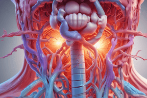
Lung Structure
- The lungs are paired but not precisely symmetrical.
- The left lung has two lobes: upper and lower, which separate at the 6th rib.
- The left lung is smaller due to the space occupied by the heart.
- The right lung has three lobes: upper, middle, and lower, with the middle lobe extending from the 4th to 6th rib.
- The apex of the lung is approximately 3 cm above the clavicle, pointing towards the substernal notch.
- The base of the lungs lies at around T10.
Palpation Techniques
- To confirm symmetrical chest expansion, stand behind the patient and place warmed hands at T10/Tenth rib.
- Point thumbs towards the spine and fingers laterally, with palms lightly contacting the posterolateral surfaces.
- Pinch a small fold of skin between the thumbs, keeping them about 5cm apart.
- Ask the patient to exhale and then inhale, noting the movement between the thumbs.
- Symmetrical movement indicates normal expansion, while unequal expansion may indicate pneumothorax, rib fractures, pneumonia, or trauma.
- For anterolateral assessment, place hands along the coastal margins with thumbs pointing towards the xiphoid process.
- Thumbs should move apart symmetrically by 3-5 cm when the patient inhales deeply.
Tactile Fremitus
- Palpation vibration felt using the palmar aspect of the hand
- Most prominent between the scapulae and around the sternum, where major bronchi are closest to the chest wall
- Decreases as you progress down the chest
- Decreased fremitus occurs when anything obstructs transmission of vibrations, such as:
- Effusions
- Pneumothorax
- Emphysema
- Increased fremitus occurs with compression or consolidation of lung tissue, such as:
- Pneumonia
- Note the correct placement of hands for palpating for a vibration
- Use the patient's vocalization of "99" or "blue moon" to detect changes in the intensity of the vibration
Breath Sounds (Normal)
- Bronchial Sounds
- High-pitched and loud
- Heard on inspiration and expiration over the trachea and larynx
- Best heard over the trachea
- Bronchovesicular Sounds
- Medium-pitched and blowing sounds of medium intensity
- Heard on inspiration and expiration over the major bronchi
- Located at the upper sternum anteriorly and posteriorly between the scapulae (especially on the right side)
- Vesicular Sounds
- Low-pitched and soft
- Heard on inspiration and expiration
- Sounds like wind rustling in the trees
- Best heard over peripheral lung fields (except over scapula)
- Located where air flows through smaller alveoli and bronchioles
Anterior Lung Landmarks
- Apex of the lungs begins 3-4 cm above the clavicle, marking the highest point
- Base of the lungs sits at the diaphragm, approximately at the 7th rib, marking the lowest point
Posterior Lung Landmarks
- Apex of the lungs is marked by the 7th cervical vertebra (C7), also known as the "big bump" at the top of the spine
- Base of the lungs sits at approximately the 10th thoracic spine (T10), extending to approximately T12 during inspiration
- Posterior lungs are primarily comprised of lower lobes
- Upper lobes are accessible from the apex at C7 to T3, where the oblique fissures begin
Breath Sounds
- Air passing through the tracheobronchial tree creates a characteristic set of audible sounds
- There are three types of breath sounds: bronchial, bronchovesicular, and vesicular
Changes in Breath Sounds
- Decreased or absent breath sounds occur when obstructed by secretions or loss of lung elasticity
- Increased (louder) breath sounds occur with consolidation or fluid (pneumonia), enhancing the bronchial sound as it moves through the lung fields
Crepitus
- A coarse crackling sensation palpable over the chest
- Caused by subcutaneous emphysema (air escapes the lungs and enters subcutaneous tissue)
High-Flow Delivery Devices
- Venturi Mask provides specific O2 concentrations (24-50%) with humidity, but has a low constant O2 flow, and may irritate skin, interfere with eating, drinking, and talking.
- High Flow Nasal Cannula offers adjustable FiO2 (21%-100%) with modifiable flow up to 60 LPM, and is suitable for adults, children, and infants, but FiO2 is dependent on patient respiratory pattern and input flow, and carries a risk of infection.
Low-Flow Delivery Devices
- Low Flow Nasal Cannula provides O2 concentrations of 24-28% at 1-2L/min, 32-36% at 3-4L/min, and 40-44% at 5-6L/min, and is safe, simple, and easily tolerated, but is not suitable for patients with nasal obstruction, and may cause drying of mucous membranes, dislodge easily, and skin irritation.
- Simple Face Mask provides O2 concentrations of 40-60% at 5-10L/min, and is useful for short periods of time, but is contraindicated for patients who retain CO2, and carries an increased risk of aspiration.
- Partial Rebreather and Partial Non-Rebreather devices provide O2 concentrations of 60-80% at 10-15L/min, and are useful for short periods of acute hypoxia, but may cause skin irritation, and the bag may twist/deflate.
- Full Non-Rebreather device provides O2 concentrations of up to 100% at 10-15L/min, and is useful for short periods of acute hypoxia, but may cause skin irritation, and the bag may twist/deflate.
Oxygen Devices
- Both high flow and low flow devices can deliver a range of inspired oxygen concentrations (FiO2) to patients.
High Flow Devices
- Control FiO2 with less dependence on patient's breathing pattern.
- Include device types:
- Venturi masks
- Large volume nebulizer
- Blender masks
- High flow nasal cannula
Low Flow Devices
- Include device types:
- Low flow nasal cannula
- Oxygen conserving nasal cannula
- Simple face masks
- Partial rebreather masks
- Nonrebreather masks
Studying That Suits You
Use AI to generate personalized quizzes and flashcards to suit your learning preferences.