Podcast
Questions and Answers
Which of the following structures is part of both the upper and the lower respiratory systems?
Which of the following structures is part of both the upper and the lower respiratory systems?
- Larynx
- Pharynx (correct)
- Trachea
- Bronchi
Which function is NOT associated with the respiratory system?
Which function is NOT associated with the respiratory system?
- Gas Exchange
- Verbal Communication
- Regulation of blood glucose levels (correct)
- Protection against pathogens
What type of epithelial cells are found in most of the respiratory tract, excluding the pharynx, smaller bronchi, and alveoli?
What type of epithelial cells are found in most of the respiratory tract, excluding the pharynx, smaller bronchi, and alveoli?
- Simple cuboidal
- Transitional
- Pseudostratified ciliated columnar (correct)
- Stratified squamous
Which of the following best describes the function of the mucus escalator?
Which of the following best describes the function of the mucus escalator?
What is the function of the vibrissae in the nasal cavity?
What is the function of the vibrissae in the nasal cavity?
In which sequence does air pass through the upper respiratory system?
In which sequence does air pass through the upper respiratory system?
Which of the following statements best describes the pharynx?
Which of the following statements best describes the pharynx?
Which of the following is NOT a region of the pharynx?
Which of the following is NOT a region of the pharynx?
At which vertebral level does the larynx begin?
At which vertebral level does the larynx begin?
The thyroid cartilage is an example of which type of cartilage in the larynx?
The thyroid cartilage is an example of which type of cartilage in the larynx?
Which laryngeal cartilage closes over the glottis during swallowing?
Which laryngeal cartilage closes over the glottis during swallowing?
Which of the following is NOT a paired cartilage of the Larynx?
Which of the following is NOT a paired cartilage of the Larynx?
What structure do the extrinsic laryngeal ligaments bind to the thyroid cartilage?
What structure do the extrinsic laryngeal ligaments bind to the thyroid cartilage?
What primarily determines the pitch of the sound produced by the vocal cords?
What primarily determines the pitch of the sound produced by the vocal cords?
What is the primary function of the intrinsic muscles of the larynx?
What is the primary function of the intrinsic muscles of the larynx?
What is the approximate length and diameter of the trachea?
What is the approximate length and diameter of the trachea?
At what level does the trachea bifurcate into the right and left bronchi?
At what level does the trachea bifurcate into the right and left bronchi?
What type of muscle is the trachealis muscle?
What type of muscle is the trachealis muscle?
Which of the following characteristics is true regarding the right primary bronchus compared to the left?
Which of the following characteristics is true regarding the right primary bronchus compared to the left?
What is the hilum of the lung?
What is the hilum of the lung?
How many lobes does the right lung have?
How many lobes does the right lung have?
The horizontal fissure divides which lobes of the right lung?
The horizontal fissure divides which lobes of the right lung?
Which surface is NOT a surface of the lungs?
Which surface is NOT a surface of the lungs?
Regarding the lungs, what are extrapulmonary bronchi?
Regarding the lungs, what are extrapulmonary bronchi?
How many tertiary bronchi are typically found in the left lung?
How many tertiary bronchi are typically found in the left lung?
Which feature distinguishes bronchioles from other structures in the respiratory tract?
Which feature distinguishes bronchioles from other structures in the respiratory tract?
What is the approximate number of alveoli in each lung?
What is the approximate number of alveoli in each lung?
What cells are associated with alveoli that secrete surfactant?
What cells are associated with alveoli that secrete surfactant?
In gas exchange within the alveoli, which vessels transport carbon dioxide to the alveolar capillaries?
In gas exchange within the alveoli, which vessels transport carbon dioxide to the alveolar capillaries?
What is the purpose of pleural fluid found between the visceral and parietal pleura?
What is the purpose of pleural fluid found between the visceral and parietal pleura?
The diaphragm contracts and lowers to cause:
The diaphragm contracts and lowers to cause:
Which of the following best describes 'eupnea'?
Which of the following best describes 'eupnea'?
Which of the following is true regarding the respiratory system prior to birth?
Which of the following is true regarding the respiratory system prior to birth?
Which respiratory center sets the respiratory pace?
Which respiratory center sets the respiratory pace?
What is the role of the apneustic and pneumotaxic respiratory centers?
What is the role of the apneustic and pneumotaxic respiratory centers?
What stimulates chemoreceptor reflexes?
What stimulates chemoreceptor reflexes?
Which of the following is a common effect of aging on the respiratory system?
Which of the following is a common effect of aging on the respiratory system?
What changes occur at birth to initiate normal respiration?
What changes occur at birth to initiate normal respiration?
Which of the following is Not a respiratory muscle?
Which of the following is Not a respiratory muscle?
What type of cells lining the alveoli are called type I pneumocytes?
What type of cells lining the alveoli are called type I pneumocytes?
Flashcards
Respiratory System
Respiratory System
Includes the nose, nasal cavity, sinuses, pharynx, larynx, trachea, bronchi, bronchioles, and alveoli.
Upper Respiratory System
Upper Respiratory System
Consists of the nose, nasal cavity, sinuses & pharynx.
Lower Respiratory System
Lower Respiratory System
Consists of the larynx, trachea, bronchi, bronchioles & alveoli.
Respiratory System Functions
Respiratory System Functions
Signup and view all the flashcards
Vibrissae
Vibrissae
Signup and view all the flashcards
Pharynx
Pharynx
Signup and view all the flashcards
Nasopharynx
Nasopharynx
Signup and view all the flashcards
Laryngopharynx
Laryngopharynx
Signup and view all the flashcards
Larynx
Larynx
Signup and view all the flashcards
Thyroid Cartilage
Thyroid Cartilage
Signup and view all the flashcards
Epiglottis
Epiglottis
Signup and view all the flashcards
Intrinsic Laryngeal Ligaments
Intrinsic Laryngeal Ligaments
Signup and view all the flashcards
Extrinsic Laryngeal Ligaments
Extrinsic Laryngeal Ligaments
Signup and view all the flashcards
Vestibular and Vocal Ligaments
Vestibular and Vocal Ligaments
Signup and view all the flashcards
Intrinsic Muscles (Larynx)
Intrinsic Muscles (Larynx)
Signup and view all the flashcards
Extrinsic Muscles (Larynx)
Extrinsic Muscles (Larynx)
Signup and view all the flashcards
Trachea
Trachea
Signup and view all the flashcards
Annular Ligaments
Annular Ligaments
Signup and view all the flashcards
Trachea Lining Sections
Trachea Lining Sections
Signup and view all the flashcards
Trachealis Muscle
Trachealis Muscle
Signup and view all the flashcards
Primary Bronchi
Primary Bronchi
Signup and view all the flashcards
Hilum
Hilum
Signup and view all the flashcards
Root
Root
Signup and view all the flashcards
Right Lung Lobes
Right Lung Lobes
Signup and view all the flashcards
Left Lung Lobes
Left Lung Lobes
Signup and view all the flashcards
Extrapulmonary Bronchi
Extrapulmonary Bronchi
Signup and view all the flashcards
Intrapulmonary Bronchi
Intrapulmonary Bronchi
Signup and view all the flashcards
Bronchopulmonary Segment
Bronchopulmonary Segment
Signup and view all the flashcards
Tertiary Bronchi
Tertiary Bronchi
Signup and view all the flashcards
Alveoli
Alveoli
Signup and view all the flashcards
Pulmonary Arteries (Alveoli)
Pulmonary Arteries (Alveoli)
Signup and view all the flashcards
Pulmonary Veins (Alveoli)
Pulmonary Veins (Alveoli)
Signup and view all the flashcards
Squamous Cells (Alveoli)
Squamous Cells (Alveoli)
Signup and view all the flashcards
Type II Pneumocytes
Type II Pneumocytes
Signup and view all the flashcards
Alveolar Macrophages
Alveolar Macrophages
Signup and view all the flashcards
Visceral Pleura
Visceral Pleura
Signup and view all the flashcards
Parietal Pleura
Parietal Pleura
Signup and view all the flashcards
Pleural Cavity
Pleural Cavity
Signup and view all the flashcards
Diaphragm
Diaphragm
Signup and view all the flashcards
External Intercostals
External Intercostals
Signup and view all the flashcards
Internal Intercostals
Internal Intercostals
Signup and view all the flashcards
Study Notes
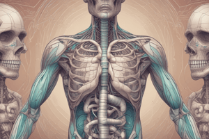
Introduction to the Respiratory System
- The respiratory system includes the nose, nasal cavity, sinuses, pharynx, larynx, trachea, bronchi, bronchioles, and alveoli.
Overview of the Respiratory System and Tract
- The upper respiratory system consists of the nose, nasal cavity, sinuses, and pharynx.
- The lower respiratory system consists of the larynx, trachea, bronchi, bronchioles, and alveoli.
- The respiratory system allows for gas exchange between the air and blood.
- It also protects respiratory surfaces from dehydration and pathogens.
- Verbal communication is enabled through sound production.
- The respiratory system assists in regulating blood volume, blood pressure, and body fluid pH.
The Respiratory Epithelium
- It consists of pseudostratified, ciliated, columnar cells, except in the pharynx, smaller bronchi, and alveoli.
- Stratified squamous cells are found in the pharynx.
- Mucus-producing cells are present in the nasal cavity and lower respiratory tract.
- Ciliated columnar cells move mucus upward to cough out debris.
- Stratified squamous cells provide protection against abrasion.
- Mucous cells produce mucus that traps inhaled debris, preventing it from entering the lungs.
Protection of the Respiratory System
- Nose hairs, known as vibrissae, block some inhaled debris.
- The nasal cavity produces mucus to trap inhaled debris, which can be removed by sneezing.
- The respiratory epithelium mucus traps inhaled debris, which can be removed by coughing.
The Upper Respiratory System - Air Pathway
- Air enters through the external nares and passes by the nasal vestibule.
- The nasal vestibule area is surrounded by two pairs of alar cartilage.
- The air then enters the nasal cavity.
- Air flows in and around the nasal conchae (inferior, middle, and superior), where debris gets stuck in the mucus.
- As air swirls around the conchae, it warms before entering the trachea.
- Air enters the internal nares, followed by entering the nasopharynx area.
The Nose and Nasal Cavity
- The nose consists of nasal bones, the nasal septum, cartilage, external nares, alar cartilage, the dorsum and apex and the nasal conchae.
- The nasal septum includes the vomer and perpendicular plate of the ethmoid.
The Pharynx
- The pharynx connects the nose to the mouth to the throat.
- The nasopharynx is located at the back of the nose area.
- The oropharynx is located at the back of the moth area and consists of the pharyngeal arch and uvula.
- The laryngopharynx is the area that has the entrance to the trachea and esophagus.
The Larynx
- The larynx is a cylindrically shaped organ.
- Its cartilaginous walls are stabilized by ligaments or skeletal muscles.
- It begins at vertebra C3 or C4 and ends at vertebra C7.
- The larynx has unpaired cartilages including the thyroid cartilage, cricoid cartilage, and epiglottis.
- The thyroid cartilage contains the laryngeal prominence.
- The cricoid and epiglottis are unpaired cartilage.
- The epiglottis closes over the glottis during swallowing of food.
- Paired laryngeal cartilages play a role in opening and closing the glottis.
- These cartilages are the arytenoid, corniculate, and cuneiform cartilages.
Laryngeal Ligaments
- Intrinsic ligaments bind the laryngeal cartilages together.
- Extrinsic ligaments bind the thyroid cartilage to the hyoid bone and cricoid cartilage.
- Vestibular and local ligaments extend between the thyroid cartilage and the arytenoids.
Sound Production and Laryngeal Musculature
- Air passing between the vocal cords creates sound.
- Pitch depends on the vocal cords' diameter, length, and tension.
- Children have slender, short vocal folds, creating a high-pitched sound.
- At puberty, males' vocal cords thicken and lengthen, creating a deeper voice than females.
- Sound amplification occurs in the sinus cavities.
- Production of sounds depends on the movement of the lips, tongue, and cheeks.
- Intrinsic muscles regulate tension of the vocal cords and the opening/closing of the glottis.
- Extrinsic muscles position and stabilize the larynx.
The Trachea
- The trachea is 11 cm long and 2.5 cm in diameter.
- It bifurcates at the carina into the right and left bronchi at T5.
- The trachea contains 15-20 C-shaped tracheal cartilages.
- Annular ligaments connect one cartilage ring to another.
- The lining consists of respiratory epithelia, lamina propria, and submucosa.
- The posterior side of the cartilage ring is the trachealis muscle.
- This allows constriction and dilation of the trachea.
The Primary Bronchi
- The trachea branches at the carina forming the left and right primary bronchi.
- Primary bronchi enter each lung at the hilum.
- The hilum is the point of entrance and exit of the pulmonary blood vessels.
- The right primary bronchus is steeper and larger in diameter than the left.
- A person can aspirate foreign objects into the right lung easier than the left lung because of this.
- The combination of the bronchus, artery, and vein is referred to as the root.
Lung Structure
- The lungs have an apex which points superiorly and the base points inferiorly.
- The right lung has superior, middle, and inferior lobes, divided by the horizontal and oblique fissures.
- The left lung has superior and inferior lobes, separated by the oblique fissure and a cardiac notch.
- Lungs have costal, mediastinal, and diaphragmatic surfaces.
- The primary bronchi branch numerous times into extrapulmonary bronchi (outside lungs) and intrapulmonary bronchi (inside lungs).
- Primary bronchus divides to form secondary and tertiary bronchi.
- Each tertiary bronchus serves a specific area called a bronchopulmonary segment.
- The right primary bronchus divides into the superior lobar, middle lobar, and inferior lobar secondary bronchi.
- The left primary bronchus divides into the superior lubar and inferior lobar secondary bronchi.
- The right lung has 10 tertiary bronchi and therefore 10 bronchopulmonary segments.
- The left lung has 9 tertiary bronchi and therefore 9 bronchopulmonary segments.
The Bronchioles
- The tertiary bronchi give rise to the bronchioles.
- Bronchioles have a diameter of 0.3–0.5 mm are self-supporting, thus not requiring cartilage plates.
- Smooth muscle enables bronchodilation (sympathetic stimulation) and bronchoconstriction (parasympathetic stimulation).
- Bronchioles terminate with clusters of alveolar sacs.
Alveolar Ducts and Alveoli
- Each lung has about 150 million alveoli, surrounded by an extensive capillary network.
- These capillaries drop off carbon dioxide and pick up oxygen.
- Elastic tissue surrounds each alveolus, maintaining its shape and position during breathing.
- The alveoli are lined with a single layer of squamous cells.
- These linings are called Type I pneumocytes.
- Type II pneumocytes secrete surfactant, which prevents alveolar collapse, as well as scattered among the Type 1 pneumocytes.
- Alveolar macrophages wander around phagocytizing particulate matter.
- Pulmonary arteries transport carbon dioxide to alveolar capillaries.
- Carbon dioxide leaves the capillaries and enters the alveolar sacs.
- Oxygen leaves the alveolar sacs and enters the lung capillaries.
- Oxygen enters the pulmonary veins which returns to the heart to be pumped to all body parts.
Pleural Cavities and Membranes
- The plural cavities are located between the visceral and parietal membranes and is filled with pleural fluid.
- The right and left pleural cavities are separated by the mediastinum.
- Visceral pleura covers the lung's outer surface, while the parietal pleura lines the thoracic wall.
- The space between the visceral and parietal pleura is the pleural cavity.
- Pleural fluid reduces friction during breathing.
- Pleurisy occurs when the membranes produce too much pleural fluid or adhere to the thoracic wall resulting in pain upon breathing.
Respiratory Muscles and Pulmonary Ventilation
- The muscles involved in pulmonary ventilation (breathing) are the diaphragm, external intercostals, and internal intercostals.
- The diaphragm contracts (lowers) to cause inhalation and relaxes (raises) to cause exhalation.
- External intercostals elevate the ribs aiding in inhalation.
- Internal intercostals depress the ribs aiding in exhalation.
- Respiratory movements can be eupnea or hyperpnea.
- Eupnea is quiet breathing, which can be diaphragmatic or costal breathing.
- Women typically use costal breathing due to pregnancy pushing upward on the diaphragm.
- Hyperpnea is forced breathing and generally requires the use of accessory breathing muscles.
Respiratory Changes at Birth
- Pulmonary arterial resistance is high and vessels are collapsed.
- The rib cage is compressed and passages contain no air, just fluid.
- At birth, air enters, forcing fluid out, which triggers the closure of Foramen ovale and ductus arteriosus.
Respiratory Centers of the Brain
- There are three pairs of nuclei in the pons and medulla oblongata that regulate the respiratory muscles.
- The respiratory rhythmicity center sets the base respiratory pace, it is located in the medulla oblongata.
- The apneustic center adjusts the respiratory pace, and is located in the pons.
- The pneumotaxic center adjusts the respiratory pace, and it is located in the pons.
- There are three different reflexes involved in respiration:
- Mechanoreceptor reflexes respond to changes in lung volume or changes in blood pressure.
- Chemoreceptor reflexes respond to changes in partial pressures of carbon dioxide and oxygen, and also changes in pH.
- Protective reflexes respond to physical injury or irritation.
Aging and the Respiratory System
- The respiratory system becomes less efficient as we age.
- Noteworthy changes include elastic tissue deterioration and restricted rib movements due to arthritis.
- Some degree of emphysema hinders breathing.
- 1 square foot of respiratory membrane is lost annually after age 30.
Studying That Suits You
Use AI to generate personalized quizzes and flashcards to suit your learning preferences.