Podcast
Questions and Answers
What effect does sympathetic stimulation have on the trachea?
What effect does sympathetic stimulation have on the trachea?
- It has no effect on the diameter.
- It causes spasms in the tracheal walls.
- It decreases the diameter.
- It increases the diameter. (correct)
How does the change in diameter of the trachea due to sympathetic stimulation affect airflow?
How does the change in diameter of the trachea due to sympathetic stimulation affect airflow?
- It restricts airflow passageways.
- It increases resistance to airflow.
- It decreases airflow velocity.
- It makes it easier to move large volumes of air. (correct)
Which physiological response is related to the increase of tracheal diameter?
Which physiological response is related to the increase of tracheal diameter?
- Parasympathetic nervous system activation.
- Oxygen depletion in tissues.
- Sympathetic nervous system activation. (correct)
- Increased carbon dioxide levels.
What is the primary purpose of increasing the tracheal diameter during sympathetic stimulation?
What is the primary purpose of increasing the tracheal diameter during sympathetic stimulation?
In the context of respiratory physiology, what does the term 'airway resistance' refer to?
In the context of respiratory physiology, what does the term 'airway resistance' refer to?
What is the typical respiratory rate range for healthy adults at rest?
What is the typical respiratory rate range for healthy adults at rest?
How does the respiratory rate in children compare to that of adults?
How does the respiratory rate in children compare to that of adults?
Which option accurately reflects normal respiratory rates for adults?
Which option accurately reflects normal respiratory rates for adults?
What is the maximum expected respiratory rate for children according to typical norms?
What is the maximum expected respiratory rate for children according to typical norms?
At what point might an adult's respiratory rate be considered abnormal based on standard ranges?
At what point might an adult's respiratory rate be considered abnormal based on standard ranges?
Flashcards
Trachea diameter
Trachea diameter
The size of the trachea (windpipe).
Sympathetic stimulation
Sympathetic stimulation
A process that makes the trachea wider.
Respiratory passageways
Respiratory passageways
The airways that carry air to the lungs.
Large air volumes
Large air volumes
Signup and view all the flashcards
Easier to move air
Easier to move air
Signup and view all the flashcards
Respiratory rate
Respiratory rate
Signup and view all the flashcards
Normal adult respiratory rate
Normal adult respiratory rate
Signup and view all the flashcards
Child's respiratory rate
Child's respiratory rate
Signup and view all the flashcards
Breathing rate
Breathing rate
Signup and view all the flashcards
Resting breathing rate
Resting breathing rate
Signup and view all the flashcards
Study Notes
The Respiratory System
- The respiratory system functions to provide a large area for gas exchange between air and circulating blood, move air to and from the gas-exchange surfaces of the lungs, protect the respiratory surfaces from dehydration and invading pathogens, produce sounds permitting speech, singing, and nonverbal auditory communication, and provide olfactory sensations.
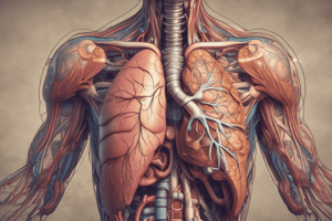
- The major structures of the respiratory system include the nose (including the nasal cavity and paranasal sinuses), pharynx (throat), larynx (voice box), trachea (wind- pipe), bronchi, bronchioles, and alveoli (exchange surfaces).
- The respiratory tract is divided into a conducting portion (begins at the entrance to the nasal cavity and continues through the pharynx, larynx, trachea, bronchi, and larger bronchioles) and a respiratory portion (includes the smallest and most delicate bronchioles and alveoli, which provide large blood-gas exchange surfaces).
- Nose: Air normally enters the respiratory system through the paired external nares, or nostrils, which open into the nasal cavity. The nasal vestibule is the space enclosed within the flexible tissues of the nose. Coarse hairs extend across the nostrils to guard the nasal cavity from large particles such as sand, dust, and insects.
- Pharynx: or throat, is a chamber shared by the digestive and respiratory systems. It extends between the internal nares and the entrances to the larynx and esophagus, and is subdivided into the nasopharynx, oropharynx, and laryngopharynx.
- Larynx: or voice box, consists of nine cartilages stabilized by ligaments, skeletal muscles, or both. The glottis is a narrow opening for entry of inhaled air.
- Trachea: or windpipe, is a tough, flexible tube. It is approximately 11 cm long, and supported by 15-20 C-shaped cartilages, that protect the airway and prevent collapse or overexpansion as pressures change in the respiratory system.
- Bronchi: within the mediastinum, the trachea branches into the right and left primary bronchi. The walls of the primary bronchi resemble those of the trachea.
- Bronchioles: Each terminal bronchiole supplies air to a lobule of the lung. Within a lobule, a terminal bronchiole divides to form several respiratory bronchioles.
- Alveolar ducts and alveoli: Respiratory bronchioles open into passageways called alveolar ducts. These ducts end at alveolar sacs, common chambers connected to multiple individual alveoli, the exchange surfaces of the lung. Alveolar epithelium is simple squamous epithelium, containing alveolar macrophages (dust cells) that patrol the epithelium. Septal cells secrete surfactant.
- Respiratory membrane: Gas exchange occurs across the respiratory membrane of the alveoli (consisting of the squamous alveolar epithelium, endothelial cells lining adjacent capillaries, and the fused basement membranes).
- Circulation to the respiratory membrane: The respiratory exchange surfaces receive blood from the arteries of the pulmonary circuit. The blood pressure in the pulmonary circuit is usually relatively low.
- The lung: Each of the two lungs has distinct lobes that are separated by deep fissures. The right lung has three lobes (superior, middle, and inferior), and the left lung has two (superior, and inferior). The lungs have a light and spongy consistency. Elastic fibers give the lungs the ability to tolerate large changes in volume.
- Pleural cavities: The two pleural cavities are separated by the mediastinum. The parietal pleura covers the outer surfaces of the lungs and extends into the fissures between the lobes. The visceral pleura covers the inner surface of the body wall.
- Respiratory physiology: The process of respiration involves three integrated steps: pulmonary ventilation, gas exchange, and gas transport.
The Respiratory Membrane
- Gas exchange occurs across the respiratory membrane of the alveoli. The respiratory membrane consists of 3 components; the squamous alveolar epithelium, the endothelial cells lining an adjacent capillary, and the fused basement membranes that lie between the alveolar and endothelial cells.
Circulation to the Respiratory Membrane
- The respiratory exchange surfaces receive blood from the arteries of the pulmonary circuit. Each lobule receives an arteriole, and a network of capillaries surrounds each alveolus beneath the alveolar epithelium.
The Organization of the Respiratory System
- The lungs, each of the two has distinct lobes (the right lung has three lobes, and the left lung has two) that are separated by deep fissures The lungs, having air-filled passageways and alveoli, have a light and spongy consistency. Elastic fibers give the lungs the ability to tolerate the large changes in volume.
The Respiratory Physiology
- Pulmonary ventilation, or breathing, is the physical movement of air in and out of the respiratory tract. A single breath, or respiratory cycle, consists of an inhalation (inspiration) and an exhalation (expiration).
- The respiratory rate, measured in breaths per minute, varies with age and health condition.
- Breathing functions to maintain adequate alveolar ventilation (the movement of air into and out of the alveoli) which prevents the buildup of carbon dioxide in the alveoli and ensures a continuous supply of oxygen that keeps pace with absorption by the bloodstream. Changes in volume of the thoracic cavity result in pressure changes within the lungs.
- Modes of breathing: Quiet breathing, when inhalation uses muscular contractions; but exhalation is passive. Forced inhalations also use accessory muscles, as well as the internal intercostal and abdominal muscles.
Gas Exchange
- During pulmonary ventilation, alveoli receive oxygen and release carbon dioxide from the bloodstream. Gas exchange occurs across the respiratory membrane. This process depends on the partial pressures of the gases involved and the diffusion of molecules between gases and fluids (gases).
- External respiration:- the diffusion of gases between the blood and alveolar air across the respiratory membrane.
- Internal respiration:- The diffusion of gases between the blood and interstitial fluid across the endothelial cells of the capillary walls.
The Control of Respiration
- The equilibrium of normal Po₂ and Pco₂ is restored through homeostatic mechanisms involving changes in blood flow and oxygen delivery under local control, and changes in the depth and rate of respiration under the control of the brain's respiratory centers.
The Larynx
- The larynx(voice box): is a tough, flexible cartilage tube about 2.5 cm in diameter and 11 cm long. The trachea begins at the 6th cervical vertebra, connecting the to cricoid cartilage of the larynx, which eventually branches into the two primary bronchi.
The Trachea
- Supported by 15-20 tracheal cartilages that help prevent the trachea from collapsing or overexpanding. The open part of the C-shaped cartilages faces posteriorly, toward the esophagus, allowing large masses of food to pass along the esophagus.
The Bronchi
- The two primary bronchi supply each lung. Each primary bronchus branches into the bronchial tree, which consists of secondary and tertiary bronchi.
Studying That Suits You
Use AI to generate personalized quizzes and flashcards to suit your learning preferences.


