Podcast
Questions and Answers
A nurse is assessing a patient with a suspected pulmonary embolism. Which finding would be considered an early symptom?
A nurse is assessing a patient with a suspected pulmonary embolism. Which finding would be considered an early symptom?
- Hypoxemia
- Hemoptysis
- Tachypnea (correct)
- Hypoxia
A patient is diagnosed with a pulmonary embolism. Which laboratory result would the nurse expect to see?
A patient is diagnosed with a pulmonary embolism. Which laboratory result would the nurse expect to see?
- Elevated potassium
- Elevated D-dimer (correct)
- Elevated aPTT
- Respiratory acidosis
A patient with a high risk for pulmonary embolism is unable to take anticoagulants. Which intervention should the nurse anticipate?
A patient with a high risk for pulmonary embolism is unable to take anticoagulants. Which intervention should the nurse anticipate?
- Chest physiotherapy
- Administration of heparin
- Insertion of an IVC filter (correct)
- Administration of tPA
A patient receiving thrombolytic therapy for a massive pulmonary embolism begins to show signs of bleeding. Which action should the nurse take first?
A patient receiving thrombolytic therapy for a massive pulmonary embolism begins to show signs of bleeding. Which action should the nurse take first?
A patient with ARDS requires mechanical ventilation with PEEP. Which finding requires the nurse's immediate action?
A patient with ARDS requires mechanical ventilation with PEEP. Which finding requires the nurse's immediate action?
A patient with ARDS is being treated with mechanical ventilation. Which ventilator setting should the nurse question?
A patient with ARDS is being treated with mechanical ventilation. Which ventilator setting should the nurse question?
A nurse is caring for a patient with ARDS who is on continuous positive airway pressure (CPAP). For which signs and symptoms should the nurse monitor the patient for the development of a tension pneumothorax?
A nurse is caring for a patient with ARDS who is on continuous positive airway pressure (CPAP). For which signs and symptoms should the nurse monitor the patient for the development of a tension pneumothorax?
The high pressure alarms keep going off on a patient's ventilator. What are some causes for high pressure alarms when providing mechanical ventilation?
The high pressure alarms keep going off on a patient's ventilator. What are some causes for high pressure alarms when providing mechanical ventilation?
A patient is scheduled for a diagnostic test with contrast. Which med could cause acute renal failure?
A patient is scheduled for a diagnostic test with contrast. Which med could cause acute renal failure?
A critical care nurse is caring for a patient who requires mechanical ventilation. What is the best way to immediately verify correct ET placement after intubation?
A critical care nurse is caring for a patient who requires mechanical ventilation. What is the best way to immediately verify correct ET placement after intubation?
A patient with mechanical ventilation starts showing signs of hypotension. Which intervention should the nurse implement?
A patient with mechanical ventilation starts showing signs of hypotension. Which intervention should the nurse implement?
What is the first thing to do if a patient's chest tube kinks?
What is the first thing to do if a patient's chest tube kinks?
What may a nurse find in a patient experiencing supraventricular tachycardia (SVT)?
What may a nurse find in a patient experiencing supraventricular tachycardia (SVT)?
For a patient with SVT, a nurse injects adenosine and explains to the patient:
For a patient with SVT, a nurse injects adenosine and explains to the patient:
A patient is started on epinephrine. What would a nurse expect since epinephrine has increased activity?
A patient is started on epinephrine. What would a nurse expect since epinephrine has increased activity?
A nurse is caring for a patient experiencing ventricular tachycardia. What is the main concern?
A nurse is caring for a patient experiencing ventricular tachycardia. What is the main concern?
When should the nurse contact the transplant team to let them know that the transplanted heart is bradycardic?
When should the nurse contact the transplant team to let them know that the transplanted heart is bradycardic?
A patient being closely monitored shows a regular heart rate of 110 bpm, a uniform P wave, PR intervals within 0.12-0.20 seconds, and a QRS measure of less than 0.11 seconds. What is the patient experiencing?
A patient being closely monitored shows a regular heart rate of 110 bpm, a uniform P wave, PR intervals within 0.12-0.20 seconds, and a QRS measure of less than 0.11 seconds. What is the patient experiencing?
A patient is scheduled for an elective cardioversion with the following diagnosis: chronic A-Fib. What needs to be monitored beforehand?
A patient is scheduled for an elective cardioversion with the following diagnosis: chronic A-Fib. What needs to be monitored beforehand?
What are some key teachings before performing a cardioversion? Select all apply.
What are some key teachings before performing a cardioversion? Select all apply.
What symptoms should a person with potential permanent pacemakers keep a look out for? Select all apply.
What symptoms should a person with potential permanent pacemakers keep a look out for? Select all apply.
A patient after an MI starts suffering from pericarditis. What should the nurse look out for?
A patient after an MI starts suffering from pericarditis. What should the nurse look out for?
A patient started having clear lungs, muffled heart sounds, hypoTN, tachycardia. What should the nurse think?
A patient started having clear lungs, muffled heart sounds, hypoTN, tachycardia. What should the nurse think?
A patien states that he had been taking medications for a long time. As a nurse, you look into his record, and see the prescription name listed as, “amiodarone". What does that medication typically treat for?
A patien states that he had been taking medications for a long time. As a nurse, you look into his record, and see the prescription name listed as, “amiodarone". What does that medication typically treat for?
What medications are used to treat supraventricular tachycardia? Select all that apply.
What medications are used to treat supraventricular tachycardia? Select all that apply.
What must a nurse do when a patient starts having Premature Atrial Contraction (PAC)?
What must a nurse do when a patient starts having Premature Atrial Contraction (PAC)?
An elderly patient is diagnosed with A-Fib. What does that put the patient at risk of?
An elderly patient is diagnosed with A-Fib. What does that put the patient at risk of?
What should a nurse monitor for when treating A-Fib?
What should a nurse monitor for when treating A-Fib?
A patient is started on tPA, also known as Tenecteplase. When should this mediction be given?
A patient is started on tPA, also known as Tenecteplase. When should this mediction be given?
What are S & S a nurse can look for for signs that reperfusion is working?
What are S & S a nurse can look for for signs that reperfusion is working?
A patient starts having chest pain post operatively when getting CABG. What does that mean will possibly happen?
A patient starts having chest pain post operatively when getting CABG. What does that mean will possibly happen?
A patient states that a doctor wants to give him digoxin. What patients typically take digoxin?
A patient states that a doctor wants to give him digoxin. What patients typically take digoxin?
What is one key point to remember to look out for post operatively ater having CABG? Select all that apply.
What is one key point to remember to look out for post operatively ater having CABG? Select all that apply.
What is the number one compication post operatively after having CABG?
What is the number one compication post operatively after having CABG?
A nurse has placed all the electrodes for the patient to have an EKG. Where to the electrodes got first?
A nurse has placed all the electrodes for the patient to have an EKG. Where to the electrodes got first?
For a patient that has percarditis, what can a nurse expect with inflammation?
For a patient that has percarditis, what can a nurse expect with inflammation?
What is a key sign of heart failure if the patient ends up developing necrosis heart tissue>
What is a key sign of heart failure if the patient ends up developing necrosis heart tissue>
If the tube is kinked and fluid accumulates around the heart, what does that entail?
If the tube is kinked and fluid accumulates around the heart, what does that entail?
What may trigger artherosclerosis? Select all that apply.
What may trigger artherosclerosis? Select all that apply.
Flashcards
Pulmonary Embolism (PE)
Pulmonary Embolism (PE)
Collection of particulate matter that enters venous circulation and lodges in pulmonary vessels.
Virchow's Triad
Virchow's Triad
Blood flow stasis, endothelial injury, and hypercoagulability that contribute to pulmonary embolism.
PE Risk Factors
PE Risk Factors
Prolonged immobilization, estrogen therapy, and genetic conditions
PE Key Features
PE Key Features
Signup and view all the flashcards
PE Lab Results
PE Lab Results
Signup and view all the flashcards
PE Treatments
PE Treatments
Signup and view all the flashcards
Massive PE
Massive PE
Signup and view all the flashcards
Submassive PE
Submassive PE
Signup and view all the flashcards
Nonmassive PE
Nonmassive PE
Signup and view all the flashcards
Why HypoTN Occurs
Why HypoTN Occurs
Signup and view all the flashcards
HypoTN Management
HypoTN Management
Signup and view all the flashcards
Low Platelet Count (<50,000)
Low Platelet Count (<50,000)
Signup and view all the flashcards
PE: Therapeutic Response
PE: Therapeutic Response
Signup and view all the flashcards
Refractory Hypoxemia
Refractory Hypoxemia
Signup and view all the flashcards
ARDS Pathophysiology
ARDS Pathophysiology
Signup and view all the flashcards
ARDS Interventions
ARDS Interventions
Signup and view all the flashcards
Mechanical Ventilation
Mechanical Ventilation
Signup and view all the flashcards
Mechanical Ventilation Indications
Mechanical Ventilation Indications
Signup and view all the flashcards
Ventilation Complications
Ventilation Complications
Signup and view all the flashcards
ET Tube Suctioning
ET Tube Suctioning
Signup and view all the flashcards
tPA Med Class
tPA Med Class
Signup and view all the flashcards
tPA Indications
tPA Indications
Signup and view all the flashcards
tPA Adverse Effects
tPA Adverse Effects
Signup and view all the flashcards
Anticoagulants Goal
Anticoagulants Goal
Signup and view all the flashcards
Anticoagulants Nursing Considerations
Anticoagulants Nursing Considerations
Signup and view all the flashcards
Anticoagulants Pt Ed
Anticoagulants Pt Ed
Signup and view all the flashcards
Antidotes
Antidotes
Signup and view all the flashcards
Coronary Artery Disease (CAD)
Coronary Artery Disease (CAD)
Signup and view all the flashcards
CAD Prevention: Exercise
CAD Prevention: Exercise
Signup and view all the flashcards
Sterm MI (ST Elevation)
Sterm MI (ST Elevation)
Signup and view all the flashcards
Medications When Having an MI
Medications When Having an MI
Signup and view all the flashcards
What you are about to do with PE?
What you are about to do with PE?
Signup and view all the flashcards
What are these?
What are these?
Signup and view all the flashcards
Defibrilation
Defibrilation
Signup and view all the flashcards
Pacemaker Education
Pacemaker Education
Signup and view all the flashcards
What is a heart?
What is a heart?
Signup and view all the flashcards
What is PEA?
What is PEA?
Signup and view all the flashcards
Study Notes
- These study notes covers respiratory emergencies, acute coronary syndrome, dysrhythmias/EKG, and shock
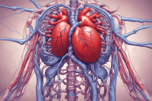
Pulmonary Embolism (PE)
- A PE is particulate matter collection that enters venous circulation and lodges in pulmonary vessels
- Virchow's triad includes blood flow stasis, endothelial injury, and hypercoagulability
- Blood flow stasis may be caused by prolonged sitting, post-operative state, fall risk, or lower extremity fractures
- Endothelial injury examples include surgery, trauma directly injuring blood vessels
- Hypercoagulability may be due to cancer, pregnancy, sickle cell disease, or polycythemia
Causes of PE
- Thrombus: blood clot (most common)
- Fat: femur fracture
- Air: improper IV priming or air bubbles from IM injections
- Oil: used for diagnostic testing
- Tumor cells
- Amniotic fluid and fetal debris
- Foreign objects: broken IV catheters
- Injected particles: mannitol
- Infected clots: from dirty or infected IV needles
Risk Factors for PE
- Prolonged immobilization
- Surgery, hip or knee replacement, trauma
- Estrogen therapy (birth control and hormone replacement)
- General and genetic conditions that increase blood clotting
- CVC, HF, and stroke
- Pregnancy up to 3 months postpartum
- Obesity
- Advancing age
- COVID-19, infection, cancer
- Smoking
- History of thromboembolism
- Atrial fibrillation (A Fib) due to stagnant blood flow
Prevention of PE
- Lifestyle changes (modifiable risk factors) can help prevent PE
- Smoking cessation is recommended, especially when using hormone replacement therapy (HRT). Nicotine increases the risk of clot formation leading to vasoconstriction
- Weight reduction is advised
- Increased physical activity is encouraged
- Drink enough water when traveling
- Change positions often while traveling
- Avoid crossing legs
- Get up from sitting position 5 minutes per hour
- Retrievable IVC filters are used for surgical patients at high risk where it prevents DVT from becoming PE as it blocks clots from moving
- Filters areplaced before any surgery requiring decreased mobility for a few days, and retrieved once the patient can walk around
- Often done for patients who cannot take VTE prophylaxis due to a history of HIT
- Those taking OCP should not smoke; OCPs are not preferred
- Prevent infections such as COVID
- Anticoagulant use for VTE prophylaxis decreases VTE by 50-80%
Clinical Probability (Wells Criteria)
- ≤4 = PE is unlikely
-
4 = PE is likely
- Signs and symptoms of DVT, extremity swelling, pain, discoloration, extremity warmth are 3 points
- Strong suspicion of PE, other diagnoses ruled out or less likely is 3 points
- Heart rate >100 is 1.5 points
- Immobilization for 3+ days or recent surgery (within 4 weeks) is 1.5 points
- History of DVT or PE is 1.5 points
- Hemoptysis is 1 point
- Malignancy is 1 point
- A high score is >6. A moderate score is 2-6. A low score is <2
- For simplified Wells criteria PE likely is >4 and PE unlikely is <4
Patient Safety for VTE
- Start passive and active range-of-motion exercises for the extremities of immobilized and postoperative patients
- Ambulate patients as soon as possible after surgery
- Use pneumatic compression devices after surgery as ordered
- Evaluate patients for the need of anticoagulant therapy
- Administer ordered prophylactic low-dose anticoagulant or antiplatelet drugs after specific surgical procedures as soon as surgical bleeding risk has subsided
- Teach patients to avoid tight garters, girdles, and constricting clothing
- Prevent pressure under the popliteal space by not placing pillows under the knees and using alternating pressure mattress
- Perform a comprehensive assessment of peripheral circulation at least once a shift
- Elevate the affected limb 20 degrees or more above the level of the heart to improve venous return, as appropriate
- Reposition the patient every 2 hours, or ambulate as tolerated
- Refrain from massaging leg muscles
- Instruct patients not to cross their legs
- Encourage smoking cessation
Key Features for PE and Lab Results
- Sudden onset of dyspnea
- Sharp, stabbing chest pain
- Apprehension, restlessness
- Feeling of impending doom
- Light-headedness
- Cough
- Hemoptysis
- Diaphoresis
- Tachypnea
- Crackles
- Pleural friction rub
- Tachycardia
- S3 or S4 heart sounds
- Fever, low grade
- Petechiae over chest and axillae
- Hypoxemia
- Tachypnea and dyspnea are early symptoms. Hypoxemia and hypoxia are late symptoms and does not occur in all patients
- Cough is dry or productive
- Pleuritic friction rub involves sharp, stabbing pain on inspiration (chest pain)
- Hemoptysis is blood cough which does not occur in all patients
- Tachycardia and hypoTN are the result of reduced tissue perfusion from pulmonary HTN
- Extra breath sounds may include clear, crackles, wheeze, or plural friction rub
- History is important to determine the cause of PE
- ABGs will be consistent with respiratory alkalosis then other changes will occur and hyperoxygenating due to tachypnea, causing low CO2
- Metabolic panel to rule out other causes of those symptoms, kidney, liver, potassium
- Troponin is elevated in MI
- BNP elevated in HF (occurs with ventricular stretch)
- D-dimer is elevated in the presence of a clot and used to rule out PE
- aPTT indicates Heparin
- Computed Tomography pulmonary angiography (CTPA) for diagnosis: can actually look at vessels and visualize clots and can rule out other mimicking conditions
Nursing Interventions for PE
- Call rapid response (RRT) for any sudden onset dyspnea and chest pain
- Apply O2 by nasal cannula or face mask if SpO2 <90%
- Raise HOB to promote lung expansion
- Reassure patient and communicate plan of action
- Apply tele monitoring and continuous pulse ox, obtain venous access
- Prepare for ABG measurement and other diagnostic testing
- Check VS q30min-2hr depending on pt, cardiac & resp assessment
- Assess chest for petechiae
- Administer anticoagulants of fibrinolytic therapy to prevent further clot formation
- Start Heparin right away because it acts quickly. This is used for 5-10 days
- Initiate Warfarin on day 1-2 of heparin & will be used concurrently. Takes several days to reach therapeutic levels
- Continue on oral anticoagulants following discharge for at least 3-6 wks or indefinitely depending on ongoing risk
- Institute bleeding precautions
PE Severity and Treatment Options
- Massive PE (high risk):
- Less than 5% of cases
- Mortality may be as high as 65%; most deaths occur due to shock within the first hour
- In severe instances: severe hypotension (SBP <90 mm Hg for at least 15 minutes), cardiac arrest, severe bradycardia, shock, RV dysfunction, distress
- CPR; inotropic or vasopressor support, intravenous fluids, systemic thrombolysis, catheter-directed treatment, surgical embolectomy, and a Vena cava filter may be used
- Submassive PE (intermediate risk):
- Mortality is 5%-15%
- Normotension, RV dysfunction, R bundle branch block, ST elevation or depression, T-wave inversion, elevated BNP or troponin
- Treatment is controversial however thrombolytic therapy surgical reperfusion strategy is an option
- Anticoagulation is optimal
- A Vena cava filter may be used also
- Nonmassive PE (low risk):
- Mortality <1%
- Normotension and no RV dysfunction, and no elevation in BNP or troponin
- Fibrinolytics not warranted
- LMWH, DOAC, Warfarin and Vena cava filter may be used
- Inpatient hospitalization not usually needed
- Age >80; HR > 110; Hx of cancer; systolic BP < 100; O2 < 90; Hx of CPD
PE Treatment
- Embolectomy: if fibrinolytic therapy cannot be used if the patient has a massive or multiple PE with shock or bleeding
- Percutaneous catheter directed therapy: used when there's contraindication to tPa to inject directly into blot
- IVC Filter: used over drugs in pts w bleeding on anticoags, have septic PE, and are ongoing surgical embolectomy
- Pharmacologic: anticoagulants (Heparin, Warfarin, DOACs) and thrombolytics
- Vasopressors (Epi/norepi/dopamine) for severe bradycardic/hypotensive patients. Positive inotropes (Dobutamine) to support cardiac function
- Non-pharmacologic: CPR if patient is in cardiac arrest; Oxygen therapy via nasal cannula or face mask; elevationg HOB
- Bleeding precaution; Home care management with lifestyle modifications (smoking cessation, weight management, activity)
Managing Complications from PE
- HypoTN occurs due to inadequate circulation in the left ventricle as results from pulmonary HTN which causes reduced forward blood flow -Increase CO and maintain BP. Administer fluids and medications -Vasopressors when hypoTN persists despite fluid resuscitation (Norepinephrine, epinephrine, dopamine) -Less vital organs suffer -Positive inotropes (dobutamine, milrinone) increase cardiac contractility with monitoring -Monitor for indicators of fluid adequacy – urine output, skin turgor, moisture of mucous membranes
- Bleeding: Ensure antidotes are always present with bleeding precautions -Ensuring correct dosage and timing of drug therapy and Assess CBC & coags -Monitor for signs of bruising/bleeding, occult blood, abdomen distention/firmness -Low platelets can suggest HIT due to the formation of anti-heparin antibodies which may cause a need for platelet transfusion
- Precaution for thrombocytopenia: Platelet <20,000 = spontaneous, uncontrolled bleeding & intracranial hemorrhage
- Avoid IM & venipunctures, and postpone procedures if PLT <50,000
- Don't forcefully blow nose, no enemas, soft bristle toothbrush and firm soles in shoes
Therapeutic Response to PE treatment
- Attains and maintains adequate gas exchange and oxygenation
- No hypovolemia and shock
- Remains free from bleeding episodes
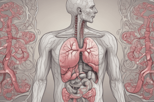
Acute Respiratory Distress Syndrome (ARDS)
- ARDS causes ARF and refractory hypoxemia
- Hypoxemia persists with 100% O2 and the vent cannot force perfusion with PaO2 still low. Decreased pulmonary compliance and Dyspnea is a hallmark of resp failure
- Bilateral pulmonary compliance NOT related to HF where an echo will show if it's not related to HF
- Dense pulmonary infiltrates on X-rays
- Direct injury comes from direct injuries to the lung (pneumonia/COVID, aspiration, trauma, fat embolisms) and indirect injuries causes inflammation such as sepsis and shock along with burns, pancreatitis, and other injuries with release of inflammatory properties
ARDS patient at risk
- Patient with direct and indirect injury
- On tube feeding and have impaired swallowing causing aspiration
- Increased WOB
- Hyperpnea: deeper breaths than normal NOT related to rate
- Noisy respiration: cyanosis, pallor, retraction intercostally of substernally
- Lower partial pressure of aterial O2 even with Remain is low even on 100% oxygen or low PaO2/FiO2 ratio
- sputum and ground glass will show on chest X-rays
Pathophysiology of ARDS
- Triggered by systemic inflammatory response
- Alveolar capillary membrane leaks separating air in lungs from blood circulation
- End result is impaired gas exchange from thick exudate
Three phases of ARDS
- first week is exudative with dyspnea, fluid may build, surfactant is reduced, and refractory hypoxemia develops
- Next 2nd week is fibrosing alveolitis for the repair by lungs and systemic inflammation and hypertension can lead to MODS,
- The lung attempts to have Epithelia restoration can lead to poor pulmonary defects
Interventions for ARDS
- Intubation with mechanical ventilation PEEP or CPAP; monitor for tension pneumothorax
- Frequent Lung Sounds assesment
- Positioning: Prone and turn pt each 2 hrs
- Drug and Therapy is to give supportive care and Conservative FLuid care to prevent excess fluid in the lungs
- Provide nutrition to reduce respirator muscle function and tube feedings ASAP
Mechanical Ventilation in ARDS patients
- Maintains patent airway, removing secretions, ventilation
- High presure can force air in lungs; must establish airway
- used for patients that are Progressive alveolar with hyperventilation or after a Surgery
- Explain procedure and have equipment ready with positioning as golden Standards
Mechanical Ventilation Indications
- PTS need Intubation or tracheostomy
- hypoxemia Progressive alveolar with hyperventilation
- chest injury or head injury
- ards and temporary surgeries
Complication with ventilator
- hypotension , fluid retention, lung problems
- GI issue. Vent dependent and needs muscle deconditioning
- Nursing Care is ventilator bundle and patient education
Key medications for respiratory issues
- TNK and other fibrinolytic, with a monitor and ICU consideration
- Heparin: monitor and patient education
Acute Coronary Syndrome (ACS)
- CAD affects the coronary arteries becoming blocked and leading to ischemia or infarction
- Ischemia is insufficient O2 supply and leads to a NSTEMI while Infarction is cell death and can lead to a STEMI
Risk factors for CAD
- High LDL, Low HDL
- Obesity
- HTN, carbon minoxide
- Sedentary or genetics
- Exercise goals for prevention or 150 min and diets low in fats and sodium
Types of Agina
- Stable: short term chest pain with reduced symptoms and is treated with rest and nitroglycerin
- UNstable: exercise does not help and the EKG is present
- Myocardial INfarction is determined with an EKG where STEMI is 100% blockage
Signs and symptoms of Myocardial Infarction
- Angina is chest pain
- MI consist of pain in jaw and lasting more than 30 min
Diagnostic for MI and Angina
- History of pain and Physical such as EKG, VS and S3 heart assessment
- Troponin levels T and I
- Stress or echoes are needed to measure activity in the heart rate
How to diagnosis a MI
- CHEST xray and blood test
- Cardiac Cath removes clots
Key interventions for MI and Agina
- Managing angina
- Nitro 2 times and then morphine can help but watch out for lung changes
- Oxygenation is needed along with decreasing afterload
Drug therapy needed are all side effective
- APlattelets, aspirin and beta blockers should reduce Heart rate with daily administration
Additional Interventions After MI
- One is prescribed ACE w/in 20 hrs to reduce Vas constriction
- Check Potassium Levels
- Reduce blood thickerness with stats an fibrin
The THromb20
- THrombolytics, Heprin, RAAS
- oxygen, morphine
- beta blockers statins and salicylate
Use of Pci needs quick implementation
- 90 min standard to insert the stain and prevent closure with EKG assesment
After care includes assessing the MI with shock
- S and s are VS change and undiagonsed check is HIGH
- Non and vas is needed for a right or left heart failure and can use pulmonary intervention for the Swan catheter
Cardiac Care
- Prevent Hypo Tn With graft rewarming with infection control
- Asses pericar and watch for low BP
Cardiac issues
- alter and infect the heart
- Key signs are Chest pressure, Friction and wbc and EKH
- Monitor BP
- treat pain, and treat infection if precent
Shock
- The lack of flow to vitals
- Stages are hypovolemic with loss of blood and leads to fluid admn
- Next is cardiogenic where the pump fails and will need Vas support
- Obstructive is pressure on the heart/ vassel
Anphlatci Showl
- This leads to constrict and swelling so needs epi and can give cpr with intubabtion
Septic issue
- With infection the system is affected
- monitor VS AND Give meds and cool
Interventions with the different shock
- Check Blood
- support Vas pressors and air
Important meds to remember
- EPI is one need if anaphylactic like
Studying That Suits You
Use AI to generate personalized quizzes and flashcards to suit your learning preferences.