Podcast
Questions and Answers
A patient with a pulmonary embolism (PE) is experiencing hypoxemia. Which mechanism primarily contributes to this reduced blood oxygen level?
A patient with a pulmonary embolism (PE) is experiencing hypoxemia. Which mechanism primarily contributes to this reduced blood oxygen level?
- Increased alveolar ventilation compensating for reduced perfusion.
- Shunting of blood from the left side of the heart to the right side.
- Excessive carbon dioxide retention causing respiratory acidosis.
- Dead space ventilation due to obstructed pulmonary blood flow. (correct)
Following a pulmonary embolism, a patient's inflammatory mediators have altered their lung surfactant. How does this alteration affect the patient's respiratory status?
Following a pulmonary embolism, a patient's inflammatory mediators have altered their lung surfactant. How does this alteration affect the patient's respiratory status?
- Increased lung compliance, resulting in respiratory acidosis.
- Increased lung recoil, leading to respiratory acidosis.
- Decreased lung compliance, potentially leading to respiratory alkalosis. (correct)
- Decreased lung recoil, potentially leading to respiratory alkalosis.
In a patient experiencing a pulmonary embolism, the release of serotonin contributes to which specific pathophysiological response?
In a patient experiencing a pulmonary embolism, the release of serotonin contributes to which specific pathophysiological response?
- Increased production of surfactant, improving alveolar gas exchange.
- Constriction of the pulmonary vasculature, reducing blood flow to the lungs. (correct)
- Decreased heart rate, reducing the workload on the right ventricle.
- Dilation of the pulmonary vasculature, increasing blood flow to the lungs.
A patient with a large pulmonary embolism is developing right ventricular dilation. Which of the following best explains the mechanism behind this complication?
A patient with a large pulmonary embolism is developing right ventricular dilation. Which of the following best explains the mechanism behind this complication?
A patient diagnosed with a pulmonary embolism is experiencing a ventilation-perfusion (V/Q) mismatch. Which statement accurately describes the underlying problem?
A patient diagnosed with a pulmonary embolism is experiencing a ventilation-perfusion (V/Q) mismatch. Which statement accurately describes the underlying problem?
A patient post-surgery develops a deep vein thrombosis and subsequent pulmonary embolism. Which of the following is the MOST important immediate physiological consequence of the embolism?
A patient post-surgery develops a deep vein thrombosis and subsequent pulmonary embolism. Which of the following is the MOST important immediate physiological consequence of the embolism?
A pulmonary embolism results in dead space ventilation. Which of the following scenarios BEST describes this condition?
A pulmonary embolism results in dead space ventilation. Which of the following scenarios BEST describes this condition?
During assessment of a patient with a suspected pulmonary embolism, the nurse anticipates which acid-base imbalance as a likely early finding?
During assessment of a patient with a suspected pulmonary embolism, the nurse anticipates which acid-base imbalance as a likely early finding?
Which of the following conditions is directly associated with the development of shunts in the pulmonary system?
Which of the following conditions is directly associated with the development of shunts in the pulmonary system?
A patient presents with a large embolism blocking the bifurcation of the main pulmonary artery. What specific type of pulmonary embolism is this?
A patient presents with a large embolism blocking the bifurcation of the main pulmonary artery. What specific type of pulmonary embolism is this?
Why does a large pulmonary embolism lead to systemic hypotension and hemodynamic instability?
Why does a large pulmonary embolism lead to systemic hypotension and hemodynamic instability?
According to Virchow's triad, which of the following factors contribute(s) to the risk of pulmonary embolism?
According to Virchow's triad, which of the following factors contribute(s) to the risk of pulmonary embolism?
A patient is diagnosed with venous thromboembolism (VTE). What does this indicate about the patient's condition?
A patient is diagnosed with venous thromboembolism (VTE). What does this indicate about the patient's condition?
A patient with suspected PE is hemodynamically unstable. Which diagnostic imaging should be performed immediately?
A patient with suspected PE is hemodynamically unstable. Which diagnostic imaging should be performed immediately?
Which of the following arterial blood gas (ABG) findings is most consistent with a patient experiencing a pulmonary embolism (PE)?
Which of the following arterial blood gas (ABG) findings is most consistent with a patient experiencing a pulmonary embolism (PE)?
Elevated troponin levels are detected in a patient suspected of a pulmonary embolism. What is the clinical significance of this finding?
Elevated troponin levels are detected in a patient suspected of a pulmonary embolism. What is the clinical significance of this finding?
A patient with a confirmed pulmonary embolism has an oxygen saturation below 90%. What is the initial treatment approach?
A patient with a confirmed pulmonary embolism has an oxygen saturation below 90%. What is the initial treatment approach?
In which clinical scenario is a thrombectomy most likely to be considered for a patient with a pulmonary embolism (PE)?
In which clinical scenario is a thrombectomy most likely to be considered for a patient with a pulmonary embolism (PE)?
Flashcards
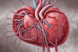
Pulmonary Embolism (PE)
Pulmonary Embolism (PE)
Blockage in the pulmonary artery, often by a blood clot.
PE's Impact on the Heart
PE's Impact on the Heart
PE blocks blood flow, increasing lung blood pressure and causing the heart to work harder.
Dead Space Ventilation
Dead Space Ventilation
Ventilation is normal, but blood flow is reduced, leading to decreased oxygen in the blood.
Hyperventilation in PE
Hyperventilation in PE
Signup and view all the flashcards
Increased Pulmonary Vascular Resistance (PVR)
Increased Pulmonary Vascular Resistance (PVR)
Signup and view all the flashcards
Ventilation-Perfusion (V/Q) Mismatch
Ventilation-Perfusion (V/Q) Mismatch
Signup and view all the flashcards
Anatomic Dead Space
Anatomic Dead Space
Signup and view all the flashcards
Shunt
Shunt
Signup and view all the flashcards
Anatomic A physiologic mismatch
Anatomic A physiologic mismatch
Signup and view all the flashcards
Saddle Embolism
Saddle Embolism
Signup and view all the flashcards
Virchow's Triad
Virchow's Triad
Signup and view all the flashcards
Pulmonary Embolism Source
Pulmonary Embolism Source
Signup and view all the flashcards
Venous Thromboembolism (VTE)
Venous Thromboembolism (VTE)
Signup and view all the flashcards
CTPA (computed tomographic pulmonary angiogram)
CTPA (computed tomographic pulmonary angiogram)
Signup and view all the flashcards
ABG Analysis in PE
ABG Analysis in PE
Signup and view all the flashcards
Troponin Levels in PE
Troponin Levels in PE
Signup and view all the flashcards
Thrombectomy
Thrombectomy
Signup and view all the flashcards
Study Notes
- Pulmonary embolism (PE) occurs when blood circulation through the pulmonary artery is blocked by a thrombus or other emboli.
- PE is a life-threatening emergency.
- Blood clots causing PE often originate from deep vein thrombosis (DVT).
- A blood clot obstructs blood supply to the lungs, increasing lung blood pressure and causing the heart to work harder, resulting in ventilation-perfusion mismatch.
Ventilation-Perfusion (V/Q) Mismatch
- When alveolar ventilation remains constant but perfusion of the capillary bed decreases, it leads to dead space ventilation and hypoxemia
- Serotonin is released, causing pulmonary vasculature contraction and reduced blood flow, hyperventilation and altered lung surfactant lead to hypocapnia and respiratory alkalosis.
- Increased pulmonary vascular resistance (PVR) raises right ventricular afterload, impairing right ventricular outflow and causing right ventricular dilation
V/Q Mismatch Types
- PE can create a V/Q mismatch due to the embolus obstructing the blood vessel
- V/Q mismatch manifestations: headache, fatigue, dizziness, shortness of breath, and wheezing
- Two classifications of V/Q mismatch: dead space and shunt.
Dead Space V/Q Mismatch
- Ventilation is adequate, but perfusion inadequate; therefore oxygen cannot enter the bloodstream
- Anatomic dead space is the area in the lung with oxygen but no blood flow
- Physiologic mismatch is when the alveoli have oxygen but blood flow is inadequate to move the oxygen
- PE is a common cause of dead space.
- V/Q mismatch is treatable with oxygen.
Shunt
- There is adequate blood flow (perfusion) but inadequate ventilation
- Relative: small amount of ventilation
- Absolute: no ventilation
- Pneumonia and pulmonary edema are conditions associated with shunts.
Saddle Embolism
- A large embolism blocks the bifurcation of the main pulmonary artery.
- Compromised cardiac output results from increased pulmonary vascular resistance increasing right ventricular afterload, impairing right ventricular outflow and causing right ventricular dilation.
- Decreased outflow from the right ventricle leads to reduced filling of the left ventricle and a reduction of cardiac output causing systemic hypotension and hemodynamic instability.
- Right ventricular failure is the main cause of death associated with pulmonary embolism.
Virchow's Triad
- Three main factors increase the risk of pulmonary embolism: hypercoagulability, vessel wall injury, and venous stasis.
- Most emboli arise from the lower extremity proximal veins (iliac, femoral, or popliteal veins).
- If a client has a pulmonary embolism and a DVT, the client is diagnosed with venous thromboembolism (VTE).
Pulmonary Embolism Risk and Diagnosis
- Can be asymptomatic
- Low-risk PE
- Intermediate risk or submassive PE
- High-risk or massive PE (saddle)
- Diagnostic tests include laboratory value assessment and diagnostic imaging.
- Bedside imaging or ultrasound may be used when the client is hemodynamically unstable
- Computed tomographic pulmonary angiogram (CTPA) is the diagnostic test of choice for a suspected PE allowing the pulmonary arteries to be visualized.
Diagnosis via Laboratory Assessment
- ABG analysis: respiratory alkalosis and hypocapnia are revealed.
- Troponin levels elevated in 30-50% of clients with moderate to large PE indicates a possibility of a severe outcome or death.
- Elevated D-dimer levels indicate concurrent activation of thrombotic pathways.
- Electrocardiogram (ECG) abnormalities such as tachycardia.
- Chest x-ray is usually normal but may identify atelectasis, effusion, or other causes for dyspnea.
Treatment
- Initial treatment is supportive.
- Supplemental oxygen is used for oxygen saturation levels below 90%.
- Mechanical ventilation may be considered for unstable clients.
- Hemodynamically unstable clients may require mechanical cardiopulmonary support devices, like extracorporeal membrane oxygenation (ECMO).
- Thrombectomy is used to remove clots if the client is unstable and thrombolysis is ineffective
Studying That Suits You
Use AI to generate personalized quizzes and flashcards to suit your learning preferences.