Podcast
Questions and Answers
What are the main functions of the nose?
What are the main functions of the nose?
- Provides an airway for respiration, moisturizes and warms air, filters inhaled air, and is a sensory organ for smell
- Provides an airway for respiration, moisturizes and warms air, filters inhaled air, and is a resonating chamber for singing
- Provides an airway for respiration, lubricates and cools air, filters inhaled air, and is a resonating chamber for speech
- Provides an airway for respiration, moisturizes and warms air, filters inhaled air, and is a resonating chamber for speech (correct)
What is the function of the carina in the respiratory system?
What is the function of the carina in the respiratory system?
- It helps regulate airflow to the lungs
- It marks the point where the trachea divides into two primary bronchi (correct)
- It prevents food from entering the trachea during swallowing
- It produces mucus to trap foreign particles in the airways
What is the primary function of the bronchioles in the respiratory system?
What is the primary function of the bronchioles in the respiratory system?
To carry air to the alveoli
Alveoli are tiny air sacs in the lungs where gas exchange occurs.
Alveoli are tiny air sacs in the lungs where gas exchange occurs.
What is the role of the diaphragm in the respiratory system?
What is the role of the diaphragm in the respiratory system?
Which of these muscles are considered accessory respiratory muscles that are typically only used when the body needs to process energy quickly?
Which of these muscles are considered accessory respiratory muscles that are typically only used when the body needs to process energy quickly?
Quiet exhalation is a passive process, whereas forced exhalation is an active process.
Quiet exhalation is a passive process, whereas forced exhalation is an active process.
What is the most important respiratory center in the brain?
What is the most important respiratory center in the brain?
What is the definition of vital capacity?
What is the definition of vital capacity?
What is the definition of tidal volume?
What is the definition of tidal volume?
Restrictive lung diseases make it harder to get air into the lungs.
Restrictive lung diseases make it harder to get air into the lungs.
Obstructive lung diseases make it harder to get air out of the lungs.
Obstructive lung diseases make it harder to get air out of the lungs.
What is the definition of diaphragmatic fatigue?
What is the definition of diaphragmatic fatigue?
What is the main nerve that supplies motor function to the diaphragm?
What is the main nerve that supplies motor function to the diaphragm?
The phrenic nerve originates from the fourth cervical ramus.
The phrenic nerve originates from the fourth cervical ramus.
An accessory phrenic nerve may sometimes arise from the subclavius muscle.
An accessory phrenic nerve may sometimes arise from the subclavius muscle.
The diaphragm is composed of 55% type I fibers, 20% type IIa fibers, and 25% type IIb fibers.
The diaphragm is composed of 55% type I fibers, 20% type IIa fibers, and 25% type IIb fibers.
What is the natural stimulus for the stretch reflex in the diaphragm?
What is the natural stimulus for the stretch reflex in the diaphragm?
The diaphragm is the major muscle of respiration.
The diaphragm is the major muscle of respiration.
The diaphragm is a flat, thin muscle.
The diaphragm is a flat, thin muscle.
The diaphragm's contraction decreases intrapleural pressure.
The diaphragm's contraction decreases intrapleural pressure.
The diaphragm's contraction expands the rib cage through its zone of apposition by generating positive intra-abdominal pressure.
The diaphragm's contraction expands the rib cage through its zone of apposition by generating positive intra-abdominal pressure.
The diaphragm's contraction expands the rib cage using the abdomen as a fulcrum.
The diaphragm's contraction expands the rib cage using the abdomen as a fulcrum.
The diaphragm is not involved in actions like laughing, singing, coughing, and sneezing.
The diaphragm is not involved in actions like laughing, singing, coughing, and sneezing.
The diaphragm is responsible for approximately 75% of the tidal volume.
The diaphragm is responsible for approximately 75% of the tidal volume.
Muscle spindles are definitely found in the human diaphragm.
Muscle spindles are definitely found in the human diaphragm.
The diaphragm is not involved in maintaining functional residual capacity.
The diaphragm is not involved in maintaining functional residual capacity.
The diaphragm's length is determined by lung volume and chest wall configuration.
The diaphragm's length is determined by lung volume and chest wall configuration.
The diaphragm's primary role during exercise is to generate pressure.
The diaphragm's primary role during exercise is to generate pressure.
The diaphragm's increase in power during exercise to 70% of maximal workload is mainly due to an increase in velocity of shortening.
The diaphragm's increase in power during exercise to 70% of maximal workload is mainly due to an increase in velocity of shortening.
The phrenic nerve is the sole motor supply to the diaphragm.
The phrenic nerve is the sole motor supply to the diaphragm.
Mechanical diaphragmatic dysfunction is caused by the disruption of respiratory nerve signals.
Mechanical diaphragmatic dysfunction is caused by the disruption of respiratory nerve signals.
Diaphragmatic fatigue is defined as the inability to develop enough force to maintain the normal lung volumes.
Diaphragmatic fatigue is defined as the inability to develop enough force to maintain the normal lung volumes.
Diaphragmatic fatigue occurs when the diaphragm generates more than 40% of its maximal pressure potential during inspiration.
Diaphragmatic fatigue occurs when the diaphragm generates more than 40% of its maximal pressure potential during inspiration.
The average human diaphragm can sustain sufficient pressure for breathing for about 60 minutes.
The average human diaphragm can sustain sufficient pressure for breathing for about 60 minutes.
About 55% of the fibers within the human diaphragm are fatigue resistant, which is a slow twitch high oxidative type I.
About 55% of the fibers within the human diaphragm are fatigue resistant, which is a slow twitch high oxidative type I.
The diaphragm is not affected by blood flow changes during strenuous breathing.
The diaphragm is not affected by blood flow changes during strenuous breathing.
Respiratory muscle fatigue is generally not affected by the sensation of dyspnea.
Respiratory muscle fatigue is generally not affected by the sensation of dyspnea.
Dyspnea can only occur due to weakness of respiratory muscles.
Dyspnea can only occur due to weakness of respiratory muscles.
Respiratory muscle fatigue is not a factor that can limit exercise capacity.
Respiratory muscle fatigue is not a factor that can limit exercise capacity.
Respiratory muscle fatigue is only a factor for people with underlying respiratory diseases.
Respiratory muscle fatigue is only a factor for people with underlying respiratory diseases.
The phrenic nerve can be affected by the level of weight in the supine position.
The phrenic nerve can be affected by the level of weight in the supine position.
Flashcards
Respiratory system
Respiratory system
The system of organs involved in breathing.
Conducting zone
Conducting zone
Part of the respiratory system that carries air to the respiratory zone.
Respiratory zone
Respiratory zone
Part of the respiratory system where gas exchange occurs.
Nose
Nose
Signup and view all the flashcards
Trachea
Trachea
Signup and view all the flashcards
Bronchi
Bronchi
Signup and view all the flashcards
Primary bronchi
Primary bronchi
Signup and view all the flashcards
Secondary bronchi
Secondary bronchi
Signup and view all the flashcards
Tertiary bronchi
Tertiary bronchi
Signup and view all the flashcards
Bronchioles
Bronchioles
Signup and view all the flashcards
Terminal bronchioles
Terminal bronchioles
Signup and view all the flashcards
Respiratory bronchioles
Respiratory bronchioles
Signup and view all the flashcards
Alveolar ducts
Alveolar ducts
Signup and view all the flashcards
Alveolar sacs
Alveolar sacs
Signup and view all the flashcards
Lungs
Lungs
Signup and view all the flashcards
Diaphragm
Diaphragm
Signup and view all the flashcards
Intercostal muscles
Intercostal muscles
Signup and view all the flashcards
Inspiration
Inspiration
Signup and view all the flashcards
Expiration
Expiration
Signup and view all the flashcards
Tidal volume
Tidal volume
Signup and view all the flashcards
Total Lung Capacity
Total Lung Capacity
Signup and view all the flashcards
Vital Capacity
Vital Capacity
Signup and view all the flashcards
Functional Residual Capacity
Functional Residual Capacity
Signup and view all the flashcards
Inspiratory Capacity
Inspiratory Capacity
Signup and view all the flashcards
Residual Volume
Residual Volume
Signup and view all the flashcards
Study Notes
Overview of Anatomy and Physiology of the Pulmonary System and Thoracic Cage
- The pulmonary system involves the organs responsible for respiration.
- The thoracic cage encloses the lungs and other vital respiratory organs.
Anatomy of the Respiratory System
- The respiratory system consists of several organs: nose, mouth (oral cavity), pharynx (throat), larynx (voice box), trachea (windpipe), bronchi, and lungs.
- These organs are divided into two zones: conducting zone and respiratory zone.
Structures of the Respiratory Zone
- The respiratory zone consists of air-exchanging structures like respiratory bronchioles, alveolar ducts, and alveolar sacs.
- Alveoli are the tiny air sacs where gas exchange occurs.
- Alveoli are surrounded by capillaries for efficient gas exchange.
- The walls of alveoli consist of type I (thin) and type II (surfactant-producing) cells
The Trachea
- The trachea is a tube that descends into the mediastinum.
- It maintains an open airway due to C-shaped cartilage rings.
- The carina marks the division of the trachea into two primary bronchi.
Bronchi
- The bronchial tree starts with the primary (main) bronchi.
- Secondary (lobar) bronchi branch out from the primary bronchi, and then tertiary (segmental) bronchi branch into each lung segment.
- Bronchioles, smaller than bronchi, are further divided up into terminal bronchioles.
Gross Anatomy of the Lungs
- The major landmarks of the lungs include the apex, base, hilum, and root.
- The left lung has two lobes (superior and inferior).
- The right lung has three lobes (superior, middle, and inferior).
- There are oblique and horizontal fissures that separate the lobes.
The Pleurae
- The pleura is a double-layered sac that surrounds each lung.
- Parietal pleura lines the thoracic cavity.
- Visceral pleura adheres to the lungs.
- The pleural cavity is the potential space between the two layers.
Mechanisms of Ventilation
- Respiration involves two phases: inhalation (inspiration) and exhalation (expiration).
Inspiration (Inhalation)
- The primary muscles involved in inhalation are the diaphragm and intercostal muscles.
- The diaphragm flattens, and the intercostal muscles contract to increase the volume of the thoracic cavity.
- This decreases internal gas pressure, pulling air into the lungs.
- Accessory muscles like scalenes, sternocleidomastoid, pectoralis major, pectoralis minor, and erector spinae, assist with forceful inhalation.
Expiration (Exhalation)
- Quiet exhalation is mostly passive due to relaxation of the inspiratory muscles.
- The diaphragm moves superiorly, and the volume of the thoracic cavity decreases.
- Forced expiration, an active process, involves contracting internal and external oblique muscles and transverse abdominal muscles.
Neural Control of Ventilation
- The most crucial respiratory center is the ventral respiratory group (VRG) in the medulla oblongata.
- Neurons here generate the respiratory rhythm.
- Pontine respiratory centers in the pons refine respiratory patterns.
Lung Volumes and Capacities
- Lung volumes are the measures of air movement in and out of the lungs.
- Types of volumes include Tidal Volume (TV), Inspiratory Reserve Volume (IRV), Expiratory Reserve Volume (ERV), Residual Volume (RV).
- Lung capacities are combinations of volumes. Examples include Total Lung Capacity (TLC), Vital Capacity (VC), Functional Residual Capacity (FRC), Inspiratory Capacity (IC).
Respiratory Diseases
- Restrictive diseases hinder inhalation, decreasing vital capacity and lung volumes.
- Examples include fibrosis, muscular diseases, chest wall deformities, pleural effusion, and pneumothorax.
- Obstructive diseases impair exhalation, leading to reduced vital capacity but increased residual volume.
- Examples include emphysema, chronic bronchitis, and asthma.
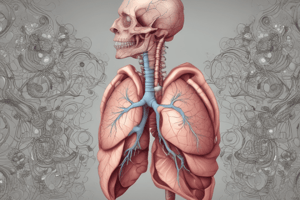
The Diaphragm
-
The diaphragm is a major respiratory muscle crucial for normal breathing.
-
It's a dome-shaped, musculotendinous septum separating the thoracic and abdominal cavities.
-
It accounts for a significant portion (approximately 75%) of tidal volume.
-
Its contraction decreases intrapleural pressure and expands the rib cage.
-
Important functions include blowing air, speaking, singing, whistling, crying, yawning, coughing, sneezing, vomiting, urination, and childbirth.
-
Diaphragm muscle spindles are present, impacting its endurance.
Phrenic Nerve
- The phrenic nerve is the sole motor nerve supplying the diaphragm.
- It arises from the fourth cervical ramus and is part of the cervical plexus.
- Accessory phrenic nerves sometimes arise from C5 and C6.
- Damage or interruption of the phrenic nerve can impede respiratory function.
Diaphragmatic Fatigue
- Diaphragmatic fatigue can lead to difficulties maintaining normal lung volumes due to lactic acid accumulation.
- This ultimately results in shortness of breath (dyspnea).
- Fatigue can also stem from high breathing load and weakness in the respiratory muscles.
Important Characteristics of the Diaphragm
- The diaphragm primarily contains high oxidative slow twitch fibers (Type I), high oxidative, rapid twitch fibers (Type IIa) and low oxidative, rapid twitch fibers, (Type IIb).
Studying That Suits You
Use AI to generate personalized quizzes and flashcards to suit your learning preferences.