Podcast
Questions and Answers
Which nerve provides cutaneous innervation to the lateral ankle and foot?
Which nerve provides cutaneous innervation to the lateral ankle and foot?
- Saphenous nerve
- Sural nerve (correct)
- Tibial nerve
- Common fibular nerve
The soleus muscle crosses both the knee and ankle joints.
The soleus muscle crosses both the knee and ankle joints.
False (B)
What is the anatomical term for the big toe?
What is the anatomical term for the big toe?
hallux
The calcaneal tendon, also known as the ______ tendon, is formed by the tendons of the gastrocnemius, soleus, and plantaris muscles.
The calcaneal tendon, also known as the ______ tendon, is formed by the tendons of the gastrocnemius, soleus, and plantaris muscles.
Match the calf muscles with their primary action:
Match the calf muscles with their primary action:
The popliteal artery branches into which two arteries?
The popliteal artery branches into which two arteries?
The small saphenous vein drains directly into the femoral vein.
The small saphenous vein drains directly into the femoral vein.
Which muscle in the deep layer of the calf helps to unlock the knee joint?
Which muscle in the deep layer of the calf helps to unlock the knee joint?
The tibial nerve innervates all the muscles of the ______ leg.
The tibial nerve innervates all the muscles of the ______ leg.
Which muscle attaches primarily to the fibula?
Which muscle attaches primarily to the fibula?
Flashcards
Posterior Leg
Posterior Leg
The anatomical term for the calf, the region between the knee and the ankle.
Hallux
Hallux
Also known as the great toe, located on the medial side of the leg.
Small Saphenous Vein
Small Saphenous Vein
A superficial vein that runs along the posterior calf, draining into deeper veins near the popliteal fossa and often implicated in varicose veins.
Sural Nerve
Sural Nerve
Signup and view all the flashcards
Gastrocnemius
Gastrocnemius
Signup and view all the flashcards
Soleus
Soleus
Signup and view all the flashcards
Calcaneal Tendon (Achilles Tendon)
Calcaneal Tendon (Achilles Tendon)
Signup and view all the flashcards
Sciatic Nerve
Sciatic Nerve
Signup and view all the flashcards
Popliteus
Popliteus
Signup and view all the flashcards
Popliteal Artery
Popliteal Artery
Signup and view all the flashcards
Study Notes
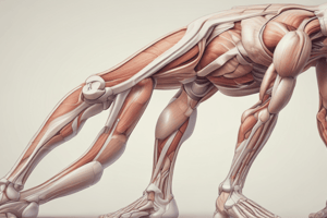
Overview of the Posterior Leg Anatomy
- The posterior leg, or calf, lies between the knee and the ankle.
- Calf anatomy includes multiple layers of muscles, nerves, arteries, and veins.
- A 100km ultra-marathon from Bath to Chilcompton is planned to raise money for Macmillan Cancer Support.
- Donations can be made via a Just Giving page (link in description).
Anatomical Terminology
- The model is of a left leg.
- The big toe is the great toe or hallux.
- Structures related to the big toe are "hallucis."
- Medial is the side of the leg where the big toe is.
- Lateral is the side of the leg where the little toe is.
- Leg is the region between the knee and the ankle.
- The lower limb is compartmentalized by fascia, with focus on the posterior compartment.
Superficial Structures
- The small (or short) saphenous vein runs along the posterior calf as a superficial vein.
- The small saphenous vein drains into deeper veins near the popliteal fossa.
- Calf varicose veins often stem from issues in the small saphenous vein's valves.
- The sural nerve accompanies the small saphenous vein, providing cutaneous innervation.
- The sural nerve innervates the lateral ankle and foot skin.
Superficial Muscles
- The gastrocnemius is the most prominent calf muscle, featuring lateral and medial heads.
- The gastrocnemius heads connect to the femur's lateral and medial condyles.
- The calcaneal tendon (Achilles tendon) comprises the tendons of the gastrocnemius, soleus, and plantaris muscles.
- The Achilles tendon is often injured by runners.
- The soleus muscle is located under the gastrocnemius.
- The soleus originates from the tibia and fibula.
- The gastrocnemius spans the knee and ankle joints.
- The soleus only spans the ankle joint.
- The gastrocnemius and soleus facilitate plantar flexion.
- The plantaris is a small muscle in the posterior leg's superficial layer.
- The plantaris crosses both the knee and ankle joints.
- The tibial nerve innervates all three superficial muscles.
Nerves
- The sciatic nerve splits into the tibial and common fibular nerves.
- The tibial nerve runs through the popliteal fossa, innervating all posterior leg muscles.
- The sural nerve branches from the tibial nerve, emerging between the heads of the gastrocnemius.
Deep Layer of Muscles
- The deep calf layer contains four muscles: popliteus, flexor hallucis longus, flexor digitorum longus, and tibialis posterior.
- The popliteus unlocks the knee joint and starts knee flexion.
- The popliteus runs from the tibia to the lateral femoral condyle.
- The flexor hallucis longus flexes the big toe.
- The flexor digitorum longus flexes the other four toes.
- Tibialis posterior is the third muscle in the deep layer.
- The flexor hallucis longus primarily attaches to the fibula.
- The flexor digitorum longus primarily attaches to the tibia.
- The tibialis posterior attaches to both the tibia and fibula, and the interosseous membrane.
- The tendons of these three muscles curve medially around the medial malleolus of the tibia.
- The tibialis posterior inserts into the medial foot bones, providing foot stabilization and inversion.
- Eccentric loading of the tibialis posterior supports the medial arch during walking and running.
Arteries and Veins
- Deep veins are in the lower limb, blood clots can form if venous blood flow is impaired.
- The popliteal artery, from the femoral artery, traverses the popliteal fossa.
- The popliteal artery branches into the anterior tibial artery and the tibioperoneal trunk.
- The anterior tibial artery moves to the leg's anterior compartment.
- The tibioperoneal trunk divides into the fibular and posterior tibial arteries.
- The fibular artery runs laterally to the fibula.
- The posterior tibial artery descends the leg and passes behind the medial malleolus.
Studying That Suits You
Use AI to generate personalized quizzes and flashcards to suit your learning preferences.