Podcast
Questions and Answers
Which artery is primarily responsible for the blood supply to the dorsum of the foot?
Which artery is primarily responsible for the blood supply to the dorsum of the foot?
- Femoral artery
- Anterior tibial artery (correct)
- Common iliac artery
- Posterior tibial artery
What is the role of the anastomoses between the gluteal and femoral circumflex arteries?
What is the role of the anastomoses between the gluteal and femoral circumflex arteries?
- To connect superficial veins to deep veins
- To drain blood from the lower limb into the abdominal aorta
- To provide collateral circulation if the femoral trunk is blocked (correct)
- To facilitate lymphatic drainage from the lower limb
Which vein is responsible for draining the lateral aspect of the foot and leg?
Which vein is responsible for draining the lateral aspect of the foot and leg?
- Anterior tibial vein
- Popliteal vein
- Short saphenous vein (correct)
- Long saphenous vein
What type of veins travel in pairs alongside smaller arteries in the lower limb?
What type of veins travel in pairs alongside smaller arteries in the lower limb?
Which structure is formed by the medial and lateral plantar arteries?
Which structure is formed by the medial and lateral plantar arteries?
Which muscle lies deep to the gastrocnemius?
Which muscle lies deep to the gastrocnemius?
What is the innervation of the gastrocnemius muscle?
What is the innervation of the gastrocnemius muscle?
Which of the following actions is NOT associated with the gastrocnemius muscle?
Which of the following actions is NOT associated with the gastrocnemius muscle?
What are the two sub-compartments of the posterior compartment of the leg?
What are the two sub-compartments of the posterior compartment of the leg?
Which of the following is a function of the soleus muscle?
Which of the following is a function of the soleus muscle?
What is the primary action of the popliteus muscle?
What is the primary action of the popliteus muscle?
Which muscle is primarily responsible for foot inversion?
Which muscle is primarily responsible for foot inversion?
Which of the following muscles does NOT insert into the achilles tendon?
Which of the following muscles does NOT insert into the achilles tendon?
What is the innervation for the flexor hallucis longus?
What is the innervation for the flexor hallucis longus?
Which muscle forms the floor of the popliteal fossa?
Which muscle forms the floor of the popliteal fossa?
Which anatomical structure does NOT pass under the flexor retinaculum?
Which anatomical structure does NOT pass under the flexor retinaculum?
What nerve provides the muscular branches to the tibialis posterior?
What nerve provides the muscular branches to the tibialis posterior?
Which artery is the primary source of blood supply to the lower limb?
Which artery is the primary source of blood supply to the lower limb?
What is the primary function of the venous drainage system in the lower limb?
What is the primary function of the venous drainage system in the lower limb?
Which nerve is responsible for supplying the medial malleolus?
Which nerve is responsible for supplying the medial malleolus?
What occurs when veins become incompetent and blood pools in the extremities?
What occurs when veins become incompetent and blood pools in the extremities?
Where does the common peroneal nerve wrap around in the lower limb?
Where does the common peroneal nerve wrap around in the lower limb?
Which statement accurately describes the lymphatic drainage system of the lower limb?
Which statement accurately describes the lymphatic drainage system of the lower limb?
What does the sural nerve primarily supply?
What does the sural nerve primarily supply?
What does 'The big 3' refer to in the context of nerves in the lower limb?
What does 'The big 3' refer to in the context of nerves in the lower limb?
What might occur due to prolonged pooling of blood in the extremities?
What might occur due to prolonged pooling of blood in the extremities?
Flashcards
What muscles form the triceps surae?
What muscles form the triceps surae?
The gastrocnemius and soleus muscles together form the triceps surae, a powerful plantar flexor of the foot.
Where does the gastrocnemius muscle originate?
Where does the gastrocnemius muscle originate?
The gastrocnemius muscle originates from the medial and lateral femoral condyles, crossing both the knee and ankle joints.
What is the role of the soleus muscle in walking?
What is the role of the soleus muscle in walking?
The soleus muscle, located deep to the gastrocnemius, plays a key role in plantarflexion during walking, working during the push-off phase.
What nerve innervates the gastrocnemius and soleus?
What nerve innervates the gastrocnemius and soleus?
Signup and view all the flashcards
What is the origin and insertion of the plantaris muscle?
What is the origin and insertion of the plantaris muscle?
Signup and view all the flashcards
Anterior and Posterior tibial arteries
Anterior and Posterior tibial arteries
Signup and view all the flashcards
Anastomoses in the lower limb
Anastomoses in the lower limb
Signup and view all the flashcards
Superficial veins (lower limb)
Superficial veins (lower limb)
Signup and view all the flashcards
Deep veins (lower limb)
Deep veins (lower limb)
Signup and view all the flashcards
Communicating veins (lower limb)
Communicating veins (lower limb)
Signup and view all the flashcards
Popliteus muscle
Popliteus muscle
Signup and view all the flashcards
Flexor Hallucis Longus (FHL)
Flexor Hallucis Longus (FHL)
Signup and view all the flashcards
Flexor Digitorum Longus (FDL)
Flexor Digitorum Longus (FDL)
Signup and view all the flashcards
Tibialis Posterior (TP)
Tibialis Posterior (TP)
Signup and view all the flashcards
Structures under Flexor Retinaculum
Structures under Flexor Retinaculum
Signup and view all the flashcards
Tibial Nerve
Tibial Nerve
Signup and view all the flashcards
Femoral Artery
Femoral Artery
Signup and view all the flashcards
Profunda Femoris Artery
Profunda Femoris Artery
Signup and view all the flashcards
Deep Venous Drainage of the Lower Limb
Deep Venous Drainage of the Lower Limb
Signup and view all the flashcards
Varicose Veins
Varicose Veins
Signup and view all the flashcards
Lymphatic Drainage of the Lower Limb
Lymphatic Drainage of the Lower Limb
Signup and view all the flashcards
Sciatic Nerve
Sciatic Nerve
Signup and view all the flashcards
Common Peroneal Nerve
Common Peroneal Nerve
Signup and view all the flashcards
Sural Nerve
Sural Nerve
Signup and view all the flashcards
Femoral Nerve
Femoral Nerve
Signup and view all the flashcards
Study Notes
Posterior Leg
- The lecture focused on the posterior leg, including a review quiz up to the current content and a review of nerves and vessels of the lower extremity.
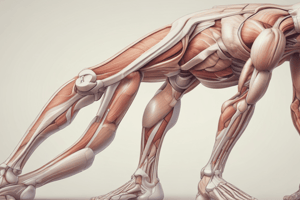
- Images of the posterior leg, highlighting bones, muscles, and nerves, were presented.
- The posterior compartment is divided into superficial and deep sub-compartments.
- Superficial compartment muscles include gastrocnemius, soleus, and plantaris.
- Gastrocnemius (two heads) and soleus comprise the triceps surae.
- Deep compartment muscles include popliteus, flexor hallucis longus (FHL), flexor digitorum longus (FDL), and tibialis posterior (TP).
Gastrocnemius
- Originates from the medial and lateral femoral condyles (above the knee joint).
- The two heads merge at the mid-leg.
- Inserts into the tendo Achilles (tendo calcaneus).
- Innervated by the tibial nerve.
- Action involves foot plantar flexion and knee flexion.
Soleus
- Lies deep to the gastrocnemius.
- Originates from the head and upper one-third of the fibula and the soleal line.
- Inserts into the tendo Achilles.
- Innervated by the tibial nerve.
- Action is plantarflexion of the foot; active in gait during loading to toe-off.
Plantaris
- A small muscle of little functional importance.
- Originates from the lateral supracondylar line (above the knee joint).
- Inserts into the Achilles tendon.
- Action is a very weak knee flexor and plantarflexor.
Deep Posterior Compartment
- Muscles: popliteus, flexor hallucis longus (FHL), flexor digitorum longus (FDL), tibialis posterior (TP).
- Popliteus forms the floor of the popliteal fossa.
- Origin/insertion: lateral condyle of the femur/lateral meniscus -> soleal line of the posterior tibia.
- Innervation: tibial nerve.
- Action: medial/lateral rotation of the knee joint.
Flexor Hallucis Longus
- Origin/insertion: lower two-thirds of the fibula/intermuscular septum--> wraps around the medial malleolus--> passes between the sesamoids (under the 1st MTPJ) --> inserts into the distal hallux phalanx.
- Innervation: tibial nerve.
- Action: 1st MTPJ/IPJ flexion and ankle joint flexion.
Flexor Digitorum Longus
- Origin/insertion: posterior tibia --> wraps around the medial malleolus under flexor retinaculum --> divides into four tendons --> insert into the distal phalanx of the lesser four toes.
- Innervation: tibial nerve.
- Action: plantarflexes the lesser four phalanges and foot; stabilises toes on ground during gait.
Tibialis Posterior
- Origin/insertion: interosseous membrane and tibia/fibula --> flexor retinaculum --> navicular tuberosity and all tarsal bones except talus.
- Nerve supply: tibial nerve.
- Action: foot inversion and plantarflexion.
- Pathology: weakness/rupture --> progressively pronated foot.
Structures under Flexor Retinaculum
- Tom, Dick and very naughty Harry: TP, FDL, Art, Vein, Nerve, and FHL (anterior-to-posterior order)
Nerve Supply: Tibial Nerve
- Larger terminal branch of the sciatic nerve.
- Passes deep to the triceps surae, wraps behind the medial malleolus.
- Divides into medial and lateral plantar nerves; muscular, cutaneous, and articular branches.
Nerve Supply: Common Peroneal Nerve
- Wraps around the lateral neck of the fibula.
- Supplies the knee joint (articular) and skin on posterior and postero-lateral aspects of the leg.
- Divides into superficial and deep peroneal nerves.
- Superficial peroneal n.: lateral compartment of leg; anterior ankle and dorsum of foot (except 1st webspace).
- Deep peroneal n.: enters anterior compartment, moves under extensor retinaculae; supplies skin (1st webspace).
Nerve Supply: Sural Nerve
- . Formed by branches of tibial and common peroneal nerves.
- Runs down the posterior calf with the short saphenous vein.
- Supplies skin on the posterior calf, heel, and lateral foot.
Femoral & Obturator Nerves
- Femoral nerve enters the inguinal ligament, sartorius, and the adductor canal.
- Motor branches supply anterior compartment muscles.
- Sensory branches supply skin over anterior and medial thigh; saphenous nerve supplies a strip of skin medially to the 1st MTPJ
- Obturator nerve passes through the obturator foramen.
- Supplies muscles of the thigh adductor compartment, and overlying skin (HJ & KJ).
Blood Supply to the Lower Limb
- External iliac --> femoral artery (enters femoral triangle) --> profunda femoris (medial & lateral circumflex and perforating a.) --> (adductor canal & adductor hiatus) --> popliteal artery --> anterior and posterior tibial arteries --> medial/lateral malleolar branches --> (AJ) dorsalis pedis --> posterior tibial --> medial and lateral plantar arteries.
- Abdominal aorta --> common iliac a. --> External iliac a. --> superior and inferior gluteal a.
Venous Drainage of the Lower Limb
- Superficial: dorsal venous arch --> short saphenous vein --> popliteal fossa —> long saphenous vein --> femoral vein —> external iliac vein—>inferior vena cava
- Deep: two venae comitantes accompanying smaller arteries --> form popliteal vein—> adductor hiatus—> femoral vein
- Connecting these two sets by fascia lata = communicating veins.
Lymphatic Drainage of the Lower Limb
- Follows the general pattern of superficial and deep venous drainage
- Little communication between superficial and deep lymphatics
- Superficial lymph glands are near the inguinal ligament and popliteal fossa.
Summary
- Students have learned about most of the lower extremity, emphasizing the importance of connecting anatomy.
- Practical exercises on family members/friends are recommended to understand anatomical variations.
Studying That Suits You
Use AI to generate personalized quizzes and flashcards to suit your learning preferences.