Podcast
Questions and Answers
Which muscle is responsible for plantar flexion of the foot?
Which muscle is responsible for plantar flexion of the foot?
- Popliteus
- Gastrocnemius (correct)
- Tibialis posterior
- Flexor digitorum longus
Which nerve supplies the muscles in the posterior compartment of the leg?
Which nerve supplies the muscles in the posterior compartment of the leg?
- Sural nerve
- Femoral nerve
- Common peroneal nerve
- Tibial nerve (correct)
Which muscle has two heads?
Which muscle has two heads?
- Tibialis posterior
- Soleus
- Plantaris
- Gastrocnemius (correct)
What is the action of the plantaris muscle?
What is the action of the plantaris muscle?
Which muscle has a long tendon deep to the lateral head of the gastrocnemius?
Which muscle has a long tendon deep to the lateral head of the gastrocnemius?
Which muscle is responsible for flexion of the knee?
Which muscle is responsible for flexion of the knee?
How many muscles are in the superficial group of the posterior compartment of the leg?
How many muscles are in the superficial group of the posterior compartment of the leg?
Which muscle is not part of the deep group of muscles in the posterior compartment of the leg?
Which muscle is not part of the deep group of muscles in the posterior compartment of the leg?
What is the function of the popliteus muscle?
What is the function of the popliteus muscle?
Which muscle originates from the posterior surface of the proximal tibia?
Which muscle originates from the posterior surface of the proximal tibia?
What is the nerve supply of the flexor hallucis longus muscle?
What is the nerve supply of the flexor hallucis longus muscle?
What is the insertion of the tibialis posterior muscle?
What is the insertion of the tibialis posterior muscle?
Which muscle is the deepest one between the flexor hallucis longus and flexor digitorum longus?
Which muscle is the deepest one between the flexor hallucis longus and flexor digitorum longus?
What is the action of the flexor digitorum longus muscle?
What is the action of the flexor digitorum longus muscle?
What is the origin of the flexor hallucis longus muscle?
What is the origin of the flexor hallucis longus muscle?
What is the terminal branch of the popliteal artery that is larger and more direct?
What is the terminal branch of the popliteal artery that is larger and more direct?
What is the attachment point of the Flexor Retinaculum above?
What is the attachment point of the Flexor Retinaculum above?
Which muscle is responsible for hip flexion?
Which muscle is responsible for hip flexion?
What is the spinal level associated with knee extension?
What is the spinal level associated with knee extension?
What is the purpose of tapping the patella tendon with a tendon hammer?
What is the purpose of tapping the patella tendon with a tendon hammer?
What is the structure that the Flexor Retinaculum is continuous with below?
What is the structure that the Flexor Retinaculum is continuous with below?
What is the muscle responsible for ankle dorsiflexion?
What is the muscle responsible for ankle dorsiflexion?
What is the spinal level associated with ankle plantarflexion?
What is the spinal level associated with ankle plantarflexion?
What is the purpose of examining muscle groups during a neurological examination?
What is the purpose of examining muscle groups during a neurological examination?
Where does the posterior tibial artery begin?
Where does the posterior tibial artery begin?
What is the largest and most important branch of the posterior tibial artery?
What is the largest and most important branch of the posterior tibial artery?
What is the primary function of the tibial nerve?
What is the primary function of the tibial nerve?
What are the two cutaneous branches of the tibial nerve?
What are the two cutaneous branches of the tibial nerve?
What is the origin of the sural nerve?
What is the origin of the sural nerve?
What is the primary region of skin supplied by the sural nerve?
What is the primary region of skin supplied by the sural nerve?
What forms the tarsal tunnel?
What forms the tarsal tunnel?
What is the terminal branch of the posterior tibial artery?
What is the terminal branch of the posterior tibial artery?
Study Notes
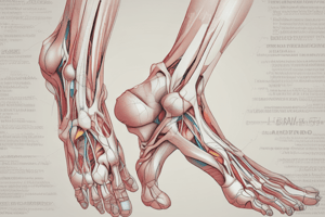
Posterior Compartment of the Leg
- Consists of muscles, blood vessels, and nerves
- Muscles:
- Organized into two groups: superficial and deep
- Blood Vessels:
- Posterior tibial vessels
- Nerves:
- Tibial nerve
Superficial Group
- Three muscles:
- Gastrocnemius: large muscle with two heads
- Plantaris: small muscle with long tendon deep to lateral head of gastrocnemius
- Soleus: large flat muscle under the gastrocnemius
Deep Group
- Four muscles:
- Popliteus: smallest and most superior of the deep muscles
- Flexor hallucis longus: located inferolaterally
- Flexor digitorum longus: located medially
- Tibialis posterior: deepest one, between FHL and FDL
Muscles and Their Actions
- Gastrocnemius: plantar flexes foot and flexes knee
- Plantaris: assists in plantar flexion
- Soleus: plantar flexes foot
- Popliteus: unlocks knee joint
- Flexor hallucis longus: flexes great toe
- Flexor digitorum longus: flexes lateral four toes
- Tibialis posterior: inverts and plantar flexes foot, supports medial arch of foot during walking
Posterior Tibial Artery
- Larger and more direct terminal branch of the popliteal artery
- Descends on the superficial surfaces of the tibialis posterior and flexor digitorum longus muscles
- Passes through the tarsal tunnel behind the medial malleolus and into the sole of the foot
- Ends by dividing into medial and lateral plantar arteries
- Branches:
- Muscular
- Nutrient
- Circumflex fibular artery
- Fibular artery
- Medial and lateral plantar arteries
Fibular Artery
- Largest and most important branch of the tibial artery
- Arises inferior to the distal border of the popliteus
- Branches:
- Muscular
- Nutrient artery of the fibula
- Perforating branches
- Terminal lateral malleolar and calcaneal branches
Tibial Nerve
- Major branch of the sciatic nerve that descends into the posterior compartment
- Leaves the posterior compartment of the leg at the ankle by passing through the tarsal tunnel behind the medial malleolus
- Enters the foot to supply most intrinsic muscles and skin
- Branches:
- Muscular
- Two cutaneous branches: sural nerve and medial calcaneal nerve
Sural Nerve
- Originates high in the leg by union of the medial sural cutaneous nerve and sural communicating branch of the common fibular nerve
- Descends superficial to the gastrocnemius muscle
- Supplies skin on the lower posterolateral surface of the leg and the lateral side of the foot and little toe
Tarsal Tunnel
- Formed on the posteromedial side of the ankle
- Formed by the medial malleolus, the medial and posterior surfaces of the talus, and the medial surface of the calcaneus
- Overlaying flexor retinaculum
Flexor Retinaculum
- Strap-like layer of connective tissue
- Attaches above to the medial malleolus and below and behind to the inferomedial margin of the calcaneus
- Continuous above with the deep fascia of the leg and below with deep fascia of the foot
- Structures deep to it from medial to lateral are:
- Tendon of tibialis posterior
- Tendon of flexor digitorum longus
- Posterior tibial artery, vein, and nerve
- Tendon of the flexor hallucis longus
Studying That Suits You
Use AI to generate personalized quizzes and flashcards to suit your learning preferences.
Description
This quiz covers the muscles, blood vessels, and nerves of the posterior compartment of the leg, including the superficial and deep muscle groups.