Podcast
Questions and Answers
Which nerve innervates all the posterior forearm muscles?
Which nerve innervates all the posterior forearm muscles?
- Ulnar nerve
- Median nerve
- Musculocutaneous nerve
- Radial nerve (correct)
Which muscle is not part of the superficial layer of the posterior forearm muscles?
Which muscle is not part of the superficial layer of the posterior forearm muscles?
- Extensor Digitorum
- Supinator (correct)
- Brachioradialis
- Extensor Carpi Radialis brevis
Which structure forms the dorsal digital expansion in the posterior forearm?
Which structure forms the dorsal digital expansion in the posterior forearm?
- Tendons of extensor muscles (correct)
- Radial artery
- Median nerve
- Tendons of flexor muscles
Which of these muscles attaches to proximal lateral surface of radius?
Which of these muscles attaches to proximal lateral surface of radius?
Which condition is caused by forceful and repeated bending of the wrist and fingers?
Which condition is caused by forceful and repeated bending of the wrist and fingers?
Which nerve is related to the anatomic snuff box?
Which nerve is related to the anatomic snuff box?
Which muscle does the deep branch of radial nerve travel under?
Which muscle does the deep branch of radial nerve travel under?
Which movement is affected by radial nerve injury?
Which movement is affected by radial nerve injury?
Which aspect of the shoulder experiences sensory deficits due to radial nerve injury?
Which aspect of the shoulder experiences sensory deficits due to radial nerve injury?
Which of these muscle is mostly responsible for locking the elbow?
Which of these muscle is mostly responsible for locking the elbow?
Which nerve innervates the superficial layer muscles of the posterior forearm?
Which nerve innervates the superficial layer muscles of the posterior forearm?
Which condition is caused by overstrain, overuse, or direct impact on the common extensor tendon?
Which condition is caused by overstrain, overuse, or direct impact on the common extensor tendon?
Which nerve innervates the thenar muscles of the hand?
Which nerve innervates the thenar muscles of the hand?
Which muscle is not part of the hypothenar muscles of the hand?
Which muscle is not part of the hypothenar muscles of the hand?
Where do the dorsal interosseous muscles insert?
Where do the dorsal interosseous muscles insert?
Which nerve innervates the hypothenar muscles of the hand?
Which nerve innervates the hypothenar muscles of the hand?
Where do the palmar interosseous muscles originate?
Where do the palmar interosseous muscles originate?
Which muscle originates from the tendons of the flexor digitorum profundus of each finger?
Which muscle originates from the tendons of the flexor digitorum profundus of each finger?
Which muscle is not part of the central group of intrinsic hand muscles?
Which muscle is not part of the central group of intrinsic hand muscles?
Which part of the hand is innervated by the ulnar nerve?
Which part of the hand is innervated by the ulnar nerve?
How many tendons does the carpal tunnel house?
How many tendons does the carpal tunnel house?
Which of these groups of muscles are capable of doing the "Ta-Ta" movement"? (flexion of MCP + extension of PIP & DIP)
Which of these groups of muscles are capable of doing the "Ta-Ta" movement"? (flexion of MCP + extension of PIP & DIP)
What is the direction of the carpal arch, forming the carpal tunnel?
What is the direction of the carpal arch, forming the carpal tunnel?
Extensor carpi radialis and Flexor carpi radialis are synergists for _______ of the wrist
Extensor carpi radialis and Flexor carpi radialis are synergists for _______ of the wrist
Which of the following muscles originate from the lateral supracondylar ridge of humerus?
Which of the following muscles originate from the lateral supracondylar ridge of humerus?
Extensor carpi radialis brevis (ECRB) attaches to base of 2nd metacarpal
Extensor carpi radialis brevis (ECRB) attaches to base of 2nd metacarpal
Both brachioradialis and ECRL originate from the ______
Both brachioradialis and ECRL originate from the ______
Extensor digiti minimi attaches to....
Extensor digiti minimi attaches to....
Dorsal digital expansions pull PIPs and DIPs into complete ________ (hint: Ta-Ta position)
Dorsal digital expansions pull PIPs and DIPs into complete ________ (hint: Ta-Ta position)
Extensor carpi ulnaris attaches to base of _____ metacarpal
Extensor carpi ulnaris attaches to base of _____ metacarpal
Where does the Aconeus muscle insert?
Where does the Aconeus muscle insert?
Supinator originates from lateral epicondyle and the ________ ________ of the ulna
Supinator originates from lateral epicondyle and the ________ ________ of the ulna
Abductor policis longus can extend the thumb at the CMC joint
Abductor policis longus can extend the thumb at the CMC joint
Extensor policis brevis provides one of the tendons that forms the ______ border of the anatomical snuff box (hint: medial or lateral)
Extensor policis brevis provides one of the tendons that forms the ______ border of the anatomical snuff box (hint: medial or lateral)
Extensor policis longus inserts onto......
Extensor policis longus inserts onto......
Which of these actions are carried out by Extensor policis longus?
Which of these actions are carried out by Extensor policis longus?
Extensor indicis originates from...
Extensor indicis originates from...
Abductor pollicis brevis originates from ....
Abductor pollicis brevis originates from ....
Abductor policis brevis and flexor policis brevis both insert onto the lateral side of the base of ________
Abductor policis brevis and flexor policis brevis both insert onto the lateral side of the base of ________
Oponens policis inserts onto the first metacarpal
Oponens policis inserts onto the first metacarpal
Palmar interosseous 1 originates from metacarpal 1
Palmar interosseous 1 originates from metacarpal 1
Palmar interosseous 2 inserts onto finger #_____
Palmar interosseous 2 inserts onto finger #_____
Lumbricals, Dorsal Interosseous, and Palmar interosseous all insert onto the dorsal digital expansions
Lumbricals, Dorsal Interosseous, and Palmar interosseous all insert onto the dorsal digital expansions
Dorsal interosseous #2 inserts onto...
Dorsal interosseous #2 inserts onto...
Which of these groups is innervated by both the ulnar nerve and the median nerve?
Which of these groups is innervated by both the ulnar nerve and the median nerve?
Oblique head of Adductor policis originates from....
Oblique head of Adductor policis originates from....
Both heads of Adductor Policis insert onto the ______ of the thumb
Both heads of Adductor Policis insert onto the ______ of the thumb
Which of these muscles originates from hook of hamate?
Which of these muscles originates from hook of hamate?
Both Flexor digiti minimi brevis and Opponens digiti minimi have the same origins
Both Flexor digiti minimi brevis and Opponens digiti minimi have the same origins
All of the hypothenar muscles act on the pinky
All of the hypothenar muscles act on the pinky
Both Opponens Pollicis and Flexor Pollicis brevis originate from the ______ and the trapezium
Both Opponens Pollicis and Flexor Pollicis brevis originate from the ______ and the trapezium
Which of these muscles originates from the pisiform?
Which of these muscles originates from the pisiform?
Flashcards are hidden until you start studying
Study Notes
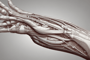
Posterior Forearm Muscles and Clinical Correlations
- Posterior forearm muscles are all innervated by the radial nerve and originate from C5, C6, C7, C8, and T1 roots.
- The superficial layer muscles include Brachioradialis (BR), Extensor Carpi Radialis Brevis (ECRB), Extensor Digitorum (ED), Flexor Carpi Radialis Longus (ECRL), Extensor Digiti Minimi (EDM), Extensor Carpi Ulnaris, and Anconeus.
- The deep layer includes Supinator, Abductor Pollicis Longus (APL), Extensor Pollicis Brevis (EPB), Extensor Pollicis Longus (EPL), and Extensor Indicis (EI).
- The dorsal digital expansion (extensor hood) is formed by extensor tendons, allowing them to work together and individually.
- Supinator originates from the supinator crest of the ulna and has two heads with a relationship with the deep branch of the radial nerve.
- Anatomic snuff box contains tendons of APL, EPB, and EPL and is related to the radial artery.
- Medial epicondylitis (golf elbow) is caused by forceful and repeated bending of the wrist and fingers, leading to tiny ruptures of the common flexor tendon.
- Lateral epicondylitis (tennis elbow) is caused by overstrain, overuse, or direct impact on the common extensor tendon.
- Radial nerve injury leads to damage to the posterior cord of the brachial plexus, affecting muscles such as deltoid, teres minor, triceps, and forearm muscles, resulting in weakness and sensory deficits.
- The clinical correlation of radial nerve injury includes weakness in abduction and external rotation of the shoulder, loss of extension in the elbow, wrist, and fingers, and sensory deficits in the lateral aspect of the shoulder and posterior forearm.
- The text provides detailed information about the origin, insertion, function, and innervation of each muscle and their clinical implications.
- The text emphasizes the importance of understanding the innervation and function of the posterior forearm muscles in clinical practice and the potential impact of nerve injuries on muscle function and sensation.
Anatomy and Function of Intrinsic Muscles of the Hand
- The hand is divided into three parts: wrist (carpus), hand proper (metacarpus), and fingers (digits)
- The long axis of the thumb has a 90° medial rotation to the rest of the fingers, resulting in movements of the thumb being at right angles to the movements of other fingers
- The carpal arch is directed anteriorly, forming the carpal tunnel via attachment of the flexor retinaculum to its sides
- The carpal tunnel houses 9 tendons (4 FDS, 4 FDP, 1 FPL) and the median nerve, and an enlargement can lead to carpal tunnel syndrome
- The flexor carpi radialis tendon does not pass through the carpal tunnel, only the flexor retinaculum
- The intrinsic muscles of the hand consist of the thenar, hypothenar, and central muscles
- The thenar muscles, innervated by the median nerve, include the abductor pollicis brevis, flexor pollicis brevis, and opponens pollicis
- The hypothenar muscles, innervated by the ulnar nerve, consist of the abductor digiti minimi, flexor digiti minimi brevis manus, and opponens digiti minimi
- The central group of intrinsic hand muscles includes the adductor pollicis, lumbricals, dorsal interosseous muscles, and palmar interosseous muscles
- The lumbricals originate from the tendons of the flexor digitorum profundus of each finger and insert into the dorsal digital expansion of the medial fingers
- The dorsal interosseous muscles originate from the metacarpals and insert into the base of the proximal phalanges and dorsal digital expansion of the fingers
- The palmar interosseous muscles originate from the anterior side of the metacarpals and insert into the base of the proximal phalanges and dorsal digital expansion of the fingers, facilitating flexion, extension, and adduction of the fingers
Studying That Suits You
Use AI to generate personalized quizzes and flashcards to suit your learning preferences.


